. 2012;7(11):e50023.
doi: 10.1371/journal.pone.0050023. Epub 2012 Nov 14.

The WSTF-ISWI chromatin remodeling complex transiently associates with the human inactive X chromosome during late S-phase prior to BRCA1 and γ-H2AX

Affiliations

The WSTF-ISWI chromatin remodeling complex transiently associates with the human inactive X chromosome during late S-phase prior to BRCA1 and γ-H2AX

Ashley E Culver-Cochran et al. PLoS One. 2012.

Abstract

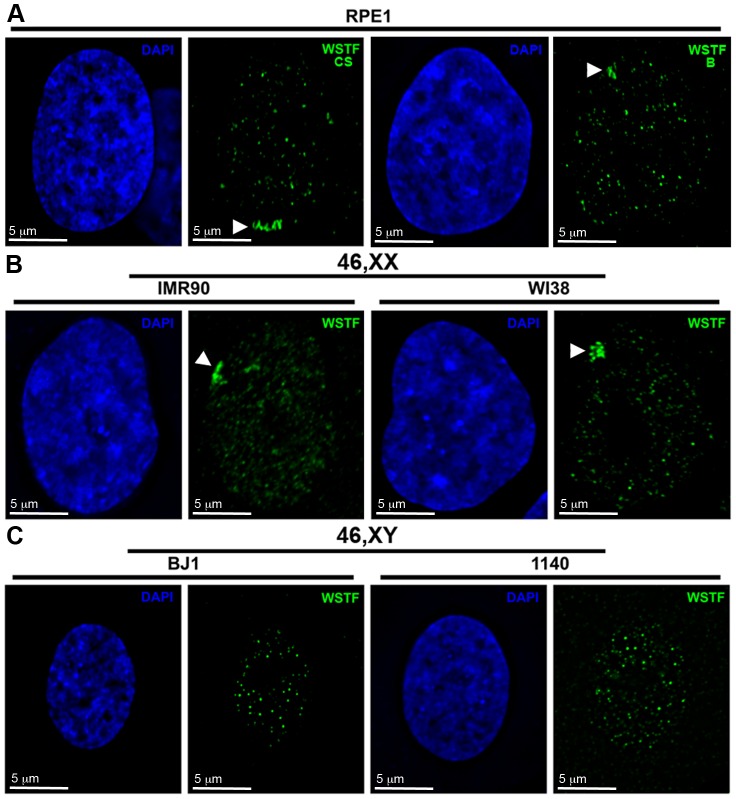
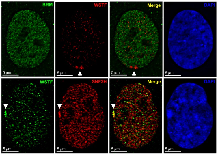
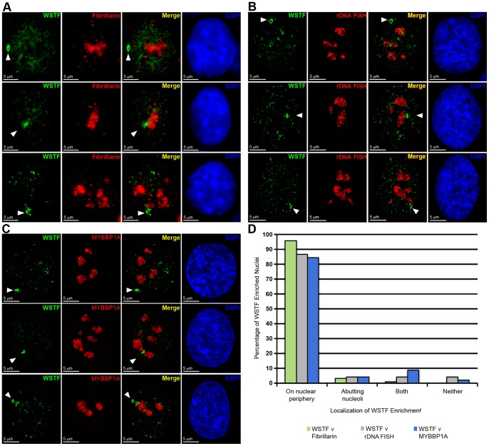
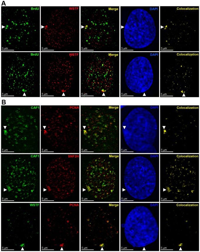
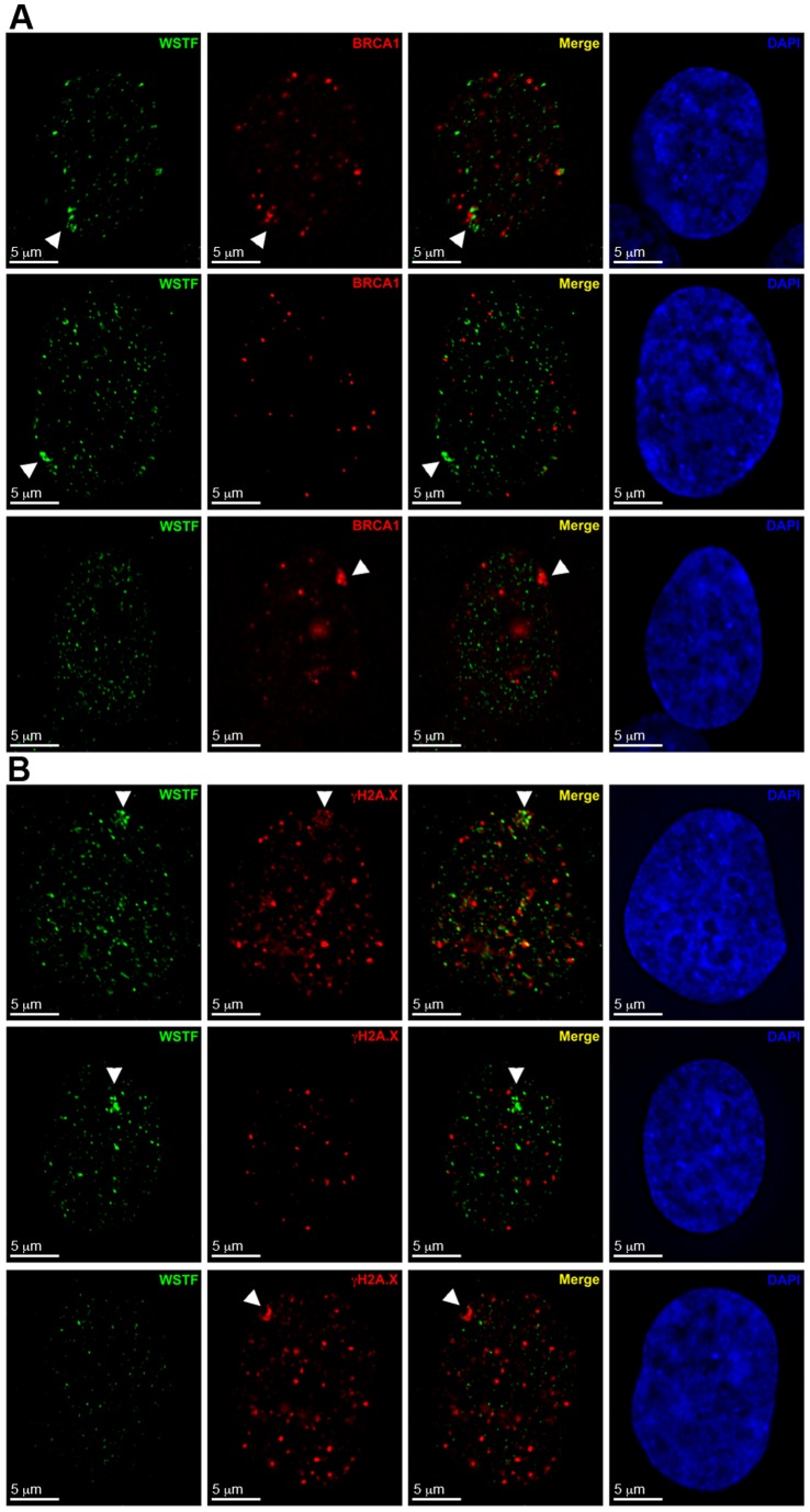
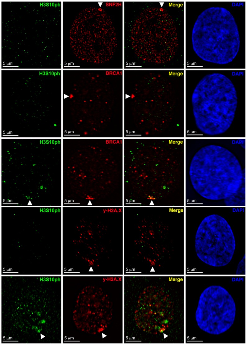
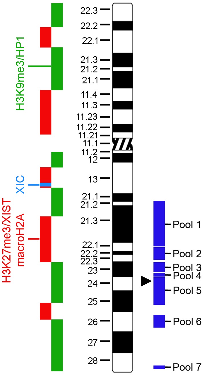
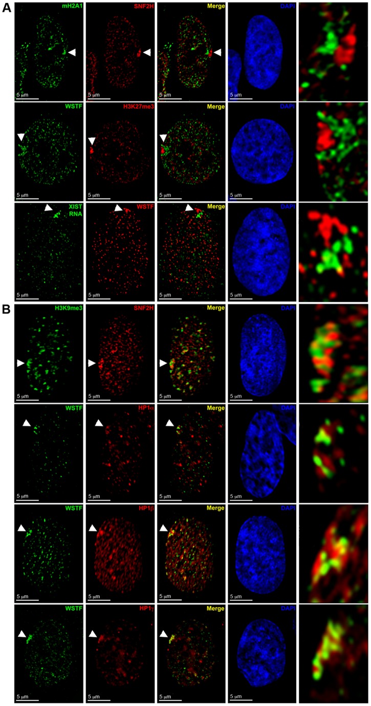
Replicating the genome prior to each somatic cell division not only requires precise duplication of the genetic information, but also accurately reestablishing the epigenetic signatures that instruct how the genetic material is to be interpreted in the daughter cells. The mammalian inactive X chromosome (Xi), which is faithfully inherited in a silent state in each daughter cell, provides an excellent model of epigenetic regulation. While much is known about the early stages of X chromosome inactivation, much less is understood with regards to retaining the Xi chromatin through somatic cell division. Here we report that the WSTF-ISWI chromatin remodeling complex (WICH) associates with the Xi during late S-phase as the Xi DNA is replicated. Elevated levels of WICH at the Xi is restricted to late S-phase and appears before BRCA1 and γ-H2A.X. The sequential appearance of WICH and BRCA1/γ-H2A.X implicate each as performing important but distinct roles in the maturation and maintenance of heterochromatin at the Xi.


Conflict of interest statement

Competing Interests: BPC serves as an academic editor for PLoS ONE. This does not alter our adherence to all the PLoS ONE policies on sharing data and materials.

Figures

Figure 1
Figure 1. WSTF is enriched in the vicinity of the Barr body in female nuclei with no comparable region of enrichment in male nuclei.
A Representative hTERT-RPE1 46,XX nuclei showing WSTF enrichment by indirect immunofluorescence with anti-WSTF from Cell Signaling Technology (CS; Green, left image) or anti-WSTF from Bethyl Laboratories (B; Green, right image). White arrowheads point to the area of enrichment. Nuclei counterstained with DAPI (Blue). B Representative female (top row; IMR90 and WI38) and male (bottom row, hTERT-BJ1 and 1140) nuclei showing WSTF distribution (Cell Signaling anti-WSTF, green). White arrowheads point to areas of enrichment. Nuclei counterstained with DAPI (Blue).
Figure 2
Figure 2. WSTF associates with the Xi as part of either WICH or B-WICH and not the WINAC complex.
Top row: Indirect immunofluorescent image of hTERT-RPE1 nucleus showing the distribution of BRM (Green) and WSTF (Red). Bottom row: Indirect immunofluorescent image of hTERT-RPE1 nucleus showing distribution of WSTF (Green) and SNF2H (Red). Overlapping signals appear yellow. White arrowheads point to the Xi. Nuclei are counterstained with DAPI (Blue).
Figure 3
Figure 3. WSTF associates with the Xi in the WICH complex and is most frequently observed at the nuclear periphery and not in the vicinity of nucleoli.
A hTERT-RPE1 nuclei showing WSTF enrichment (Green) and the location of nucleoli based on Fibrillarin distribution (Red) by indirect immunofluorescence. Examples show WSTF enrichment at the nuclear periphery (Top row), abutting a nucleoli (Middle row), and both peripheral and adjacent to a nucleoli (Bottom row). White arrowheads point to the Xi. Nuclei counterstained with DAPI (Blue). B Representative examples, as above, of hTERT-RPE1 nuclei showing WSTF enrichment (Green) and the location of nucleoli based on the hybridization pattern of a direct-labeled rDNA PAC probe RP5-1174A5 (Red). C Representative examples, as above, of hTERT-RPE1 nuclei showing WSTF enrichment (Green) and the location of nucleoli based on MYBBP1A distribution (Red) by indirect immunofluorescence. D Graph showing the percentage of nuclei for which the location of WSTF enrichment was comparable to one of the three examples depicted in parts A (green, n = 93 from five independent experiments), B (gray, n = 46 from two independent experiments), and C (blue, n = 45 from two independent experiments).
Figure 4
Figure 4. The WICH complex is enriched at the Xi during late S-phase as the Xi is replicated.
A hTERT-RPE1 cells showing the distribution of WSTF (Red) by indirect immunofluorescence relative to DNA replicated in the last 10 minutes indicated by BrdU incorporation detected by direct immunofluorescence (Green). White arrowheads point to the Xi. Nuclei counterstained with DAPI (Blue). The last panel in the series highlights the overlap between WSTF and BrdU (Yellow). B WSTF and SNF2H enrichment relative to the replication fork and sites of chromatin assembly, designated by PCNA and CAF1. Images show examples of indirect immunofluorescence in hTERT-RPE1 nuclei. Top row: CAF1 (Green) and PCNA (Red). Middle row: CAF1 (Green) and SNF2H (Red). Bottom row: WSTF (Green) and PCNA (Red). White arrowheads point to the Xi. Nuclei counterstained with DAPI (Blue). The last panel in the series highlights the overlap between the red and green channels (Yellow).
Figure 5
Figure 5. WICH enrichment at the Xi is spatially and temporally distinct from BRCA1 and γ-H2A.X association.
A Examples of hTERT-RPE1 nucleus showing the distribution by indirect immunofluorescence of WSTF (Green) and BRCA1 (Red). Nuclei counterstained with DAPI (Blue). White arrowheads point to the Xi. Top row: WSTF enrichment with BRCA1 enrichment. Middle row: WSTF enrichment without BRCA1 enrichment. Bottom row: both BRCA1 enrichment without WSTF. B Representative examples as above, but showing γ-H2A.X (Red) instead of BRCA1. White arrowheads point to the Xi.
Figure 6
Figure 6. WICH associates with the Xi prior to BRCA1 and γ-H2A.X.
Panels show representative images of indirect immunofluorescence to H3S10ph (Green) and SNF2H, BRCA1, or γ-H2A.X (Red) in hTERT-RPE1 nuclei. Top row: SNF2H (Red) and H3S10ph (Green). Second and third rows: BRCA1 (Red) and H3S10ph (Green). Fourth and firth rows: γ-H2A.X (Red) and H3S10ph (Green). Nuclei counterstained with DAPI (Blue). White arrowheads point to the Xi.
Figure 7
Figure 7. Ideogram of the X chromosome (Center, black and white) detailing the approximate locations of H3K9me3/HP1 (Left, green) and H3K27me3/XIST RNA/macroH2A (Left, red) territories.
The X inactivation center (XIC) is depicted in light blue (Left). The groups of BAC clones used are shown as blocks (Right, dark blue) which span the approximate location of all clones in the pool. The location of BAC clone 423G05 is indicated by the black arrowhead. The X chromosome ideogram was adapted from David Adler, University of Washington (www.pathology.washington.edu/research/cytopages/ideograms/human). For a detailed list of the BAC clones, see the Supporting Information, Table S1.
Figure 8
Figure 8. WICH enrichment at the Xi does not overlap with H3K27me3, macroH2A1 or XIST, but does show limited overlap with the HP1 and H3K9me3 territories.
A WSTF or SNF2H enrichment relative to facultative-like heterochromatin. Images show examples for the distribution of SNF2H (Red) relative to macroH2A.1 (Green, top row), and WSTF (Green) relative to H3K27me3 (Red, middle row) in hTERT-RPE1 nuclei by indirect immunofluorescence. Bottom panels show WSTF distribution by indirect immunofluorescence (Red) relative to hybridization pattern of a direct-labeled XIST probe (Green) in a hTERT-RPE1 nucleus. White arrowheads point to the Xi. Overlapping signals appear yellow. Nuclei counterstained with DAPI (Blue). The last panel in the series shows an enlarged view of the area indicated by white arrowheads. B WSTF or SNF2H enrichment relative to constitutive-like heterochromatin marks. Images show examples for the distribution of SNF2H (Red) relative to H3K9me3 (Green, top row) and WSTF (Green) relative to HP1α (Red, second row), HP1β (Red, third row) and HP1γ (Red, bottom row) in hTERT-RPE1 nuclei by indirect immunofluorescence. Overlapping signals appear yellow. Panels to the right show nuclei counterstained with DAPI (Blue). White arrowheads point to the Xi. The last panel in the series shows an enlarged view of the area indicated by white arrowheads.
Figure 9
Figure 9. WICH is enriched in the vicinity of the Xi as defined by the hypo-H3K9Ac and hypo-H3K4me2 territory of the Xi at interphase.
Representative images showing SNF2H distribution (Red) relative to H3K4me2 (Green, top row) or H3K9Ac (Green, bottom row) in hTERT-RPE1 nuclei by indirect immunofluorescence. White arrowheads point to the Xi. Nuclei were counterstained with DAPI (Blue, far right panels).
Figure 10
Figure 10. WICH does not associate with all Xi DNA equally as defined by FISH with X-linked BAC probes.
Examples show the distribution of WSTF (Red) by indirect immunofluorescence relative to the hybridization pattern of direct-labeled 423G05 BAC probe (Green) in hTERT-RPE1 nuclei. White arrowheads point to the Xi FISH signal, and white arrows point to the FISH signal on the active X (Xa). Nuclei were counterstained with DAPI (Blue).

Similar articles

Cited by

References

    1. Alabert C, Groth A (2012) Chromatin replication and epigenome maintenance. Nat Rev Mol Cell Biol 13: 153–167. - PubMed
    1. Portela A, Esteller M (2010) Epigenetic modifications and human disease. Nat Biotechnol 28: 1057–1068. - PubMed
    1. Lyon MF (1961) Gene action in the X-chromosome of the mouse (Mus musculus L.). Nature 190: 372–373. - PubMed
    1. Carrel L, Willard HF (2005) X-inactivation profile reveals extensive variability in X-linked gene expression in females. Nature 434: 400–404. - PubMed
    1. Yang F, Babak T, Shendure J, Disteche CM (2010) Global survey of escape from X inactivation by RNA-sequencing in mouse. Genome Res 20: 614–622. - PMC - PubMed

Publication types

MeSH terms