[Prognostic value of lymphangiogenesis and lymphatic vessel invasion in non-small cell lung cancer]
- PMID: 23164352
- PMCID: PMC6000040
- DOI: 10.3779/j.issn.1009-3419.2012.11.09
[Prognostic value of lymphangiogenesis and lymphatic vessel invasion in non-small cell lung cancer]
Abstract
Background and objective: Studies have shown that tumor metastasis in a variety of tumors is associated with lymphangiogenesis and lymphatic vessel invasion (LVI). Tumor metastasis is an important factor that affects the prognosis of patients. The aim of this study is to determine the prognostic value of lymphangiogenesis and LVI in non-small cell lung cancer (NSCLC).
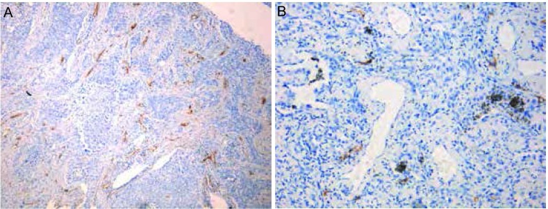
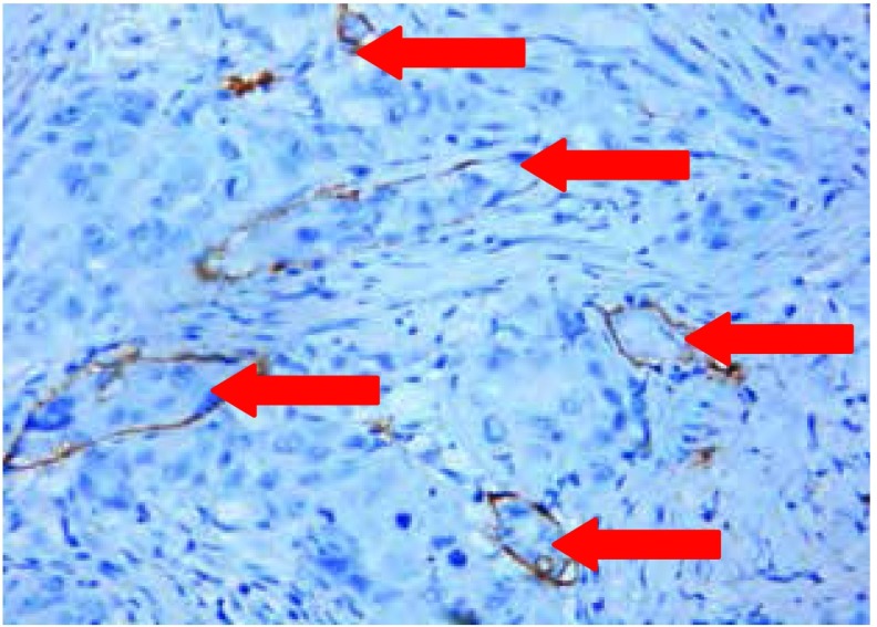
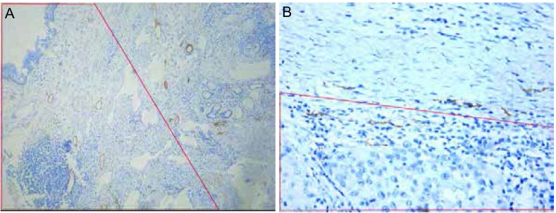
Methods: We marked the endothelial cells of lymph vessels in lymphangiogenesis with specific monoclonal antibody D2-40. Immunohistochemistry was used to detect the expression of lymphangiogenesis and LVI in 79 cases of stage I-III NSCLC.
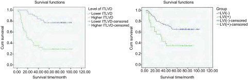
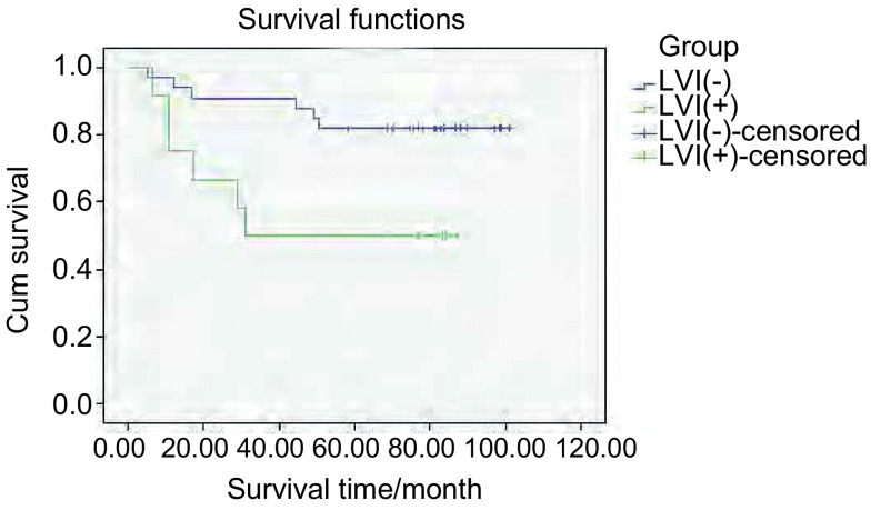
Results: The intratumoral lymphatic vessel density (ITLVD) was significantly higher in patients with N2 disease than those with N0 disease (P=0.015). The ITLVD was significantly higher in patients with LVI+ than that in those with LVI- (P = 0.009). The ITLVD was also remarkably higher in poorly differentiated tumors than that in highly differentiated ones (P = 0.007). The ITLVD was remarkably higher in adenocarcinoma than that in squamous cell carcinomas (P = 0.025). Kaplan-Meier revealed that the survival rates of patients with higher ITLVD were remarkably poorer than those with lower ITLVD (P = 0.007). Thus, the ITLVD is an important prognostic factor of NSCLC. The peritumoral lymphatic vessel density is not correlated with the prognosis.
Conclusions: The ITLVD level is an important prognostic factor of NSCLC.
背景与目的: 已有的研究表明新生淋巴管与多种肿瘤的进展和淋巴转移相关,本研究拟探讨非小细胞肺癌病灶内(旁)新生淋巴管密度及其受肿瘤侵袭状况对非小细胞肺癌的预后价值。
方法: 选择特异性单克隆抗体D2-40标记新生淋巴管内皮细胞,以免疫组化方法检测79例Ⅱ期-Ⅲ期非小细胞肺癌病灶内及其中部分病灶旁新生淋巴管表达及其受侵状况,结合患者临床、病理及随访资料,判断其对患者预后的影响和评估价值。
结果: ① 病灶内新生淋巴管密度:N2患者高于N0患者(P=0.015),新生淋巴管受侵患者明显高于未受侵患者(P=0.009),肺癌低分化明显高于高分化肺癌(P=0.007),腺癌高于鳞癌(P=0.025),患者生存率与之呈负性相关(P=0.007);②病灶旁新生淋巴管密度与预后无明显相关性;③N2肺癌病灶内新生淋巴管受侵多于N0肺癌;④病灶内和病灶旁新生淋巴管受侵患者的生存率低于未受侵患者。
结论: 病灶内新生淋巴管密度是影响非小细胞肺癌患者预后的重要因素,可作为判断预后的指标。新生淋巴管受侵的预后意义值得关注。
Figures
Similar articles
-
Detection of lymphangiogenesis in non-small cell lung cancer and its prognostic value.J Exp Clin Cancer Res. 2009 Feb 16;28(1):21. doi: 10.1186/1756-9966-28-21. J Exp Clin Cancer Res. 2009. PMID: 19216806 Free PMC article.
-
Lymphangiogenesis occurs in upper tract urothelial carcinoma and correlates with lymphatic tumour dissemination and poor prognosis.BJU Int. 2009 Apr;103(8):1040-6. doi: 10.1111/j.1464-410X.2008.08135.x. Epub 2008 Oct 17. BJU Int. 2009. PMID: 18990139
-
The lymphatic system in clinically localized urothelial carcinoma of the bladder: morphologic characteristics and predictive value.Urol Oncol. 2013 Nov;31(8):1606-14. doi: 10.1016/j.urolonc.2012.02.012. Epub 2012 Apr 13. Urol Oncol. 2013. PMID: 22503575
-
[Lymphatic extension and lymphangiogenesis in non-small cell lung cancer].Rev Pneumol Clin. 2014 Feb-Apr;70(1-2):26-31. doi: 10.1016/j.pneumo.2013.09.008. Epub 2014 Feb 22. Rev Pneumol Clin. 2014. PMID: 24566036 Review. French.
-
Blood vessel invasion as a strong independent prognostic indicator in non-small cell lung cancer: a systematic review and meta-analysis.PLoS One. 2011;6(12):e28844. doi: 10.1371/journal.pone.0028844. Epub 2011 Dec 14. PLoS One. 2011. PMID: 22194927 Free PMC article. Review.
References
-
- Weidner N. Tumor angiogenesis: review of current applications in tumor prognostication. http://med.wanfangdata.com.cn/Paper/Detail/PeriodicalPaper_PM7511250. Semin Diagn Pathol. 1993;10(4):302–313. - PubMed
Publication types
MeSH terms
LinkOut - more resources
Full Text Sources
Medical