. 2012 Nov 27;109(48):19739-44.
doi: 10.1073/pnas.1208927109. Epub 2012 Nov 12.

Tissue-resident memory CD8+ T cells continuously patrol skin epithelia to quickly recognize local antigen

Affiliations

Tissue-resident memory CD8+ T cells continuously patrol skin epithelia to quickly recognize local antigen

Silvia Ariotti et al. Proc Natl Acad Sci U S A. .

Abstract

Recent work has demonstrated that following the clearance of infection a stable population of memory T cells remains present in peripheral organs and contributes to the control of secondary infections. However, little is known about how tissue-resident memory T cells behave in situ and how they encounter newly infected target cells. Here we demonstrate that antigen-specific CD8(+) T cells that remain in skin following herpes simplex virus infection show a steady-state crawling behavior in between keratinocytes. Spatially explicit simulations of the migration of these tissue-resident memory T cells indicate that the migratory dendritic behavior of these cells allows the detection of antigen-expressing target cells in physiologically relevant time frames of minutes to hours. Furthermore, we provide direct evidence for the identification of rare antigen-expressing epithelial cells by skin-patrolling memory T cells in vivo. These data demonstrate the existence of skin patrol by memory T cells and reveal the value of this patrol in the rapid detection of renewed infections at a previously infected site.


Conflict of interest statement

The authors declare no conflict of interest.

Figures
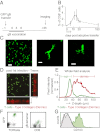
Fig. 1.
Fig. 1.
Morphology, localization, and phenotype of skin-resident memory CD8+ T cells. (A) Recipients of GFP+gB T cells (day −1) were vaccinated with a DNA vaccine encoding the gB498–505 epitope (days 0, 3, and 6) and imaged 1 mo later. (B) The percentage of GFP+gB498–505tetramer+ cells was measured in blood over time (arrows indicate vaccination timepoints). (C) Top view confocal maximum intensity projections of GFP+gB T cells infiltrating skin 1 mo after local HSVTOM infection. Images are representative of five experiments. (Scale bars: Left, 50 µm; Middle and Right, 10 µm.) (D and E) Areas of skin containing infiltrating GFP+gB T cells were imaged by intravital multiphoton microscopy 3 mo after infection; the position of the dermis was determined by exploiting second harmonic generation properties of collagen type I. Arrows indicate tissue direction, from outside to inside the animal. (F) Cell suspensions from skin biopsies 1 mo after local HSVTOM infection were analyzed for the indicated markers and GFP expression. Left plot is gated on live cells; right plot is gated on TCRβ+ live cells. GFP+ cells are depicted in green and GFP cells are depicted in black. (G) Surface expression of CD103 by TCRβ+CD8+GFP+ cells from previously infected skin (green), TCRβ+CD8+GFP+ splenocytes (gray), and TCRβ+CD8+GFP splenocytes (black line).
Fig. 2.
Fig. 2.
Steady-state, skin-resident memory CD8+ T cells migrate in between epithelial cells. (A and B) Recipients of GFP+OTI T cells (day −1) were vaccinated with a DNA vaccine encoding the Ova257–264 epitope (days 0, 3, and 6), infected with HSVTOM (day 10), and imaged at the site of HSVTOM infection 1 mo later. Top view confocal maximum intensity projections of GFP+OTI T cells infiltrating skin 1 mo after local HSVTOM infection are depicted. (C) Migration of a representative GFP+gB T cell residing in epidermis recovered from a previous HSVTOM infection; each snapshot is the superimposition of five consecutive images taken at 1-min intervals. (Scale bar, 10 μm.) (D and E, Upper) First image of a 4-h time-lapse imaging session of GFP+gB T cells (D) or GFP+ LCs cells (E) migrating through skin 1 mo after local HSVTOM infection. (Lower) Superimposition of all images from the 4-h time-lapse imaging session, indicating the area explored by GFP+gB T cells and GFP+ LCs cells during that time interval. Images are representative of two experiments. (Scale bar, 20 µm.)
Fig. 3.
Fig. 3.
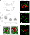
Skin-resident memory CD8+ T cells are morphologically different from γδ T cells. (A and B) Skin biopsies containing tissue-resident memory T cells were stained to detect γδ T cells and memory T cells. Two-dimensional masks of the cellular bodies were used to calculate dendrite projection angles. The angles were measured based on side projections of the single cells and calculated between two vectors starting from the cell’s center of mass, one vector being parallel to the stratum corneum, the other going through the dendrite tip, as depicted in A. The results from 16 Tmem (28 dendrites) and 18 γδ T cells (92 dendrites) are shown. Bars indicate median. (C) Representative image of a skin-resident memory CD8+ T cell and Langerhans cell with dendrites protruding sideways and upward, respectively.
Fig. 4.
Fig. 4.
Aspects of local migration and dendricity of memory T cells. (A and B) Recipients of GFP+OTI T cells (day −1) were vaccinated with a DNA vaccine encoding the Ova257–264 peptide (days 0, 3, and 6). Subsequently, mice were infected with HSVTOM at day 10 postvaccination and bystander T cells were monitored at the site of HSVTOM infection the next day (B, Upper); alternatively, mice were infected with HSVTOM at the site of former DNA vaccination at day 34 postvaccination and locally resident Tmem were monitored the next day (B, Lower). Images are confocal maximum intensity projections representative of five different experiments. (Scale bar, 50 µm.) (C) Circularity of epidermal effector T cells and skin-resident Tmem was determined from maximum intensity projections of recorded confocal Z-stacks, with a value of 1.0 reflecting a perfectly circular shape. (D) Detail of a Tmem crawling in between epithelial cells. H-2B-GFP transgenic recipients of Kaede+gB T cells were vaccinated with a DNA vaccine encoding the gB498–505 epitope and infected with HSVTOM, according to the scheme in Fig. 1A. One month postinfection, Kaede was photoconverted and imaging was performed. Top and bottom view of the same flipped Z-stack are shown. Representative of two experiments. (Scale bar, 10 µm.)
Fig. 5.
Fig. 5.
Dendricity and motility of skin-resident memory T cells allow skin patrol and are regulated by TCR triggering. (A) Simulations (CPM) of Tmem in a field of keratinocytes were performed for 4 h in “memory T-cell time” (i.e., with the in silico cells covering a similar distance as Tmem in experimental measurements). Examples of images at the final time point of simulation for dendritic-motile simulated T cells (with motility, persistence, and dendricity matched to the experimental Tmem), roundish-motile simulated T cells (with motility and persistence matched to the experimental Tmem, but with the circular appearance of effector T cells), and dendritic-still simulated T cells (nonmotile cells that extend and retract dendrites) in a field of 500 × 500 pixels. Keratinocytes contacted by simulated T cells are depicted in different shades of blue, with darker blue indicating more intense contact (based on cumulative contacted area of the skin cell). (B) The time to find antigen-expressing “targets” in silico, using either the experimentally observed densities of simulated T cells (230 T cells/field, equivalent to 430 T cells/mm2) or the indicated lower densities. (C) Time-lapse imaging of GFP+gB T cells at 9 h after peptide injection. Recipients of GFP+gB cells were infected with HSVTOM. Three months after local infection, the skin was injected with gB498–505 peptide, CMV pp65495–503 peptide, Ova257–264 peptide, or left untreated. Time-lapse imaging of the area was conducted for 30 min at 0, 3, 6, and 9 h after peptide delivery. Left: initial image of each time-lapse. (Right) Superimposition of all images comprising the 30-min time-lapse imaging session. Representative of three experiments. (Scale bar, 50 µm.) (D) Mean ± SEM circularity of the indicated Tmem was determined by imaging at the indicated time points after peptide delivery. Dashed line indicates mean circularity of skin-resident memory T cells in untreated animals (see also Fig. 3C). (E) Tmem motility, depicted as a mean square displacement ± SEM plot, as determined by imaging at 9 h after peptide delivery.
Fig. 6.
Fig. 6.
Patrolling skin-resident memory T cells can quickly identify rare targets. (A) Cell migration tracks of individual GFP+gB Tmem either in contact or not in contact with an antigen-expressing target, normalized for their origin. (B) Circularity of GFP+gB Tmem was analyzed from maximum intensity projections of individual cells. Cells were categorized based on contact with Katushka-gB498–505–expressing cells at any time point during the imaging session, derived from one experiment. Bars indicate mean ± SEM. (C) Sequential images of migrating skin-resident memory T cells that either stop upon contact with specific target (asterisk) or keep migrating without target contact (arrow).

Similar articles

Cited by

References

    1. Bevan MJ. Memory T cells as an occupying force. Eur J Immunol. 2011;41(5):1192–1195. - PMC - PubMed
    1. Gebhardt T, et al. Different patterns of peripheral migration by memory CD4+ and CD8+ T cells. Nature. 2011;477(7363):216–219. - PubMed
    1. Gebhardt T, et al. Memory T cells in nonlymphoid tissue that provide enhanced local immunity during infection with herpes simplex virus. Nat Immunol. 2009;10(5):524–530. - PubMed
    1. Wakim LM, Woodward-Davis A, Bevan MJ. Memory T cells persisting within the brain after local infection show functional adaptations to their tissue of residence. Proc Natl Acad Sci USA. 2010;107(42):17872–17879. - PMC - PubMed
    1. Masopust D, et al. Dynamic T cell migration program provides resident memory within intestinal epithelium. J Exp Med. 2010;207(3):553–564. - PMC - PubMed

Publication types