Spinal transection induces widespread proliferation of cells along the length of the spinal cord in a weakly electric fish
- PMID: 23147638
- PMCID: PMC3706082
- DOI: 10.1159/000342485
Spinal transection induces widespread proliferation of cells along the length of the spinal cord in a weakly electric fish
Abstract
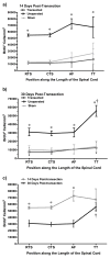
The ability to regenerate spinal cord tissue after tail amputation has been well studied in several species of teleost fish. The present study examined the proliferation and survival of cells following complete spinal cord transection rather than tail amputation in the weakly electric fish Apteronotus leptorhynchus. To quantify cell proliferation along the length of the spinal cord, fish were given a single bromodeoxyuridine (BrdU) injection immediately after spinal transection or sham surgery. Spinal transection significantly increased the density of BrdU⁺ cells along the entire length of the spinal cord at 1 day posttransection (dpt), and most newly generated cells survived up to 14 dpt. To examine longer-term survival of the newly proliferated cells, BrdU was injected for 5 days after the surgery, and fish were sacrificed at 14 or 30 dpt. Spinal transection significantly increased cell proliferation and/or survival, as indicated by an elevated density of BrdU⁺ cells in the spinal cords of spinally transected compared to sham-operated and intact fish. At 14 dpt, BrdU⁺ cells were abundant at all levels of the spinal cord. By 30 dpt, the density of BrdU⁺ cells had decreased at all levels of the spinal cord except at the tip of the tail. Thus, newly generated cells in the caudal-most segment of the spinal cord survived longer than those in more rostral segments. Our findings indicate that spinal cord transection stimulates widespread cellular proliferation; however, there were regional differences in the survival of the newly generated cells.
Copyright © 2012 S. Karger AG, Basel.
Figures


Similar articles
-
Effect of temperature on spinal cord regeneration in the weakly electric fish, Apteronotus leptorhynchus.J Comp Physiol A Neuroethol Sens Neural Behav Physiol. 2010 May;196(5):359-68. doi: 10.1007/s00359-010-0521-9. Epub 2010 Mar 26. J Comp Physiol A Neuroethol Sens Neural Behav Physiol. 2010. PMID: 20339850
-
Dynamics of caspase-3-mediated apoptosis during spinal cord regeneration in the teleost fish, Apteronotus leptorhynchus.Brain Res. 2009 Dec 22;1304:14-25. doi: 10.1016/j.brainres.2009.09.071. Epub 2009 Sep 24. Brain Res. 2009. PMID: 19782669
-
Structural and functional regeneration after spinal cord injury in the weakly electric teleost fish, Apteronotus leptorhynchus.J Comp Physiol A Neuroethol Sens Neural Behav Physiol. 2009 Jul;195(7):699-714. doi: 10.1007/s00359-009-0445-4. Epub 2009 May 10. J Comp Physiol A Neuroethol Sens Neural Behav Physiol. 2009. PMID: 19430939
-
Transplants and neurotrophic factors increase regeneration and recovery of function after spinal cord injury.Prog Brain Res. 2002;137:257-73. doi: 10.1016/s0079-6123(02)37020-1. Prog Brain Res. 2002. PMID: 12440372 Review.
-
Stem-Cell-Driven Growth and Regrowth of the Adult Spinal Cord in Teleost Fish.Dev Neurobiol. 2019 May;79(5):406-423. doi: 10.1002/dneu.22672. Epub 2019 Mar 18. Dev Neurobiol. 2019. PMID: 30829442 Review.
Cited by
-
The central nervous system transcriptome of the weakly electric brown ghost knifefish (Apteronotus leptorhynchus): de novo assembly, annotation, and proteomics validation.BMC Genomics. 2015 Mar 11;16(1):166. doi: 10.1186/s12864-015-1354-2. BMC Genomics. 2015. PMID: 25879418 Free PMC article.
-
Radial Glia and Neuronal-like Ependymal Cells Are Present within the Spinal Cord of the Trunk (Body) in the Leopard Gecko (Eublepharis macularius).J Dev Biol. 2022 Jun 1;10(2):21. doi: 10.3390/jdb10020021. J Dev Biol. 2022. PMID: 35735912 Free PMC article.
References
-
- Anderson MJ, Waxman SG. Morphology of regenerated spinal cord in Sternarchus albifrons. Cell Tissue Res. 1981;219:1–8. - PubMed
-
- Anderson MJ, Waxman SG. Caudal spinal cord of the teleost Sternarchus albifrons resembles regenerating cord. Anat Rec. 1983a;205:85–92. - PubMed
-
- Anderson MJ, Waxman SG. Regeneration of spinal neurons in inframammalian vertebrates: morphological and developmental aspects. J Hirnforsch. 1983b;24:371–398. - PubMed
-
- Anderson MJ, Waxman SG. Neurogenesis in adult vertebrate spinal cord in situ and in vitro: a new model system. Ann N Y Acad Sci. 1985;457:213–233. - PubMed
-
- Becker T, Wullimann MF, Becker CG, Bernhardt RR, Schachner M. Axonal regrowth after spinal cord transection in adult zebrafish. J Comp Neurol. 1997;377:577–595. - PubMed
Publication types
MeSH terms
Grants and funding
LinkOut - more resources
Full Text Sources
Other Literature Sources
Medical
Miscellaneous

