Comparative Study
. 2012 Dec 15;189(12):5622-31.
doi: 10.4049/jimmunol.1200709. Epub 2012 Nov 9.

A combination of local inflammation and central memory T cells potentiates immunotherapy in the skin

Affiliations
Comparative Study

A combination of local inflammation and central memory T cells potentiates immunotherapy in the skin

Salvatore Fiorenza et al. J Immunol. .

Abstract

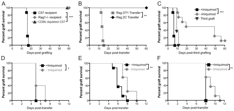
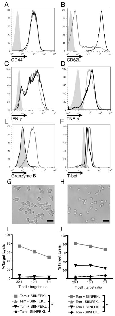
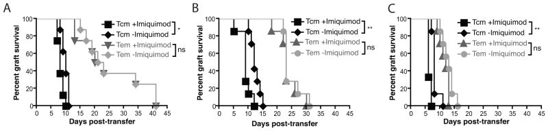
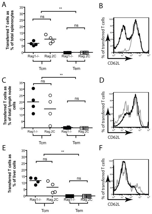
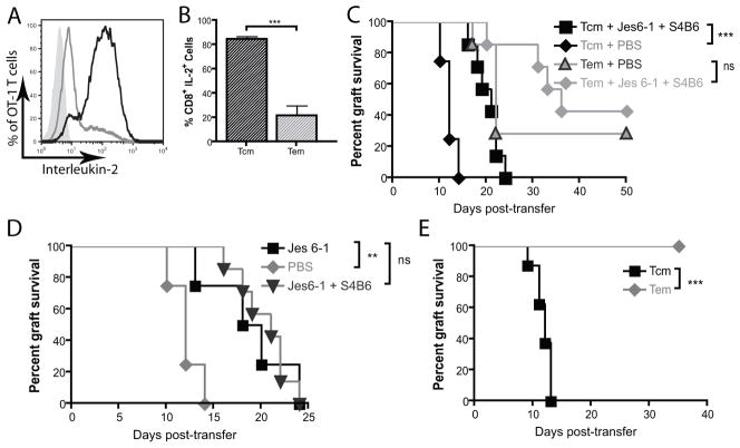
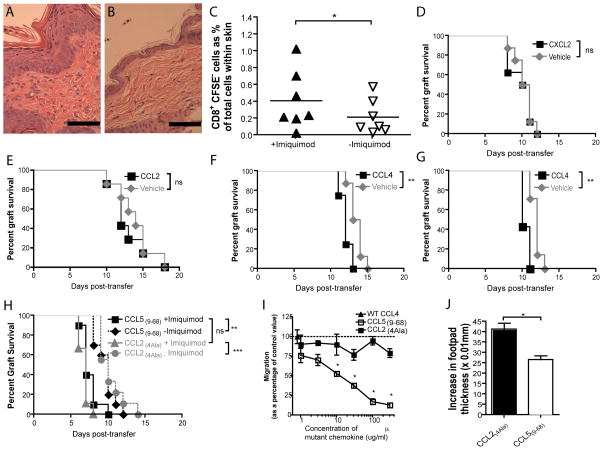
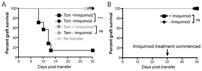
Adoptive T cell therapy uses the specificity of the adaptive immune system to target cancer and virally infected cells. Yet the mechanism and means by which to enhance T cell function are incompletely described, especially in the skin. In this study, we use a murine model of immunotherapy to optimize cell-mediated immunity in the skin. We show that in vitro-derived central but not effector memory-like T cells bring about rapid regression of skin-expressing cognate Ag as a transgene in keratinocytes. Local inflammation induced by the TLR7 receptor agonist imiquimod subtly yet reproducibly decreases time to skin graft rejection elicited by central but not effector memory T cells in an immunodeficient mouse model. Local CCL4, a chemokine liberated by TLR7 agonism, similarly enhances central memory T cell function. In this model, IL-2 facilitates the development in vivo of effector function from central memory but not effector memory T cells. In a model of T cell tolerogenesis, we further show that adoptively transferred central but not effector memory T cells can give rise to successful cutaneous immunity, which is dependent on a local inflammatory cue in the target tissue at the time of adoptive T cell transfer. Thus, adoptive T cell therapy efficacy can be enhanced if CD8(+) T cells with a central memory T cell phenotype are transferred, and IL-2 is present with contemporaneous local inflammation.


Figures

Figure 1
Figure 1. K5mOVA skin graft rejection is CD8+ T cell dependent and is enhanced by local TLR7 ligation
Kaplan-Meier survival curves of membrane-bound ovalbumin-expressing skin grafts (K5mOVA). A. C57BL/6, Rag1−/− mice and C57BL/6 mice treated with anti-CD8β monoclonal antibody were grafted with K5mOVA expressing skin and grafts monitored for rejection. n=8, ***p<0.001 by log rank test. B. Rag1−/− mice bearing K5mOVA skin grafts received an intravenous transfer of 106 SIINFEKL-specific Rag.OT-I splenocytes or SIYRYYGL-specific Rag.2C splenocytes. Grafts were then monitored for rejection. n=7, **p<0.01 by log rank test. C. C57BL/6 mice were depleted of CD8β+ T cells by treatment with the monoclonal antibody 53–5.8 and grafted with two K5mOVA skin grafts. 10 weeks later, animals received a third, contralateral graft, and one of the two original grafts were treated for 10 days with imiquimod whilst the other received control cream. n=7, ***p<0.001 by log rank test. D–F. Rag1−/− mice bearing bilateral, well-healed K5mOVA skin graft were adoptively transferred with 107 (D), 106 (E), or 105 (F) naïve Rag.OT-I splenocytes. At the same time all animals received subcutaneous immunisation with the adjuvant OVA in Quil A, whilst one of the two grafts was treated with imiquimod and the other with the control cream. n=8 for all groups, *, p < 0.05, **p<0.01 by log rank test.
Figure 2
Figure 2. Ex vivo differentiated TCM and TEM exhibit extracellular markers, intracellular molecules and killing capacity characteristic of TCM and TEM
A&B. CD44 and CD62L staining of OT-I in vitro derived TCM (black lines) and TEM (grey lines) cultured as per materials and methods. Isotype control shown in filled grey histograms. C–E. Intracellular Interferon-γ, tumour necrosis factor-α, granzyme B expression in in vitro differentiated TCM and TEM following stimulation with PMA/ionomycin. TCM shown as black lines, TEM as grey lines and unstimulated cells shown as filled grey histograms. F. Intracellular expression of the T-box transcription factor T-bet. TCM shown as black lines, TEM as grey lines and naïve cells shown as filled grey histograms. All vertical axes in histograms are frequency of cells as percentage of total cells analysed. Flow cytometry plots represent one three individual experiments conducted with pooled lymphocytes from OT-I mice. G&H. Micrograph of OT-I TEM (G) and TCM (H). Photos represent one of two individual experiments using pooled lymphocytes. Scale bar = 20μm. I & J. In vitro cytotoxic killing assays of TCM (black lines) and TEM (grey lines) demonstrating chromium release following 2.5 hours (K) and 5 hours (L) of exposure to SIINFEKL-pulsed (sold lines) and non-pulsed targets (broken lines). **p<0.01 by one-way ANOVA test with Bonferroni post-hoc analysis. Target lysis calculated by [(sample − spontaneous)/(maximum − spontaneous)] × 100%.
Figure 3
Figure 3. In vitro differentiated central but not effector memory CD8+ T cell rejection of skin grafts is enhanced by a local inflammatory stimulus
Rag1−/−mice bearing two well-healed K5mOVA grafts received either in vitro differentiated TCM (black lines) or TEM (grey lines). Grafts were then treated with either imiquimod or control cream and graft rejection plotted on a Kaplan-Meier survival curve. A. 104 adoptively transferred TCM and TEM CD8 T cells. Data analysed by log rank test, n=7 for each group, *p<0.05, ns = not significant. B. 105 adoptively transferred TCM and TEM CD8 T cells. Data analysed by log rank test, n=7 for each group, **p<0.01, ns = not significant. C. 106 adoptively transferred TCM and TEM CD8 T cells. Data analysed by log rank test, n=8 for each group, **p<0.01, ns = not significant.
Figure 4
Figure 4. TCM persist to greater extent than TEM in both Rag1−/− and Rag.2C mice
Rag1−/− (filled symbols) and Rag.2C (open symbols) mice bearing well-healed K5mOVA grafts were adoptively transferred with 106 TCM (circles, black lines on histogram) or TEM (squares, grey lines on histogram). 45 days following transfer spleen (panels A&B), pooled inguinal, brachial, axillary and mesenteric lymph nodes (panels C&D) and liver (panels E&F) were harvested and cells stained for CD8, Vα2, and CD62L. A, C and E. CD8+ Vα2+ cells as percent of total cells in each organ. n=4per group, **p<0.01 when comparing Rag1−/− to Rag1−/− and Rag.2C to Rag.2C, ns = not significant; analysed by one-way ANOVA analysis with post-hoc multiple comparison test. B, D and F. Representative CD62L expression of CD8+ Vα2+ CD44+ in various organs analysed by flow cytometry 45 days following transfer into Rag1−/− mice.
Figure 5
Figure 5. IL-2 inhibition delays TCM mediated-rejection of well-healed K5mOVA grafts
A. Intracellular expression of interleukin-2 by flow cytometry following stimulation of in vitro differentiated central memory T cells (T CM, black line) and effector memory T cells (TEM, grey line) with PMA/Ionomycin. B. Quantitation of percent of IL-2-expressing in vitro differentiated TCM and TEM. Data from three individual experiments. Data analysed by Student’s t-test. Error bars represent S.E.M. ***p<0.001. C & D. Kaplan-Meier survival curve of well-healed K5mOVA skin grafts on Rag1−/− mice which received an adoptive transfer of either 105 TCM or TEM treated for 10 days with intraperitoneal injections of PBS or 100μg of the IL-2-blocking monoclonal antibodies Jes6-1 and S4B6 (C); or Jes6-1 alone or PBS (D). Animals were then monitored for graft rejection. n=8 for TCM and n=7 for TEM. **p<0.001, ns = not significant. Data analysed by log rank test. E. Rag.2C mice bearing well-healed K5mOVA skin grafts were adoptively transferred with 105 TCM or TEM. Following transfer, grafts were monitored daily for graft rejection. n=8 per group, ***p<0.001. Data analysed by log rank test.
Figure 6
Figure 6. Imiquimod enhances inflammatory infiltrate and traffic of adoptively transferred CD8+ T cells into inflamed grafts. The effects of imiquimod on TCM-mediated graft rejection are recapitulated by the chemotactic molecule, CCL4
A & B. Haematoxylin and eosin micrograph of flank skin of C57BL/6 mice treated for 5 days with imiquimod (A) or control cream (B). Scale bars represent 50μm. C. Rag1−/− mice bearing two, bilateral K5mOvA received an adoptive transfer of 105 CFSE-labelled, in vitro-derived TCM 6 weeks following grafting. At time of transfer one of the two grafts was treated with imiquimod daily for 5 days, whilst the other received control cream. Grafts were removed, processed into single cell suspension. Cells were then analysed by flow cytometry for CD3, CD8 and CFSE staining. n=7 per group, *p<0.05 by paired Student’s t test. D–G. Rag1−/− bearing two well-healed K5mOVA skin grafts were adoptively transferred with 105 TCM and received intradermal injections of 200ng of recombinant murine CCL2 (D) or CXCL2 (E) or CCL4 (F) or 1μg of CCL4 (G) or vehicle control on days 4, 5, 7 and 9 post-transfer. Grafts were then monitored for rejection. n=8 for each experiment, **p<0.01, ns. not significant by log rank test. H. Two groups of Rag1−/− mice bearing bilateral, well-healed K5mOVA skin grafts were adoptively transferred with TCM and treated with the CCR5 inhibiting peptide CCL5(–68) or the non-functional mutated chemokine CCL2(4Ala) whilst one of the two grafts was treated with imiquimod. Grafts were then monitored for rejection and plotted on a Kaplan-Meier survival curve. n=8 for all groups, **p<0.01, ***p<0.001, ns = not significant by log rank test. I. Transwell migration assay of purified CD8+ T cells stimulated with CD3 and CD28 monoclonal antibodies, migrating in response to CCL4. Inhibition of T cell migration by CCL59–68 was evaluated by increasing concentration of CCL59–68 and assessing percent of migrated cells relative to wild-type CCL4. Non-functional CCL24Ala peptide was used as control. n=2, *p<0.05 by one-way ANOVA. J. Delayed-type hypersensitivity reaction was established by tail-base immunisation of C57BL/6 mice with Ovalbumin in complete Freund’s adjuvant. 7 days later, recall was conducted in footpad and increase in footpad thickness was assessed by callipers. To assess for in vivo activity of CCL59–68, one day prior to recall animals were treated with CCL59–68 or control peptide CCL24Ala. Data are presented as mean + S.E.M. Data analysed by Student’s t-test. n = 12 per group, *p<0.05.
Figure 7
Figure 7. Local TLR7 ligation and TCM transfer is able to overcome tolerogenic environments and instate cutaneous immunotherapy
A. CD11c.OVA mice bearing two well-healed K5mOVA skin grafts were adoptively transferred with 105 TEM or TCM 6 weeks following grafting. At time of transfer one of the two grafts was treated with imiquimod whilst the other was treated with control cream. Data analysed by log rank test, n=7 for TCM, n=8 for TEM. ***p<0.001, ns = not significant. B. CD11c.OVA mice bearing well-healed K5mOVA skin grafts were adoptively transferred with 105 TCM. 30 days later grafts were treated with either imiquimod or control cream for 10 days. Data analysed by log rank test, n=8, ns = not significant.

Similar articles

Cited by

References

    1. Frazer IH. Prevention of cervical cancer through papillomavirus vaccination. Nat Rev Immunol. 2004;4:46–54. - PubMed
    1. Rosenberg SA, Restifo NP, Yang JC, Morgan RA, Dudley ME. Adoptive cell transfer: a clinical path to effective cancer immunotherapy. Nat Rev Cancer. 2008;8:299–308. - PMC - PubMed
    1. Rosenberg SA, Dudley ME. Adoptive cell therapy for the treatment of patients with metastatic melanoma. Curr Opin Immunol. 2009;21:233–240. - PMC - PubMed
    1. Dudley ME, Yang JC, Sherry R, Hughes MS, Royal R, Kammula U, Robbins PF, Huang J, Citrin DE, Leitman SF, Wunderlich J, Restifo NP, Thomasian A, Downey SG, Smith FO, Klapper J, Morton K, Laurencot C, White DE, Rosenberg SA. Adoptive cell therapy for patients with metastatic melanoma: evaluation of intensive myeloablative chemoradiation preparative regimens. J Clin Oncol. 2008;26:5233–5239. - PMC - PubMed
    1. Besser MJ, Shapira-Frommer R, Treves AJ, Zippel D, Itzhaki O, Hershkovitz L, Levy D, Kubi A, Hovav E, Chermoshniuk N, Shalmon B, Hardan I, Catane R, Markel G, Apter S, Ben-Nun A, Kuchuk I, Shimoni A, Nagler A, Schachter J. Clinical responses in a phase II study using adoptive transfer of short-term cultured tumor infiltration lymphocytes in metastatic melanoma patients. Clin Cancer Res. 2010;16:2646–2655. - PubMed

Publication types

MeSH terms