

Regulatory T-lymphocytes mediate amyotrophic lateral sclerosis progression and survival
- PMID: 23143995
- PMCID: PMC3569654
- DOI: 10.1002/emmm.201201544
Regulatory T-lymphocytes mediate amyotrophic lateral sclerosis progression and survival
Erratum in
- EMBO Mol Med. 2013 Feb;5(2):326
Abstract
In amyotrophic lateral sclerosis (ALS) mice, regulatory T-lymphocytes (Tregs) are neuroprotective, slowing disease progression. To address whether Tregs and FoxP3, a transcription factor required for Treg function, similarly influence progression rates of ALS patients, T-lymphocytes from patients were assessed by flow cytometry. Both numbers of Tregs and their FoxP3 protein expressions were reduced in rapidly progressing ALS patients and inversely correlated with progression rates. The mRNA levels of FoxP3, TGF-β, IL4 and Gata3, a Th2 transcription factor, were reduced in rapidly progressing patients and inversely correlated with progression rates. Both FoxP3 and Gata3 were accurate indicators of progression rates. No differences in IL10, Tbx21, a Th1 transcription factor or IFN-γ expression were found between slow and rapidly progressing patients. A 3.5-year prospective study with a second larger cohort revealed that early reduced FoxP3 levels were indicative of progression rates at collection and predictive of future rapid progression and attenuated survival. Collectively, these data suggest that Tregs and Th2 lymphocytes influence disease progression rates. Importantly, early reduced FoxP3 levels could be used to identify rapidly progressing patients.
Copyright © 2013 The Authors. Published by John Wiley and Sons, Ltd on behalf of EMBO.
Figures
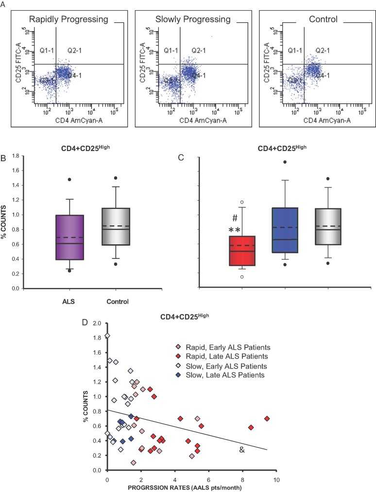
Representative flow diagrams showing CD4+CD25High Tregs from a rapidly progressing ALS patient, a slowly progressing ALS patient, and a control volunteer.
Box and whisker plots indicating that percent of CD4+CD25High Tregs in total leukocytes from ALS patients (mean = 0.692%, median = 0.610%) were not different when compared with control volunteers using the t-test (mean = 0.845%, median = 0.800%).
When the ALS patients were separated based on the rate of disease progression into rapidly (AALS points per month ≥1.5; 26 patients) versus slowly (AALS points per month <1.5; 28 patients) progressing ALS patients, the percent of CD4+CD25High Tregs were reduced in rapidly progressing patients (mean = 0.573%, median = 0.495%) compared with slowly progressing patients (mean = 0.825%, median = 0.660%) and reduced compared with control volunteers (mean = 0.845%, median = 0.800%); slowly progressing patients were not different than controls. #p = 0.018 versus slowly progressing ALS patients; **p = 0.003 versus controls.
Scatter plot with regression line demonstrating that the percent of CD4+CD25High T cells were inversely correlated with rate of ALS progression (R = 0.301; linear regression). Slowly progressing ALS patients = AALS points/month <1.5; rapidly progressing ALS patients = AALS points/month >1.5, at the time of collection. ALS patients early in disease = AALS score < 100; ALS patients late in disease = AALS score ≥100, at the time of collection. &p = 0.028.

There was a trend toward reduced numbers of CD4+FoxP3+ Tregs in the blood of rapidly progressing patients compared with both slowly progressing patients and control volunteers.
FoxP3 intensity in CD4+FoxP3+ Tregs is reduced in rapidly progressing patients compared with slowly progressing patients and compared with control volunteers; slowly progressing patients were not different than controls. #p = 0.049 versus slowly progressing ALS patients; **p = 0.015 versus controls.

A,B. FoxP3 mRNA expression levels were down-regulated in rapidly progressing ALS patients (t-test) and negatively correlated with disease progression rates (R = 0.419; linear regression).
C,D. CD25 mRNA expression levels were reduced in rapidly progressing ALS patients (t-test) and inversely correlated with rate of disease progression (R = 0.444; linear regression).
E. FoxP3 and CD25 mRNA expression levels of ALS patients directly correlated with each other (R = 0.815; linear regression). Note that slowly progressing patients early in their disease expressed the highest FoxP3 and CD25 mRNA levels, whereas rapidly progressing patients late in their course of disease expressed the lowest levels of FoxP3 and CD25 mRNAs. ##p ≤ 0.005 versus slowly progressing ALS patients. ***p ≤ 0.001 versus controls. &&p ≤ 0.005, &&&p ≤ 0.001.

A,B. Gata3 mRNA expression was decreased in rapidly progressing ALS patients (t-test) and inversely correlated with rate of disease progression (R = 0.405; linear regression).
C. Gata3 and FoxP3 levels correlated (R = 0.737; linear regression).
D. Gata3 and CD25 mRNA levels correlated (R = 0.810; linear regression). Note that slowly progressing patients early in their disease expressed the highest Gata3 mRNA levels, whereas rapidly progressing patients late in their course of disease expressed the lowest levels of Gata3 levels. ###p = 0.003 versus slowly progressing ALS patients; ***p = 0.00001 versus controls; &&&p ≤ 0.004.

A,B. IL4 mRNA expression was decreased in rapidly progressing ALS patients (t-test) and inversely correlated with rate of disease progression (R = 0.388; linear regression).
C,D. TGF-β mRNA expression was reduced in rapidly progressing ALS patients (t-test) and inversely correlated with rate of disease progression (R = 0.475; linear regression). Note that slowly progressing patients early in their disease expressed the highest IL4 and TGF-β mRNA levels, whereas rapidly progressing patients late in their course of disease expressed the lowest levels of IL4 and TGF-β levels. ###p ≤ 0.0007 versus slowly progressing ALS patients; ***p ≤ 0.0002 versus controls; &&p ≤ 0.006, &&&p ≤ 0.001.


FoxP3 expression had a 78.9% accuracy (95% CI: 0.658–0.921), 73.9% sensitivity and 73.1% specificity, using a fold-increase over 0.66 as positive and a rate of progression of less than 1.5 points per month as slow.
ALS patients with low FoxP3 mRNA expression levels (based on the 0.66-fold cutoff determined by the ROC analysis) progressed more rapidly than patients with high FoxP3 levels.
Gata3 expression had a 81.1% accuracy (95% CI: 0.651–0.902), 76.9% sensitivity and 69.9% specificity, using a fold-increase over 0.52 as positive and a rate of progression of less than 1.5 points per month as slow.
ALS patients with low Gata3 mRNA expression levels (based on the 0.52-fold cutoff determined by the ROC analysis) progressed more rapidly than patients with high Gata3 levels.
Also shown for comparison is the ROC analysis of the time from first symptom to first exam in the same 54 patients compared to their progression rates. Time from first symptom to first exam had a 65.7% accuracy (95% CI: 0.473–0.786), 60.9% sensitivity and 61.5% specificity, using a fold-increase over 14.3 months as positive and a rate of progression of less than 1.5 points per month as slow.
ALS patients with short times from first symptom to first exam (based on the 14.3 months cutoff determined by the ROC analysis) progressed more rapidly than patients with longer times from first symptom to first exam. *p = 0.04; ***p = 0.003.

With this new ‘test’ set of 102 patients, ALS patients with low FoxP3 mRNA expression levels (based on cutoffs determined by the ROC analysis of the original 54 patients) early in disease progressed more rapidly than patients with high FoxP3 levels, both at the time of collection and at the end of the evaluation period. *p < 0.05 versus high FoxP3 levels, **p < 0.01 versus high FoxP3 levels.
FoxP3 levels of this new ‘test’ set of patients were reflective of progression rates at the time of collection and low FoxP3 levels were predictive of future rapid progression rates. At the time of collection, low FoxP3 levels correctly predicted rapid progression rates 69% of the time, and high FoxP3 levels correctly predicted slow progression rates 75% of the time. At the end of the analysis period, low FoxP3 levels correctly predicted rapid progression rates 82% of the time, and high FoxP3 levels correctly predicted slow progression rates 53% of the time. aCutoff levels were defined by the prior ROC curve analyses (Fig 7; FoxP3 = 0.66). bLevels were compared with progression rates both at the time of collection and at the end of the evaluation period. cEvaluation period = 3.5 years.

A. Sixty-six percent of patients with FoxP3 expression levels below the cutoff were above 100 AALS points at the end of the 3.5 years, while only 36.7% of the patients with FoxP3 expression levels above the cutoff were above 100 AALS points.
B,C. Thirty-five percent of patients with FoxP3 expression levels below the cutoff were placed on a ventilator or were deceased during the 3.5 years, while only 13% of the patients with FoxP3 expression levels above the cutoff were on a ventilator or deceased. Optimum cutoff level was defined by the ROC curve analysis in Fig 7 (FoxP3 = 0.66). *p = 0.013, **p = 0.0072, log-rank tests; #p = 0.023, Chi square test.
Similar articles
-
Endogenous regulatory T lymphocytes ameliorate amyotrophic lateral sclerosis in mice and correlate with disease progression in patients with amyotrophic lateral sclerosis.Brain. 2011 May;134(Pt 5):1293-314. doi: 10.1093/brain/awr074. Brain. 2011. PMID: 21596768 Free PMC article.
-
Evaluation of regulatory T lymphocytes and IL2Ra and FOXP3 gene expression in peripheral mononuclear cells from patients with amyotrophic lateral sclerosis.Ir J Med Sci. 2018 Nov;187(4):1065-1071. doi: 10.1007/s11845-018-1793-2. Epub 2018 Mar 25. Ir J Med Sci. 2018. PMID: 29574662
-
ALS patients' regulatory T lymphocytes are dysfunctional, and correlate with disease progression rate and severity.JCI Insight. 2017 Mar 9;2(5):e89530. doi: 10.1172/jci.insight.89530. JCI Insight. 2017. PMID: 28289705 Free PMC article.
-
Neuroinflammatory mechanisms in amyotrophic lateral sclerosis pathogenesis.Curr Opin Neurol. 2018 Oct;31(5):635-639. doi: 10.1097/WCO.0000000000000599. Curr Opin Neurol. 2018. PMID: 30048339 Review.
-
Amyotrophic Lateral Sclerosis, a Multisystem Pathology: Insights into the Role of TNFα.Mediators Inflamm. 2017;2017:2985051. doi: 10.1155/2017/2985051. Epub 2017 Sep 10. Mediators Inflamm. 2017. PMID: 29081600 Free PMC article. Review.
Cited by
-
Friend or foe: the dichotomous impact of T cells on neuro-de/re-generation during aging.Oncotarget. 2017 Jan 24;8(4):7116-7137. doi: 10.18632/oncotarget.12572. Oncotarget. 2017. PMID: 27738345 Free PMC article. Review.
-
Phase I trial of repeated intrathecal autologous bone marrow-derived mesenchymal stromal cells in amyotrophic lateral sclerosis.Stem Cells Transl Med. 2015 Jun;4(6):590-7. doi: 10.5966/sctm.2014-0212. Epub 2015 May 1. Stem Cells Transl Med. 2015. PMID: 25934946 Free PMC article. Clinical Trial.
-
T cell responses at diagnosis of amyotrophic lateral sclerosis predict disease progression.Nat Commun. 2022 Nov 8;13(1):6733. doi: 10.1038/s41467-022-34526-9. Nat Commun. 2022. PMID: 36347843 Free PMC article.
-
Bacillus Calmette-Guerin vaccine-mediated neuroprotection is associated with regulatory T-cell induction in the 1-methyl-4-phenyl-1,2,3,6-tetrahydropyridine mouse model of Parkinson's disease.J Neurosci Res. 2013 Oct;91(10):1292-302. doi: 10.1002/jnr.23253. Epub 2013 Aug 1. J Neurosci Res. 2013. PMID: 23907992 Free PMC article.
-
Evidence for fungal infection in cerebrospinal fluid and brain tissue from patients with amyotrophic lateral sclerosis.Int J Biol Sci. 2015 Apr 2;11(5):546-58. doi: 10.7150/ijbs.11084. eCollection 2015. Int J Biol Sci. 2015. PMID: 25892962 Free PMC article.
References
Publication types
MeSH terms
Substances
LinkOut - more resources
Full Text Sources
Other Literature Sources
Medical
Miscellaneous
