. 2013 Feb 1;288(5):3359-69.
doi: 10.1074/jbc.M112.384289. Epub 2012 Nov 6.

p21-Activated kinase 6 (PAK6) inhibits prostate cancer growth via phosphorylation of androgen receptor and tumorigenic E3 ligase murine double minute-2 (Mdm2)

Affiliations

p21-Activated kinase 6 (PAK6) inhibits prostate cancer growth via phosphorylation of androgen receptor and tumorigenic E3 ligase murine double minute-2 (Mdm2)

Tong Liu et al. J Biol Chem. .

Abstract

The androgen receptor (AR) signaling pathway plays a crucial role in the development and growth of prostate malignancies. Regulation of AR homeostasis in prostate tumorigenesis has not yet been fully characterized. In this study, we demonstrate that p21-activated kinase 6 (PAK6) inhibits prostate tumorigenesis by regulating AR homeostasis. First, we demonstrated that in normal prostate epithelium, AR co-localizes with PAK6 in the cytoplasm and translocates into the nucleus in malignant prostate. Furthermore, AR phosphorylation at Ser-578 by PAK6 promotes AR-E3 ligase murine double minute-2 (Mdm2) association, causing AR degradation upon androgen stimuli. We also showed that PAK6 phosphorylates Mdm2 on Thr-158 and Ser-186, which is critical for AR ubiquitin-mediated degradation. Moreover, we found that Thr-158 collaborates with Ser-186 for AR-Mdm2 association and AR ubiquitin-mediated degradation as it facilitates PAK6-mediated AR homeostasis. PAK6 knockdown promotes prostate tumor growth in vivo. Interestingly, we found a strong inverse correlation between PAK6 and AR expression in the cytoplasm of prostate cancer cells. These observations indicate that PAK6 may be important for the maintenance of androgen-induced AR signaling homeostasis and in prostate malignancy, as well as being a possible new therapeutic target for AR-positive and hormone-sensitive prostate cancer.


Figures

FIGURE 1.
FIGURE 1.
PAK6 involvement in AR localization and expression. A, PAK6 and AR are co-localized in the cytoplasm of normal prostate cells, and AR is localized in the nucleus in prostate cancer cells. Tissue specimens were subjected to immunohistochemical analysis as usual. After antigen retrieval, specimens were fixed and incubated with anti-PAK6 antibody followed by Alexa 594 Fluor (red) antibody and anti-AR antibody followed by Alexa 488 Fluor (green) antibody. Nucleus was stained by DAPI (blue). The 2nd and 4th lines are the 25-fold enlarged pictures of the 1st and 3rd lines, respectively. The 4th row was merged with red and green images, and 5th row was merged with red, green, and blue images. The white arrows in the 2nd line indicate AR co-localizes with PAK6 in the cytoplasm, and arrows in 4th line indicate AR translocates in the nucleus. B, starvation experiment showing that PAK6 expression increased and AR decreased upon starvation. CW22Rv1 cells were starved with Hanks' buffered saline solution (Invitrogen) at different time points as indicated. Western blot analysis was performed, and endogenous proteins were detected with the indicated antibodies. C, CWR22Rv1 cells transfected with Myc-PAK6 show decreased AR levels upon DHT activation. CWR22Rv1 cells were transfected with indicated amounts of pcDNA3.1-Myc-His-PAK6. Endogenous AR and exogenous PAK6 were detected with indicated antibodies.
FIGURE 2.
FIGURE 2.
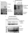
PAK6 regulates AR localization through phosphorylation of AR on Ser-578. A, PAK6 phosphorylates AR between amino acids 559 and 624. HEK293 cells were transfected with pcDNA3.1Myc-His-PAK6 and lysed for immunoprecipitation with anti-Myc antibody, and the immunoprecipitated PAK6 kinase was incubated with GST-AR deletions (505–559, 559–624, 624–676, and 505–676 amino acids) for in vitro kinase assay. B, AR Ser-578 is the phosphorylation site by PAK6. In vitro kinase assay was performed using commercialized PAK6 kinase and GST-AR WT (amino acids 505–676) or GST-AR single-site mutants as indicated. Histone H4 served as a positive control. C, wild-type PAK6 phosphorylated AR in the cytoplasm and reduced its nuclear translocation, leading to decreased prostate-specific antigen (PSA) levels. HEK293 cells were co-transfected with pcDNA3.1-His-PAK6 WT/KA and pcDNA3.1-His-AR in the presence or absence of DHT, and cytoplasmic and nuclear proteins were subjected to SDS-PAGE separately. Western blot analysis was performed as indicated. p-AR is phosphorylated AR Ser-578 antibody. Poly(ADP-ribose) polymerase (PARP) served as a nuclear loading control, and paxillin served as a cytoplasmic loading control. D, inhibition of AR nuclear translocation by PAK6 relied on Ser-578 phosphorylation. HEK293 cells were co-transfected with pcDNA-EGFP-PAK6 WT/KA and pcDNA3.1-His-AR. Cells were starved with steroid hormone for 16 h, stimulated with 10 nm DHT for 0, 30, and 60 min as indicated, fixed, and incubated with anti-AR antibody followed by Alexa Fluor 546 (red) antibody. Nucleus was stained with Topro3 (blue). The white arrows in the 4th rows indicate the subcellular co-localization of PAK6 WT/KA and AR.
FIGURE 3.
FIGURE 3.
PAK6-mediated AR phosphorylation promotes its ubiquitin-mediated degradation. A, PAK6 reduced AR levels under DHT stimulation. HEK293 cells were transfected with pcDNA3.1-Myc-His-PAK6 and pcDNA 3.1-His-AR and treated with 10 nm DHT and 10 μm cycloheximide at the indicated times. PAK6 and AR protein levels were measured by Western blot analysis as indicated. B, PAK6 reduced wild-type AR under DHT stimulation, while AR S578A mutant was unaffected. HEK293 cells were co-transfected with pcDNA-FLAG-AR wild-type/S578A (505–919 amino acids) (including DNA binding domain and ligand binding domain), and pcDNA3.1-Myc-His-PAK6 was treated with 10 nm DHT and 10 μm cycloheximide. C, ubiquitination assay showed that PAK6 markedly enhanced AR WT but not AR S578A ubiquitination. HEK293 cells were transfected with Myc-ubiquitin and pcDNA-FLAG-AR WT/S578A (505–919 amino acids) in combination with pcDNA3.1-Myc-His PAK6/vector-treated or untreated with 5 μm MG132 for 6 h in the presence of 10 nm DHT. 30 μg of total lysates were set aside as input; equal amounts lysates of protein were then harvested for ubiquitination assay. IP, immunoprecipitation; IB, immunoblot. D, stable knockdown of PAK6 by shRNA in CWR22Rv1 cells showed impairment of the endogenous ubiquitination of AR when treated with MG132 and DHT (7th lane from left). CWR22Rv1 cells infected with lentiviruses harboring shRNA control/PAK6 were treated with or without 5 μm MG132 for 6 h in the presence/absence of 10 nm DHT. Equal amounts lysates of protein were then harvested for ubiquitination assay.
FIGURE 4.
FIGURE 4.
PAK6-mediated phosphorylation of AR enhances AR association with Mdm2. A, Mdm2 interacted strongly with AR WT but much more weakly with AR S578A mutant. In vitro translated pcDNA-FLAG-AR WT/S578A (505–919 amino acids) was incubated with GST-Mdm2 as indicated and analyzed with anti-FLAG antibody. GST served as control. B, Mdm2 associated stronger with AR WT than AR S578A mutant in the presence of PAK6. COS7 cells were co-transfected with pcDNA-FLAG-AR WT/S578A (505–919 amino acids) in combination with pcDNA-EGFP-PAK6 and Mdm2 as indicated, treated with 10 nm DHT. 30 μg of total lysates were set aside as input; equal amounts of lysates of protein were immunoprecipitated (IP) with anti-FLAG antibody and immunoblotted (IB) with anti-Mdm2 antibody. C, GST pulldown assay showed that PAK6 and Mdm2 interact with each other in vitro. In vitro translated pcDNA3.1-Myc-His-PAK6 was incubated with GST-Mdm2 and analyzed with anti-Myc antibody. GST served as control. D, immunoprecipitation assay confirmed that PAK6 associates with Mdm2 in CWR22Rv1 cells. Equal amounts of CWR22Rv1 cell lysates were immunoprecipitated with anti-PAK6 or IgG. Endogenous PAK6 and Mdm2 were immunoblotted with anti-PAK6 and anti-Mdm2 antibodies.
FIGURE 5.
FIGURE 5.
PAK6 phosphorylates Mdm2 on Thr-158 and Ser-186. A, PAK6 phosphorylates Mdm2. HEK293 cells were transfected with pcDNA3.1-Myc-His-PAK6 and lysed for immunoprecipitated with anti-Myc antibody; the immunoprecipitated PAK6 kinase was incubated with GST-Mdm2 and analyzed by autoradiography. Histone H3 served as positive control. B, PAK6 phosphorylates Mdm2 between amino acids (aa) 121 and 300. In vitro kinase assay was performed using commercialized PAK6 kinase and GST-Mdm2 full-length and deletions as indicated and analyzed by autoradiography (auto). C, Mdm2 Thr-158 and Ser-186 are the phosphorylation sites by PAK6. In vitro kinase assay was performed using commercialized PAK6 kinase and GST-Mdm2 (121–300 amino acids) or GST-Mdm2 single-site mutants as indicated and analyzed by autoradiography. Histone H3 (H3) served as a positive control.
FIGURE 6.
FIGURE 6.
Functional roles of Mdm2 Thr-158 and Ser-186 in AR degradation. A, PAK6 induces AR ubiquitination dependent on Mdm2 phosphorylation. COS7 cells were transfected with pcDNA3.1-His-AR in combination with vector, pcDNA3.1-His-PAK6, or pcDNA3.1-His-Mdm2 mutants in the presence or absence of Myc-ubiquitin (Myc-Ub) in 10% charcoal-stripped medium for 16 h, followed by treatment with 10 nm DHT for 16 h. The cells were then harvested for ubiquitination assay. IP, immunoprecipitation; IB, immunoblot. B, AR interacted strongly with Mdm2 WT and T158A but weakly with Mdm2 S186A mutant. In vitro translated pcDNA3.1-His-AR was incubated with GST-Mdm2 mutants as indicated and analyzed with anti-AR antibody. GST served as control. C, PAK6-mediated phosphorylation of Mdm2 Thr-158 promotes Ser-186 association with AR. In vitro translated pcDNA3.1-His-AR was incubated with equal amounts of GST-Mdm2 S186A or T158A mutant. Meanwhile, increasing amounts of in vitro translated T158A or S186A (5, 10, and 20 μl) were added and analyzed with anti-AR antibody. GST served as control. D, Mdm2 S186A but not T158A mutant weakened PAK6-AR association. In vitro translated pcDNA3.1-His-AR was incubated with equal amounts of GST-PAK6; meanwhile, increasing amounts of in vitro translated Mdm2 mutants (5, 10, and 20 μl) were added and analyzed with anti-His and anti-AR antibodies. GST served as control.
FIGURE 7.
FIGURE 7.
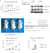
PAK6 inhibits prostate cancer growth in vivo. A, stable knockdown of PAK6 by shRNA in CWR22Rv1 cells showed a significant enhancement of proliferation in vitro. CWR22Rv1 cells infected with lentiviruses harboring shRNA control/PAK6 were plated in 12-well plates with equal amounts and counted every day for 1 week. B, proliferating marker gene cyclin D1 in AR signaling was up-regulated in shRNA PAK6 group upon DHT stimuli. shRNA control/PAK6 in CWR22Rv1 cells was treated with or without DHT for 16 h. Equal amounts of protein lysates were then harvested for Western blot with antibodies as indicated. C, effect of shRNA control (left dorsal side) or shRNA PAK6 (right dorsal side) on the growth of CWR22Rv1 cells inoculated into nude mice. NOD SCID nude male mice were subcutaneously injected with 2 × 10⁶ CWR22Rv1 cells infected with lentiviruses harboring shRNA PAK6/control. Tumor volume was monitored over time as indicated, and the tumor was excised and weighed after 49 days. PAK6 depletion causes a high ratio of tumor development (table) and an increase in tumor volume and weight (graphs).
FIGURE 8.
FIGURE 8.
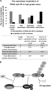
A, relationship between PAK6 and AR in prostate cancer. 14 normal prostate, 6 well differentiated, 9 moderately differentiated, and 8 poorly differentiated prostate tumor specimens were collected and subjected to immunofluorescence analysis. The score of immunostaining co-localization of PAK6 and AR in cytoplasm or localization of AR in nucleus was counted from 30 independent visions and determined by intensity (0 to 3) and fraction of stained cells (0 to 4). A total score (ranging from 0 to 12) was obtained by multiplying the staining intensity and fraction scores. ** means p value less than or equal to 0.001 was considered statistically significant according to unpaired t test. B, proposed mechanism for the relation between PAK6, AR, Mdm2, and ubiquitin-proteasome degradation. Upon DHT stimulation, PAK6 obstructs AR nuclear translocation through phosphorylation on AR Ser-578; this phosphorylation enhances the binding between AR and Mdm2. The further phosphorylation of Mdm2 by PAK6 promotes proteasome to recognize the polyubiquitylated AR, leading to AR degradation.

Comment in

Similar articles

Cited by

References

    1. Agoulnik I. U., Weigel N. L. (2008) Androgen receptor coactivators and prostate cancer. Adv. Exp. Med. Biol. 617, 245–255 - PubMed
    1. Buchanan G., Irvine R. A., Coetzee G. A., Tilley W. D. (2001) Contribution of the androgen receptor to prostate cancer predisposition and progression. Cancer Metastasis Rev. 20, 207–223 - PubMed
    1. Hobisch A., Culig Z., Radmayr C., Bartsch G., Klocker H., Hittmair A. (1996) Androgen receptor status of lymph node metastases from prostate cancer. Prostate 28, 129–135 - PubMed
    1. Ruizeveld de Winter J. A., Janssen P. J., Sleddens H. M., Verleun-Mooijman M. C., Trapman J., Brinkmann A. O., Santerse A. B., Schröder F. H., van der Kwast T. H. (1994) Androgen receptor status in localized and locally progressive hormone refractory human prostate cancer. Am. J. Pathol. 144, 735–746 - PMC - PubMed
    1. Saraon P., Jarvi K., Diamandis E. P. (2011) Molecular alterations during progression of prostate cancer to androgen independence. Clin. Chem. 57, 1366–1375 - PubMed

Publication types

MeSH terms

LinkOut - more resources