. 2012 Dec 21;287(52):43995-4005.
doi: 10.1074/jbc.M112.417501. Epub 2012 Nov 5.

Cysteine 70 of ankyrin-G is S-palmitoylated and is required for function of ankyrin-G in membrane domain assembly

Affiliations

Cysteine 70 of ankyrin-G is S-palmitoylated and is required for function of ankyrin-G in membrane domain assembly

Meng He et al. J Biol Chem. .

Abstract

Ankyrin-G (AnkG) coordinates protein composition of diverse membrane domains, including epithelial lateral membranes and neuronal axon initial segments. However, how AnkG itself localizes to these membrane domains is not understood. We report that AnkG remains on the plasma membrane in Madin-Darby canine kidney (MDCK) cells grown in low calcium, although these cells lack apical-basal polarity and exhibit loss of plasma membrane association of AnkG partners, E-cadherin and β(2)-spectrin. We subsequently demonstrate using mutagenesis and mass spectrometry that AnkG is S-palmitoylated exclusively at Cys-70, which is located in a loop of the first ankyrin repeat and is conserved in the vertebrate ankyrin family. Moreover, C70A mutation abolishes membrane association of 190-kDa AnkG in MDCK cells grown in low calcium. C70A 190-kDa AnkG fails to restore biogenesis of epithelial lateral membranes in MDCK cells depleted of endogenous AnkG. In addition, C70A 270-kDa AnkG fails to cluster at the axon initial segment of AnkG-depleted cultured hippocampal neurons and fails to recruit neurofascin as well as voltage-gated sodium channels. These effects of C70A mutation combined with evidence for its S-palmitoylation are consistent with a requirement of palmitoylation for targeting and function of AnkG in membrane domain biogenesis at epithelial lateral membranes and neuronal axon initial segments.


Figures
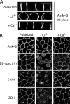
FIGURE 1.
FIGURE 1.
AnkG remains membrane-associated in MDCK cells following calcium switch. A and B, fully polarized MDCK cells (polarized) were trypsinized and replated in Ca2+-free medium with 5% FBS overnight to reach a steady state. Cells were then either fixed (−Ca2+) or allowed 24 h to recover in normal growth medium (+Ca2+) and then fixed and processed for immunofluorescence to stain against AnkG, β2-spectrin, E-cadherin, and ZO-1. A, the xz planes showing AnkG staining in MDCK cells following calcium switch described above. B, the xy planes showing immunostaining of AnkG, β2-spectrin, E-cadherin (E-cad), and ZO-1 (a marker of tight junction) in MDCK cells following calcium switch. Scale bars, 10 μm. Images are representative of at least three independent repeated experiments.
FIGURE 2.
FIGURE 2.
AnkG is S-palmitoylated at cysteine 70. A, endogenous AnkG was immunoprecipitated (IP) from MDCK cell lysates and processed for the biotin switch assay (see “Experimental Procedures”) to detect protein S-acylation. Four major AnkG polypeptides are detected in MDCK cells using an antibody against the C-terminal region. The blot (IB) shows that only the ∼200-kDa isoform, which has ankyrin repeats, is S-acylated (blots are representative of three independent experiments). B, GFP-tagged wild-type AnkG and cysteine-to-alanine mutants (C70A, C315A, C357A, C385A, C416A, and C746A) were transfected into HEK293 cells and processed for the biotin switch assay. The blot shows that C70A completely abolishes AnkG S-acylation. (Blots are representative of two independent experiments.) C, HEK293 cells transfected with AnkG-GFP were lysed and blocked with N-ethylmaleimide. Following immunoisolation, AnkG was incubated with activated thiol-Sepharose 4B in the presence of hydroxylamine (or Tris buffer as a negative control). Sepharose samples were then digested and eluted after extensive washes. Eluted samples were analyzed using LC/MS, which identified an S-acylated AnkG peptide KNGVDVNICNQNGLNALHLASKE containing cysteine 70 (for details, see “Experimental Procedures”). C, a mass chromatogram of the selected AnkG peptide. D, stable MDCK cell lines of WT or C70A AnkG-GFP were labeled with 100 μCi/ml [3H]palmitic acids. AnkG was then immunoprecipitated and analyzed by SDS-PAGE. The blot shows that radioactive palmitic acids can be incorporated into WT AnkG but not C70A in a hydroxylamine-sensitive way (the experiment was performed once).
FIGURE 3.
FIGURE 3.
The conservation of cysteine 70 and its position in the predicted ankyrin repeat structure. A, a predicted structure of ankyrin repeats shows that cysteine 70 (C70) locates in the loop connecting the first two repeats, highlighted in red. B, cysteine 70 is conserved among three human ankyrin members and also conserved in AnkG across different species.
FIGURE 4.
FIGURE 4.
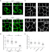
C70A mutation prevents AnkG retention in MDCK cells grown in low calcium. A, stable MDCK cells expressing WT or C70A AnkG-GFP were grown in low calcium medium overnight and then fixed and immunostained against GFP. B, stable MDCK cells expressing WT or C70A AnkG-GFP were grown in normal medium to reach a polarized monolayer and then fixed and immunostained against GFP. C, 60 ng of WT, C70A, DAR999AAA (DAR), and DAR999AAA/C70A double mutant (C70A, DAR) AnkG-GFP cDNA were transfected into MDCK cells in 14-mm MatTek plates. Cells were then fixed and immunostained against GFP 24 h post-transfection (green channel in grayscale). Scale bars, 10 μm for A–C. D, quantitative results of the fluorescence intensity ratio of plasma membrane versus cytoplasmic staining in AnkG stable MDCK cell lines. Data were compared using one-way ANOVA followed by a Tukey post hoc test (p < 0.001 for ANOVA, n = 26–31 from two independent experiments). *, p < 0.05; ns, not significant. E, quantitative results of the fluorescence intensity ratio of plasma membrane versus cytoplasmic staining of WT, C70A, DAR999AAA (DAR), and DAR999AAA/C70A double mutant (C70A, DAR) transfected into MDCK cells. Data were compared using one-way ANOVA followed by a Tukey post hoc test (p < 0.001 for ANOVA, n = 26–28 from three independent experiments). *, p < 0.05. Error bars, S.E.
FIGURE 5.
FIGURE 5.
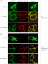
Cys-70 mutation does not affect AnkG interactions with E-cadherin or neurofascin. The membrane recruitment assay was used to monitor AnkG interactions with the interacting partners, E-cadherin and neurofascin. 80 ng of WT AnkG-GFP (A) or C70A/DAR999AAA AnkG-GFP (B) was transfected into HEK293 cells with or without (Control) 100 ng of binding partner (V5-E-cadherin or HA-neurofascin). Cells were then fixed and double-stained with anti-HA or -V5 (red) and anti-GFP (green) antibodies (scale bar, 10 μm).
FIGURE 6.
FIGURE 6.
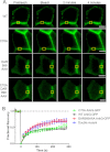
Effects of C70A mutation on AnkG dynamics at the lateral membrane. A, 300 ng of WT, C70A, DAR999AAA, and C70A/DAR999AAA double mutant AnkG-GFP were transfected into MDCK cells plated in 14-mm MatTek plates. FRAP was performed using an LSM780 laser-scanning confocal microscope. The representative graphs show the fluorescence recovery within 5 min; yellow boxes indicate the regions bleached. B, quantitative results of n = 10 repetitive measurements. The normalized fluorescence intensities were plotted against time and regressed against the one-phase exponential equation, y = y0 + Aek × t, using GraphPad Prism version 5 (scale bar, 10 μm). Error bars, S.E.
FIGURE 7.
FIGURE 7.
C70A mutation abolishes AnkG function in lateral membrane biosynthesis. A, doxycycline-inducible AnkG shRNA MDCK cell line was generated using an engineered Tet-pLKO-mCherry vector. Two days of doxycycline induction could selectively knock down 90% of ∼200-kDa AnkG. (Blot is representative of at least three independent experiments). B, XZ planes of lateral membrane under different conditions (green, AnkG; white, ZO-1, scale bar, 10 μm). AnkG knockdown cells show a defect in lateral membrane height compared with luciferase-shRNA cells. WT AnkG, but not the C70A mutant, is able to restore the height of lateral membrane. C, quantitative results of the height of lateral membranes. The height was defined as the distance between the bottom of ZO-1 staining and the end of the lateral membrane. Mean lateral membrane height was 9.8 ± 0.3 μm for luciferase shRNA, 3.4 ± 0.4 μm for AnkG shRNA, 9.1 ± 0.6 μm for WT-AnkG rescue, and 4.1 ± 0.5 μm for rescue with C70A AnkG-GFP. Conditions were compared using one-way ANOVA followed by a Tukey post hoc test (p < 0.001 for ANOVA, n = 24–30/condition). *, p < 0.05. Error bars, S.E.
FIGURE 8.
FIGURE 8.
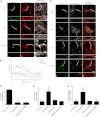
C70A mutation abolishes 270-kDa AnkG clustering and function at axon initial segments in hippocampal neurons. Neonatal mouse-derived hippocampal neurons were grown in tissue culture for 4–5 days following a standard protocol. Cells in MatTek plates were co-transfected with 500 ng of AnkG shRNA plasmids and 500 ng of wild type, C70A, or DAR999AAA 270-kDa AnkG-GFP plasmids or just 500 ng of AnkG shRNA plasmid alone (No Rescue). Two days after transfection, cells were fixed with 4% PFA in PBS buffer at room temperature for 15 min, permeabilized by methanol at −20 °C for 7 min, and processed for immunofluorescence. A, anti-GFP staining for 270-kDa AnkG-GFP shown in green, mCherry marking transfected neurons shown in red, and the axonal marker neurofilament shown in white (scale bar, 50 μm). Brackets mark the axon initial segment, which is shown at higher magnification below each image (scale bar, 10 μm). B, quantification of fluorescence intensity along the axons. The immunofluorescence intensity in the soma was normalized to 1. Average data from 10 neurons per condition are shown. C, quantification of the anti-GFP fluorescence intensity ratio of axons to dendrites in cells depleted of endogenous 270-kDa AnkG and rescued with WT, C70A, or DAR999AAA AnkG-GFP (n = 3; *, p < 0.05). D, anti-GFP staining for 270-kDa AnkG-GFP shown in green, anti-neurofascin staining shown in white, and mCherry marking transfected neurons shown in red. Axons were identified by morphology (scale bar, 50 μm). Brackets mark the axon initial segment, which is shown at higher magnification below each image (scale bar, 10 μm). E, quantification of the anti-endogenous neurofascin fluorescence intensity ratio of axons to dendrites in cells depleted of endogenous 270-kDa AnkG and rescued with mock (No Rescue), WT, C70A, or DAR999AAA AnkG-GFP (n = 3–4; *, p < 0.05). F, quantification of the anti-endogenous pan-sodium channel fluorescence intensity ratio of axons to dendrites in cells depleted of endogenous 270-kDa AnkG and rescued with mock (No Rescue), WT, C70A, or DAR999AAA AnkG-GFP (n = 5; *, p < 0.05). Error bars, S.E.

Similar articles

Cited by

References

    1. Bennett V., Healy J. (2008) Organizing the fluid membrane bilayer. Diseases linked to spectrin and ankyrin. Trends Mol. Med. 14, 28–36 - PubMed
    1. Jenkins S. M., Bennett V. (2001) Ankyrin-G coordinates assembly of the spectrin-based membrane skeleton, voltage-gated sodium channels, and L1 CAMs at Purkinje neuron initial segments. J. Cell Biol. 155, 739–746 - PMC - PubMed
    1. Zhou D., Lambert S., Malen P. L., Carpenter S., Boland L. M., Bennett V. (1998) AnkyrinG is required for clustering of voltage-gated Na channels at axon initial segments and for normal action potential firing. J. Cell Biol. 143, 1295–1304 - PMC - PubMed
    1. Pan Z., Kao T., Horvath Z., Lemos J., Sul J. Y., Cranstoun S. D., Bennett V., Scherer S. S., Cooper E. C. (2006) A common ankyrin-G-based mechanism retains KCNQ and NaV channels at electrically active domains of the axon. J. Neurosci. 26, 2599–2613 - PMC - PubMed
    1. Hedstrom K. L., Xu X., Ogawa Y., Frischknecht R., Seidenbecher C. I., Shrager P., Rasband M. N. (2007) Neurofascin assembles a specialized extracellular matrix at the axon initial segment. J. Cell Biol. 178, 875–886 - PMC - PubMed

Publication types

MeSH terms