Pansynaptic enlargement at adult cortical connections strengthened by experience
- PMID: 23118196
- PMCID: PMC3888373
- DOI: 10.1093/cercor/bhs334
Pansynaptic enlargement at adult cortical connections strengthened by experience
Abstract
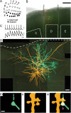
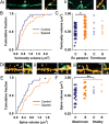
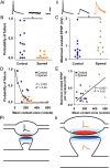
Behavioral experience alters the strength of neuronal connections in adult neocortex. These changes in synaptic strength are thought to be central to experience-dependent plasticity, learning, and memory. However, it is not known how changes in synaptic transmission between neurons become persistent, thereby enabling the storage of previous experience. A long-standing hypothesis is that altered synaptic strength is maintained by structural modifications to synapses. However, the extent of synaptic modifications and the changes in neurotransmission that the modifications support remain unclear. To address these questions, we recorded from pairs of synaptically connected layer 2/3 pyramidal neurons in the barrel cortex and imaged their contacts with high-resolution confocal microscopy after altering sensory experience by whisker trimming. Excitatory connections strengthened by experience exhibited larger axonal varicosities, dendritic spines, and interposed contact zones. Electron microscopy showed that contact zone size was strongly correlated with postsynaptic density area. Therefore, our findings indicate that whole synapses are larger at strengthened connections. Synaptic transmission was both stronger and more reliable following experience-dependent synapse enlargement. Hence, sensory experience modified both presynaptic and postsynaptic function. Our findings suggest that the enlargement of synaptic contacts is an integral part of long-lasting strengthening of cortical connections and, hence, of information storage in the neocortex.
Keywords: barrel cortex; confocal microscopy; electrophysiology; experience-dependent plasticity; structural plasticity.
Figures


Similar articles
-
Altered sensory experience induces targeted rewiring of local excitatory connections in mature neocortex.J Neurosci. 2008 Sep 10;28(37):9249-60. doi: 10.1523/JNEUROSCI.2974-08.2008. J Neurosci. 2008. PMID: 18784305 Free PMC article.
-
Imaging of experience-dependent structural plasticity in the mouse neocortex in vivo.Behav Brain Res. 2008 Sep 1;192(1):20-5. doi: 10.1016/j.bbr.2008.04.005. Epub 2008 Apr 18. Behav Brain Res. 2008. PMID: 18501438
-
Quantal analysis reveals a functional correlation between presynaptic and postsynaptic efficacy in excitatory connections from rat neocortex.J Neurosci. 2010 Jan 27;30(4):1441-51. doi: 10.1523/JNEUROSCI.3244-09.2010. J Neurosci. 2010. PMID: 20107071 Free PMC article.
-
Anatomical and physiological plasticity of dendritic spines.Annu Rev Neurosci. 2007;30:79-97. doi: 10.1146/annurev.neuro.30.051606.094222. Annu Rev Neurosci. 2007. PMID: 17280523 Review.
-
Nanoscale analysis of structural synaptic plasticity.Curr Opin Neurobiol. 2012 Jun;22(3):372-82. doi: 10.1016/j.conb.2011.10.019. Epub 2011 Nov 14. Curr Opin Neurobiol. 2012. PMID: 22088391 Free PMC article. Review.
Cited by
-
Differences in motor learning-related structural plasticity of layer 2/3 parvalbumin-positive interneurons of the young and aged motor cortex.Geroscience. 2024 Sep 30. doi: 10.1007/s11357-024-01350-6. Online ahead of print. Geroscience. 2024. PMID: 39343864
-
Sleep, synaptic homeostasis and neuronal firing rates.Curr Opin Neurobiol. 2017 Jun;44:72-79. doi: 10.1016/j.conb.2017.03.016. Epub 2017 Apr 8. Curr Opin Neurobiol. 2017. PMID: 28399462 Free PMC article. Review.
-
Three-Dimensional Synaptic Organization of Layer III of the Human Temporal Neocortex.Cereb Cortex. 2021 Aug 26;31(10):4742-4764. doi: 10.1093/cercor/bhab120. Cereb Cortex. 2021. PMID: 33999122 Free PMC article.
-
Evidence for sleep-dependent synaptic renormalization in mouse pups.Sleep. 2019 Oct 21;42(11):zsz184. doi: 10.1093/sleep/zsz184. Sleep. 2019. PMID: 31374117 Free PMC article.
-
Neurons in the barrel cortex turn into processing whisker and odor signals: a cellular mechanism for the storage and retrieval of associative signals.Front Cell Neurosci. 2015 Aug 21;9:320. doi: 10.3389/fncel.2015.00320. eCollection 2015. Front Cell Neurosci. 2015. PMID: 26347609 Free PMC article.
References
-
- Barnes SJ, Finnerty GT. Sensory experience and cortical rewiring. Neuroscientist. 2010;16:186–198. - PubMed
-
- Becker N, Wierenga CJ, Fonseca R, Bonhoeffer T, Nagerl UV. LTD induction causes morphological changes of presynaptic boutons and reduces their contacts with spines. Neuron. 2008;60:590–597. - PubMed
-
- Berning S, Willig KI, Steffens H, Dibaj P, Hell SW. Nanoscopy in a living mouse brain. Science. 2012;335:551. - PubMed

