

Microfluidic investigation of BDNF-enhanced neural stem cell chemotaxis in CXCL12 gradients
- PMID: 23109183
- PMCID: PMC3984949
- DOI: 10.1002/smll.201202208
Microfluidic investigation of BDNF-enhanced neural stem cell chemotaxis in CXCL12 gradients
Abstract
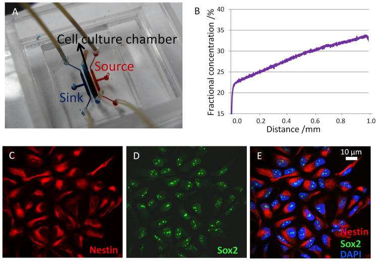
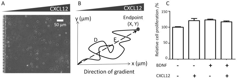
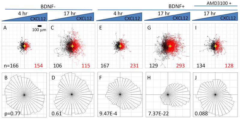
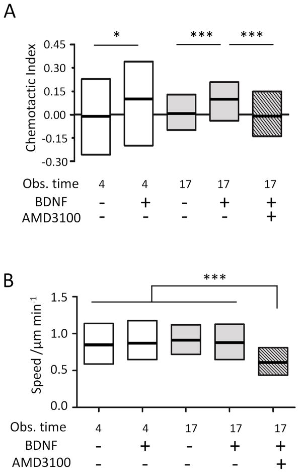
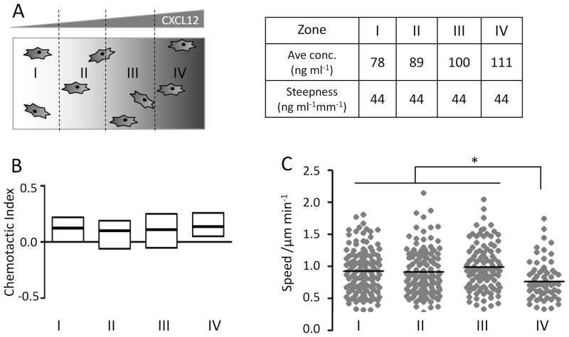
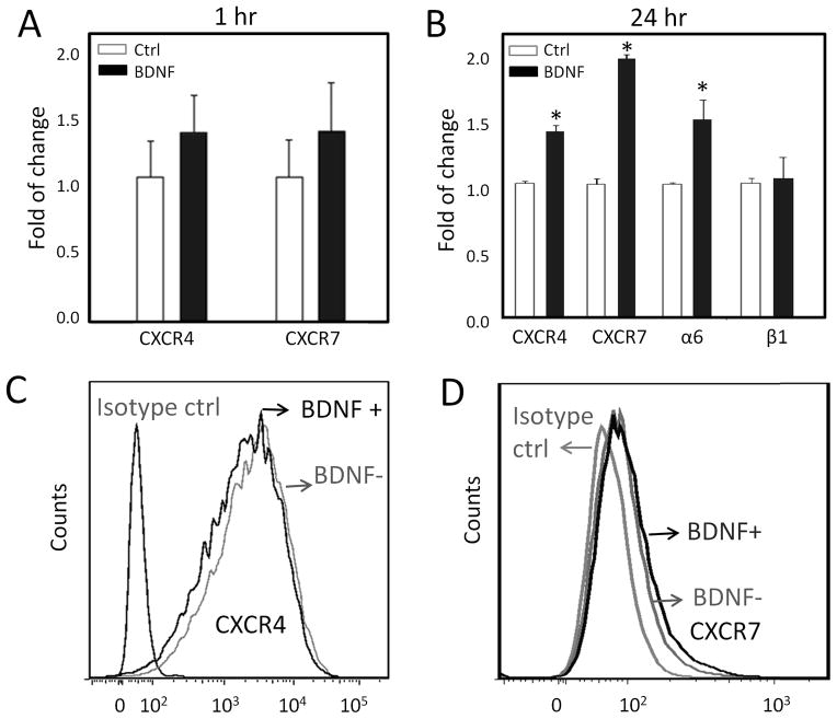
In vivo studies have suggested that gradients of CXCL12 (aka stromal cell-derived factor 1α) may be critical for neural stem cell (NSC) migration during brain development and neural tissue regeneration. However, traditional in vitro chemotaxis tools are limited by unstable concentration gradients and the inability to decouple cell migration directionality and speed. These limitations have restricted the reproducible and quantitative analysis of neuronal migration, which is required for mechanism-based studies. Using a microfluidic gradient generator, nestin and Sox-2 positive human embryonic NSC chemotaxis is quantified within a linear and stable CXCL12 gradient. While untreated NSCs are not able to chemotax within CXCL12 gradients, pre-treatment of the cells with brain-derived neurotrophic factor (BDNF) results in significant chemotactic, directional migration. BDNF pre-treatment has no effect on cell migration speed, which averages about 1 μm min(-1). Quantitative analysis determines that CXCL12 concentrations above 9.0 nM are above the minimum activation threshold, while concentrations below 14.7 nM are below the saturation threshold. Interestingly, although inhibitor studies with AMD 3100 revealed that CXCL12 chemotaxis requires receptor CXCR4 activation, BDNF pre-treatment is found to have no profound effects on the mRNA levels or surface presentation of CXCR4 or the putative CXCR7 scavenger receptor. The microfluidic study of NSC migration within stable chemokine concentration profiles provides quantitative analysis as well as new insight into the migratory mechanism underlying BDNF-induced chemotaxis towards CXCL12.
Copyright © 2013 WILEY-VCH Verlag GmbH & Co. KGaA, Weinheim.
Figures
Similar articles
-
CXCR7 Mediates Neural Progenitor Cells Migration to CXCL12 Independent of CXCR4.Stem Cells. 2015 Aug;33(8):2574-85. doi: 10.1002/stem.2022. Epub 2015 May 13. Stem Cells. 2015. PMID: 25833331 Free PMC article.
-
Microfluidic platform for chemotaxis in gradients formed by CXCL12 source-sink cells.Integr Biol (Camb). 2010 Nov;2(11-12):680-6. doi: 10.1039/c0ib00041h. Epub 2010 Sep 27. Integr Biol (Camb). 2010. PMID: 20871938 Free PMC article.
-
Chemotactic responses of neural stem cells to SDF-1α correlate closely with their differentiation status.J Mol Neurosci. 2014;54(2):219-33. doi: 10.1007/s12031-014-0279-6. Epub 2014 Mar 22. J Mol Neurosci. 2014. PMID: 24659235
-
Chemotaxis during neural crest migration.Semin Cell Dev Biol. 2016 Jul;55:111-8. doi: 10.1016/j.semcdb.2016.01.031. Epub 2016 Jan 25. Semin Cell Dev Biol. 2016. PMID: 26820523 Review.
-
Microfluidic technologies for temporal perturbations of chemotaxis.Annu Rev Biomed Eng. 2010 Aug 15;12:259-84. doi: 10.1146/annurev-bioeng-070909-105241. Annu Rev Biomed Eng. 2010. PMID: 20450351 Free PMC article. Review.
Cited by
-
Neuroprotective effects of physical activity on the brain: a closer look at trophic factor signaling.Front Cell Neurosci. 2014 Jun 20;8:170. doi: 10.3389/fncel.2014.00170. eCollection 2014. Front Cell Neurosci. 2014. PMID: 24999318 Free PMC article. Review.
-
The brain, sirtuins, and ageing.Nat Rev Neurosci. 2017 May 18;18(6):362-374. doi: 10.1038/nrn.2017.42. Nat Rev Neurosci. 2017. PMID: 28515492 Review.
-
Study on chemotaxis and chemokinesis of bone marrow-derived mesenchymal stem cells in hydrogel-based 3D microfluidic devices.Biomater Res. 2016 Aug 2;20:25. doi: 10.1186/s40824-016-0070-6. eCollection 2016. Biomater Res. 2016. PMID: 27489724 Free PMC article.
-
A Concentration Gradients Tunable Generator with Adjustable Position of the Acoustically Oscillating Bubbles.Micromachines (Basel). 2020 Aug 31;11(9):827. doi: 10.3390/mi11090827. Micromachines (Basel). 2020. PMID: 32878158 Free PMC article.
-
Prenatal Environment That Affects Neuronal Migration.Front Cell Dev Biol. 2019 Jul 17;7:138. doi: 10.3389/fcell.2019.00138. eCollection 2019. Front Cell Dev Biol. 2019. PMID: 31380373 Free PMC article. Review.
References
-
- Martino G, Pluchino S. Nat Rev Neurosci. 2006;7(5):395–406. - PubMed
- Imitola J, Raddassi K, Park KI, Mueller FJ, Nieto M, Teng YD, Frenkel D, Li J, Sidman RL, Walsh CA, Snyder EY, Khoury SJ. Proceedings of the National Academy of Sciences of the United States of America. 2004;101(52):18117–22. - PMC - PubMed
Publication types
MeSH terms
Substances
Grants and funding
LinkOut - more resources
Full Text Sources
Other Literature Sources
Miscellaneous
