. 2013 Feb;12(1):112-20.
doi: 10.1111/acel.12024. Epub 2012 Nov 23.

Caenorhabditis elegans HSF-1 is an essential nuclear protein that forms stress granule-like structures following heat shock

Affiliations

Caenorhabditis elegans HSF-1 is an essential nuclear protein that forms stress granule-like structures following heat shock

Elizabeth A Morton et al. Aging Cell. 2013 Feb.

Abstract

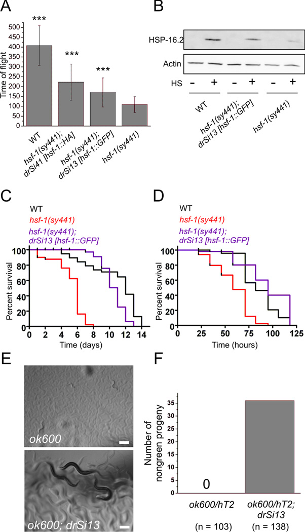
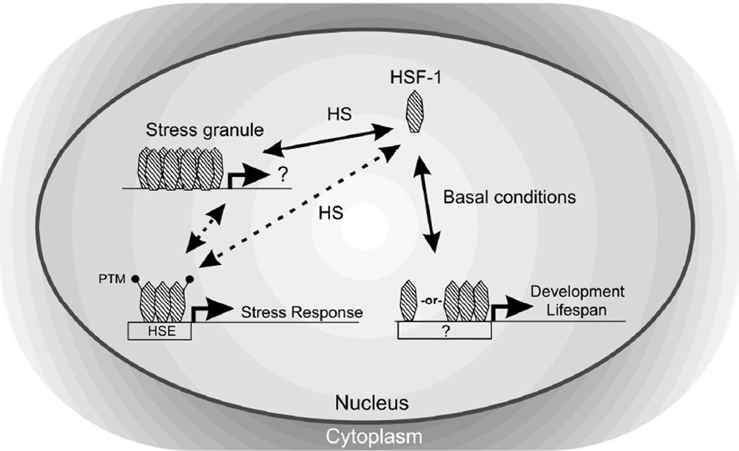
The heat shock transcription factor (HSF) is a conserved regulator of heat shock-inducible gene expression. Organismal roles for HSF in physiological processes such as development, aging, and immunity have been defined largely through studies of the single Caenorhabditis elegans HSF homolog, hsf-1. However, the molecular and cell biological properties of hsf-1 in C. elegans are incompletely understood. We generated animals expressing physiological levels of an HSF-1::GFP fusion protein and examined its function, localization, and regulation in vivo. HSF-1::GFP was functional, as measured by its ability to rescue phenotypes associated with two hsf-1 mutant alleles. Rescue of hsf-1 development phenotypes was abolished in a DNA-binding-deficient mutant, demonstrating that the transcriptional targets of hsf-1 are critical to its function even in the absence of stress. Under nonstress conditions, HSF-1::GFP was found primarily in the nucleus. Following heat shock, HSF-1::GFP rapidly and reversibly redistributed into dynamic, subnuclear structures that share many properties with human nuclear stress granules, including colocalization with markers of active transcription. Rapid formation of HSF-1 stress granules required HSF-1 DNA-binding activity, and the threshold for stress granule formation was altered by growth temperature. HSF-1 stress granule formation was not induced by inhibition of IGF signaling, a pathway previously suggested to function upstream of hsf-1. Our findings suggest that development, stress, and aging pathways may regulate HSF-1 function in distinct ways, and that HSF-1 nuclear stress granule formation is an evolutionarily conserved aspect of HSF-1 regulation in vivo.


Figures

Figure 1
Figure 1. HSF-1::GFP is broadly expressed and condenses into nuclear granules following heat shock
(A) Diagram of the hsf-1p::hsf-1(cDNA)::GFP::unc-54 3’UTR expression construct (drSi13) used in this study. hsf-1p is 4 kb of sequence upstream of the hsf-1 start ATG. Notations above the construct diagram indicate changes made to the transgene (R to A at residue 145 and HA insertion after residue 370); notations below indicate relative positions of mutations in the endogenous gene (diagram not to scale.) (B) qRT-PCR comparing wild type (N2) and HSF-1::GFP hsf-1 mRNA levels relative to actin mRNA, normalized to N2. Shown is the mean relative expression ± SEM in drSi13 for three independent experiments. hsf-1 mRNA in a wild-type background is less than double control wild type, suggesting compensatory mechanisms acting on hsf-1 expression. (C) HSF-1::GFP localizes primarily to the nucleus at 20°C. (D) After 1 min of 35°C heat shock, HSF-1::GFP collects into nuclear puncta (arrows). Shown are four merged (Z-dimension) deconvolved slices depicting hypodermal nuclei. Scale bar = 5μm. (E) HSF-1::GFP granule size in hypodermal cells after 1 min 35°C heat shock (N = 349 granules). (F) Number of HSF-1::GFP granules per cell in hypodermal cells after 1 min 35°C heat shock. (N = 100 nuclei). (G-I) drSi41, a single copy line expressing hsf-1p::hsf-1::HA::unc-54 3’UTR, in which the HA tag is inserted into the region between the putative trimerization and transactivation domains of the hsf-1 cDNA. drSi41 worms were either heat shocked (G, 30 min at 35°C) or not (F, 30 min at 25°C), dissected, fixed, probed for HA (red, G-I), and stained with Hoechst dye (blue, G’-I’). Heat shocked N2 worms are shown as a control (I). Shown are nine merged (Z-dimension) deconvolved slices. Dotted line indicates outline of nuclei as determined by Hoechst. Scale bar = 5 µm.
Figure 2
Figure 2. HSF-1::GFP is functional
(A) Wild type (WT), hsf-(sy441);drSi41[hsf-1p::hsf-1::HA::unc-54 3’UTR], hsf-(sy441);drSi13[hsf-1p::hsf-1::GFP::unc-54 3’UTR], and hsf-1(sy441) eggs were placed at 25°C and allowed to grow until wild type was L4/young adult. Worms were analyzed for size (time of flight) in a COPAS Biosort (N ≥ 89 animals. Mean ± SD, *** - p < 0.001 as compared to hsf-1(sy441)). (B) Representative Western against HSP-16.2 (top panel) and β-actin (bottom panel) on WT, hsf-1(sy441);drSi13, and hsf-1(sy441) worms ± a 35°C 3h heat shock followed by 3 hr recovery at 16°C. Relative HSP-16.2:actin ratio for WT : hsf-1(sy441);drSi13 : hsf-1(sy441) is 1.0:0.67:0.22. (C) Lifespan of WT, hsf-1(sy441), and hsf-1(sy441);drSi13 animals at 25°C (N = 50 for all). (p < 0.0001 between hsf-1(sy441) and hsf-1(sy441);drSi13; p = 0.0036 between WT and hsf-1(sy441);drSi13) (D) Survival of WT, hsf-1(sy441), and hsf-1(sy441); drSi13 animals on P. aeruginosa PA14 at 25°C (N = 50 for all) (p < 0.0001 between hsf-1(sy441) and hsf-1(sy441);drSi13; p = 0.498 between WT and hsf-1(sy441);drSi13). (E) Images of ok600 homozygous animals with (lower) or without (upper) the drSi13 HSF-1::GFP transgene. Scale bar = 100μm. (F) Quantification of the number of ok600 homozygous animals that reach L4 stage or later within 3 days at 20°C with or without the drSi13 HSF-1::GFP transgene.
Figure 3
Figure 3. Nuclear granules form in response to heat shock and sodium azide
Worms expressing drSi13 HSF-1::GFP were anesthetized 30 min in 1 mM levamisole at either room temperature (A), 35°C (HS) (B), 5 mM sodium azide (C), 219 mM NaCl (D), 100 μM CdCl2 (E), or 100 mM ethanol (F). Scale bar = 5μm. (G) Percent of hypodermal nuclei with ≥ one visible granule were quantified for each condition (N ≥ 10 worms per condition, representing ≥ 85 nuclei. Mean ± SEM, *** - p < 0.001 vs. control, n.s. - not significant). (H-M) TJ375 (hsp-16.2p::GFP) worms were subjected to 30 min of the same conditions as in A-F, followed by recovery at 20°C for 4 hr before imaging. Scale bar = 100μm.
Figure 4
Figure 4. HSF-1 DNA binding promotes stress granule formation and developmental rescue of hsf-1(sy441)
(A) Alignment of the region of the DNA binding domain containing R145 (red) from the indicated species. (B,C) Images of drSi13 HSF-1::GFP (WT) and drSi28 HSF-1(R145A)::GFP (R145A) taken after a 1 min 35°C heat shock, showing granule formation (arrow). Scale bar = 5μm. Shown are four merged (Z-dimension) deconvolved slices. (D) Number of granules per nucleus was quantified for WT and R145A. (N ≥ 18 worms, representing ≥ 140 nuclei for each line. Mean ± SD, *** - p < 0.001). (E) N2 wild type (WT), hsf-1(sy441), and hsf-1(sy441);drSi28 eggs were placed at 25°C and allowed to grow until wild type was L4/young adult. Worms were analyzed for size (time of flight) in a COPAS Biosort (N ≥ 30 animals. Mean ± SD, *** - p < 0.001, n.s. - not significant). (F) Representative Western against HSP-16.2 (top panel) and β-actin (bottom panel) on young adult wild type, hsf-1(sy441);drSi28, and hsf-1(sy441) worms ± a 3 hr 35°C heat shock followed by 3 hr recovery at 16°C.
Figure 5
Figure 5. HSF-1::GFP granules colocalize with markers of active transcription
drSi13 HSF-1::GFP worms grown at 25°C were heat shocked for 1.5 hr at 35°C (HS) or put at 25°C 1.5 hr (no HS) and intestinal nuclei were probed for GFP (green, B,F,J,N) histone H2A acetylated on Lysine 5 (red, A,E), or RNA polymerase II phosphorylated on Serine 2 (red, I,M. Exposure times in I and M were different because we observed substantially reduced RNA polII Ser2p staining post-heat shock). Nuclei (intestinal) were detected by Hoechst staining (D,H,L,P). Nuclear staining showed puncta of fluorescence, some of which show colocalization of GFP and an active transcription marker (arrows). Other puncta exhibit GFP-only (carrot) or active transcription marker-only (open arrowhead) staining. Scale bar = 5μm.
Figure 6
Figure 6. Model for HSF-1 regulation in C. elegans.
HSF-1 is a predominately nuclear protein in C. elegans, and its modes of activity under basal conditions and stress conditions (HS) differ. Stress-inducible activity is distinguished by stress granule formation, oligomerization, and post-translational modification of HSF-1. Due to its oligomeric nature, we hypothesize that physiological levels of the DNA binding-deficient HSF-1(R145A) can still associate with the active HSF-1 complex and contribute transactivation function to stress-inducible targets in trans. The observation that HSF-1(R145A) cannot rescue developmental defects in the sy441 transactivation-deficient background suggests that HSF-1 activity in the context of development may not operate in trans. Basal targets of HSF-1, including genes involved in development and possibly lifespan, require DNA binding activity, but may not involve stress granule formation or oligomerization.

Similar articles

Cited by

References

    1. Akerfelt M, Morimoto RI, Sistonen L. Heat shock factors: integrators of cell stress, development and lifespan. Nat Rev Mol Cell Biol. 2010;11:545–555. - PMC - PubMed
    1. Alavez S, Vantipalli MC, Zucker DJ, Klang IM, Lithgow GJ. Amyloid-binding compounds maintain protein homeostasis during ageing and extend lifespan. Nature. 2011;472:226–229. - PMC - PubMed
    1. Baler R, Dahl G, Voellmy R. Activation of human heat shock genes is accompanied by oligomerization, modification, and rapid translocation of heat shock transcription factor HSF1. Molecular and cellular biology. 1993;13:2486–2496. - PMC - PubMed
    1. Biamonti G, Vourc'h C. Nuclear stress bodies. Cold Spring Harbor perspectives in biology. 2010;2:a000695. - PMC - PubMed
    1. Chiang WC, Ching TT, Lee HC, Mousigian C, Hsu AL. HSF-1 regulators DDL-1/2 link insulin-like signaling to heat-shock responses and modulation of longevity. Cell. 2012;148:322–334. - PMC - PubMed

Publication types

Substances

LinkOut - more resources