

Diurnal variations in articular cartilage thickness and strain in the human knee
- PMID: 23102493
- PMCID: PMC3552039
- DOI: 10.1016/j.jbiomech.2012.09.013
Diurnal variations in articular cartilage thickness and strain in the human knee
Abstract
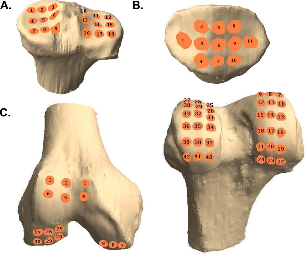
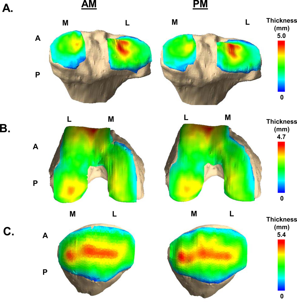
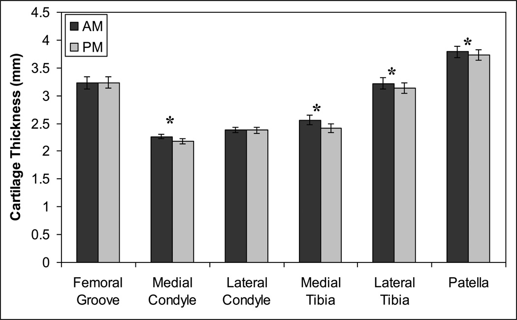
Due to the biphasic viscoelastic nature of cartilage, joint loading may result in deformations that require times on the order of hours to fully recover. Thus, cartilaginous tissues may exhibit cumulative strain over the course of each day. The goal of this study was to assess the magnitude and spatial distribution of strain in the articular cartilage of the knee with daily activity. Magnetic resonance (MR) images of 10 asymptomatic subjects (six males and four females) with mean age of 29 years were obtained at 8:00 AM and 4:00 PM on the same day using a 3T magnet. These images were used to create 3D models of the femur, tibia, and patella from which cartilage thickness distributions were quantified. Cartilage thickness generally decreased from AM to PM in all areas except the patellofemoral groove and was associated with significant compressive strains in the medial condyle and tibial plateau. From AM to PM, cartilage of the medial tibial plateau exhibited a compressive strain of -5.1±1.0% (mean±SEM) averaged over all locations, while strains in the lateral plateau were slightly lower (-3.1±0.6%). Femoral cartilage showed an average strain of -1.9±0.6%. The findings of this study show that human knee cartilage undergoes diurnal changes in strain that vary with site in the joint. Since abnormal joint loading can be detrimental to cartilage homeostasis, these data provide a baseline for future studies investigating the effects of altered biomechanics on diurnal cartilage strains and cartilage physiology.
Copyright © 2012 Elsevier Ltd. All rights reserved.
Conflict of interest statement
Figures


Similar articles
-
High body mass index is associated with increased diurnal strains in the articular cartilage of the knee.Arthritis Rheum. 2013 Oct;65(10):2615-22. doi: 10.1002/art.38062. Arthritis Rheum. 2013. PMID: 23818303 Free PMC article.
-
In vivo measurement of localized tibiofemoral cartilage strains in response to dynamic activity.Am J Sports Med. 2015 Feb;43(2):370-6. doi: 10.1177/0363546514559821. Epub 2014 Dec 10. Am J Sports Med. 2015. PMID: 25504809 Free PMC article.
-
Knee Cartilage Thickness, T1ρ and T2 Relaxation Time Are Related to Articular Cartilage Loading in Healthy Adults.PLoS One. 2017 Jan 11;12(1):e0170002. doi: 10.1371/journal.pone.0170002. eCollection 2017. PLoS One. 2017. PMID: 28076431 Free PMC article.
-
In vivo morphometry and functional analysis of human articular cartilage with quantitative magnetic resonance imaging--from image to data, from data to theory.Anat Embryol (Berl). 2001 Mar;203(3):147-73. doi: 10.1007/s004290000154. Anat Embryol (Berl). 2001. PMID: 11303902 Review.
-
Bony and cartilaginous anatomy of the patellofemoral joint.Knee Surg Sports Traumatol Arthrosc. 2006 Mar;14(3):235-40. doi: 10.1007/s00167-005-0683-0. Epub 2005 Oct 28. Knee Surg Sports Traumatol Arthrosc. 2006. PMID: 16254736 Review.
Cited by
-
The effects of a 6-month weight loss intervention on physical function and serum biomarkers in older adults with and without osteoarthritis.Osteoarthr Cartil Open. 2023 May 26;5(3):100376. doi: 10.1016/j.ocarto.2023.100376. eCollection 2023 Sep. Osteoarthr Cartil Open. 2023. PMID: 37719442 Free PMC article.
-
[Limits of kinematic alignment and recommendations for its safe application].Orthopade. 2020 Jul;49(7):617-624. doi: 10.1007/s00132-020-03931-7. Orthopade. 2020. PMID: 32494904 Review. German.
-
Femoral cartilage ultrasound echo-intensity is a valid measure of cartilage composition.J Orthop Res. 2024 Apr;42(4):729-736. doi: 10.1002/jor.25722. Epub 2023 Nov 4. J Orthop Res. 2024. PMID: 37874323
-
Effects of cartilage impact with and without fracture on chondrocyte viability and the release of inflammatory markers.J Orthop Res. 2013 Aug;31(8):1283-92. doi: 10.1002/jor.22348. Epub 2013 Apr 25. J Orthop Res. 2013. PMID: 23620164 Free PMC article.
-
The Influence of Weather Conditions on the Diurnal Variation in Range of Motion in Older Adults with Knee Osteoarthritis.J Clin Med. 2024 Jan 1;13(1):254. doi: 10.3390/jcm13010254. J Clin Med. 2024. PMID: 38202261 Free PMC article.
References
-
- Aaboe J, Bliddal H, Messier SP, Alkjaer T, Henriksen M. Effects of an intensive weight loss program on knee joint loading in obese adults with knee osteoarthritis. Osteoarthritis and cartilage / OARS, Osteoarthritis Research Society. 2011;19:822–828. - PubMed
-
- Abebe ES, Moorman CT, 3rd, Dziedzic TS, Spritzer CE, Cothran RL, Taylor DC, Garrett WE, Jr, DeFrate LE. Femoral tunnel placement during anterior cruciate ligament reconstruction: an in vivo imaging analysis comparing transtibial and 2-incision tibial tunnel-independent techniques. Am J Sports Med. 2009;37:1904–1911. - PubMed
-
- Anandacoomarasamy A, Leibman S, Smith G, Caterson I, Giuffre B, Fransen M, Sambrook PN, March L. Weight loss in obese people has structure-modifying effects on medial but not on lateral knee articular cartilage. Annals of the rheumatic diseases. 2012;71:26–32. - PubMed
-
- Andriacchi TP, Lang PL, Alexander EJ, Hurwitz DE. Methods for evaluating the progression of osteoarthritis. J Rehabil Res Dev. 2000;37:163–170. - PubMed
-
- Andriacchi TP, Mundermann A, Smith RL, Alexander EJ, Dyrby CO, Koo S. A framework for the in vivo pathomechanics of osteoarthritis at the knee. Annals of biomedical engineering. 2004;32:447–457. - PubMed
Publication types
MeSH terms
Grants and funding
LinkOut - more resources
Full Text Sources
Other Literature Sources
