. 2013 Jan;81(1):154-65.
doi: 10.1128/IAI.01080-12. Epub 2012 Oct 22.

Genome expression analysis of nonproliferating intracellular Salmonella enterica serovar Typhimurium unravels an acid pH-dependent PhoP-PhoQ response essential for dormancy

Affiliations

Genome expression analysis of nonproliferating intracellular Salmonella enterica serovar Typhimurium unravels an acid pH-dependent PhoP-PhoQ response essential for dormancy

Cristina Núñez-Hernández et al. Infect Immun. 2013 Jan.

Abstract

Genome-wide expression analyses have provided clues on how Salmonella proliferates inside cultured macrophages and epithelial cells. However, in vivo studies show that Salmonella does not replicate massively within host cells, leaving the underlying mechanisms of such growth control largely undefined. In vitro infection models based on fibroblasts or dendritic cells reveal limited proliferation of the pathogen, but it is presently unknown whether these phenomena reflect events occurring in vivo. Fibroblasts are distinctive, since they represent a nonphagocytic cell type in which S. enterica serovar Typhimurium actively attenuates intracellular growth. Here, we show in the mouse model that S. Typhimurium restrains intracellular growth within nonphagocytic cells positioned in the intestinal lamina propria. This response requires a functional PhoP-PhoQ system and is reproduced in primary fibroblasts isolated from the mouse intestine. The fibroblast infection model was exploited to generate transcriptome data, which revealed that ∼2% (98 genes) of the S. Typhimurium genome is differentially expressed in nongrowing intracellular bacteria. Changes include metabolic reprogramming to microaerophilic conditions, induction of virulence plasmid genes, upregulation of the pathogenicity islands SPI-1 and SPI-2, and shutdown of flagella production and chemotaxis. Comparison of relative protein levels of several PhoP-PhoQ-regulated functions (PagN, PagP, and VirK) in nongrowing intracellular bacteria and extracellular bacteria exposed to diverse PhoP-PhoQ-inducing signals denoted a regulation responding to acidic pH. These data demonstrate that S. Typhimurium restrains intracellular growth in vivo and support a model in which dormant intracellular bacteria could sense vacuolar acidification to stimulate the PhoP-PhoQ system for preventing intracellular overgrowth.


Figures
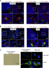
Fig 1
Fig 1
S. Typhimurium attenuates growth inside nonphagocytic cells positioned in the lamina propria of intestinal villi. (A) Tissue sections of the intestinal ileum corresponding to Peyer's patch areas were labeled with antibodies recognizing S. Typhimurium lipopolysaccharide (LPS) and the panphagocytic marker CD18 or CD45. To-pro3 was used to stain nuclei. Samples were collected at 6 or 24 h postchallenge of BALB/c mice with the SV5015 (wild-type) and MD1120 (phoP mutant) strains. Areas in boxes in upper panels are magnified in the lower panels. (B) Tissue sections showing bacterium-containing cells in the lamina propria of intestinal villi. Samples were collected at 6 or 24 h postinfection as described for panel A and were labeled with antibodies against S. Typhimurium LPS, CD18, or CD45. Note the presence of nonphagocytic stromal cells containing large numbers of intracellular phoP mutant bacteria. Areas in boxes in upper panels are magnified in the lower panels. (C) Morphology of primary intestinal fibroblasts isolated from intestinal tissue. These primary fibroblasts were infected with the SV5015 (wild-type) or MD1120 (phoP mutant) strain. In parallel, NRK-49F fibroblasts were also infected with the same strains. Bacteria were detected with anti-S. Typhimurium LPS antibodies, and nuclei were stained with 4′,6′-diamidino-2-phenylindole (DAPI). Note the similar bacterial phenotypes in both types of fibroblasts. L, intestinal lumen.
Fig 2
Fig 2
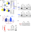
Genome-wide expression analyses reveal unique signatures in nongrowing intracellular wild-type S. Typhimurium. (A) Number of genes displaying expression changes higher than 4-fold (log2 = M value of ≤−2 or ≥2) in intracellular nongrowing bacteria (wild-type strain SV5015) or intracellular proliferating bacteria (phoP mutant strain MD1120) at 24 h postinfection of NRK-49F fibroblasts. A third sample, corresponding to extracellular bacteria grown overnight to stationary phase in LB medium in shaking conditions, was included for comparison. The three samples are referenced to the expression pattern displayed by actively growing extracellular bacteria grown in LB medium to an OD of 0.2 (exponential phase) (see the text for details). The number of genes that displayed up- and downregulation compared to bacteria grown to exponential phase are also indicated. (B) Heat map showing the upregulation in intracellular bacteria of genes mapping in the virulence plasmid pSLT. None that, relative to extracellular bacteria, pSLT plasmid genes are upregulated to a greater extent in nongrowing intracellular wild-type bacteria than in intracellular phoP mutant bacteria (also see Table S2 in the supplemental material). A representative case confirming this difference is shown for the TlpA (PSLT048) plasmid protein. (C) Heat map showing the expression changes of gene clusters encoding distinct fimbriae or flagellar proteins. Note the downregulation in the expression of flagellar and chemotaxis genes in intracellular wild-type and mutant bacteria (both wild-type and phoP mutant strains) (see Table S2). A Western assay demonstrating the marked drop in flagellin (FliC/FljB) relative levels in intracellular bacteria is shown.
Fig 3
Fig 3
Characterization of the S. Typhimurium PhoP-PhoQ regulon in dormant nongrowing intracellular bacteria. (A) Venn diagram showing the overlapping among transcriptomes obtained from intracellular nongrowing bacteria (wild-type strain SV5015), intracellular proliferating bacteria (phoP mutant strain MD1120), and extracellular wild-type bacteria grown to stationary phase in LB medium. Each of these transcriptomes refers to the expression of extracellular bacteria grown to exponential phase (see Table S2 in the supplemental material). Numbers of genes differing in expression among transcriptomes by more than 4-fold are indicated. Highlighted in boxes are some of the genes displaying differential expression among transcriptomes (intra-WT versus extra-WT and intra-WT versus intra-phoP), which respond to both the intracellular environment and the functional status of the PhoP-PhoQ system. Genes previously reported to be regulated by PhoP-PhoQ are indicated in blue (see Tables S6 and S7 for details). (B) Validation data obtained by RT-qPCR for two PhoP-PhoQ-regulated genes, mgtC and pagC. Two other genes hitherto not assigned to the PhoP-PhoQ regulon, ushA and glpK, were also validated at the transcript and protein levels, respectively. mgtC and pagC data refer to different postinfection times (1, 4, 8, and 24 h) upon invasion of NRK-49F fibroblasts and are relative to expression levels detected in extracellular bacteria growing to exponential phase. ushA data are relative to the expression levels registered at 24 h postinfection in nongrowing intracellular bacteria. In the RT-qPCR assays, expression values were normalized to those obtained for the ompA gene used as an internal control. (C) Relative levels of the MgtC-3×FLAG- and PagC-3×FLAG-tagged proteins detected in intracellular nongrowing wild-type bacteria and in the overgrowing phoP mutant at the indicated postinfection times. As a control of canonical PhoP-PhoQ regulation, these two proteins were also monitored in extracellular bacteria grown in inducing (8 μM Mg2+) or repressing (10 mM Mg2+) conditions. (D) Levels of the alternative sigma factor RpoS detected in intracellular bacteria at different postinfection times upon entry into NRK-49F fibroblasts. DnaK, OmpA (bacterial proteins), and calnexin (eukaryotic protein) were used as loading controls.
Fig 4
Fig 4
Regulation exerted by the PhoP-PhoQ system in nongrowing dormant intracellular bacteria matches the regulatory pattern observed in extracellular bacteria incubated in acidified growth medium. Western assays showing the relative levels of three distinct 3× FLAG-tagged proteins regulated by the PhoP-PhoQ system (VirK, PagN, and PagP) in extracellular and intracellular bacteria. Induction in intracellular bacteria was monitored by analysis of protein levels in extracellular bacteria used to infect the NRK-49F fibroblasts (inoculum, nonshaking growth conditions) and intracellular bacteria collected at 24 h postinfection. These samples are marked as extra and intra, respectively. The positive regulation of these three proteins by PhoP-PhoQ was tested in low Mg2+ concentrations and acid pH using the N and PCN media, respectively (see Materials and Methods). Shown are the levels of VirK, PagN, and PagP detected in extracellular bacteria grown in inducing (either 8 μM Mg2+ or pH 5.8) or repressing (either 10 mM Mg2+ or pH 7.4) conditions. Note that the response observed in acidified PCN medium matches, to a large extent, that observed in intracellular bacteria. However, the marked increase of PagP levels observed in 8 μM Mg2+ is not observed in nonproliferating intracellular bacteria. Loading controls based on DnaK are shown for the pagN::3×FLAG-tagged strains with equivalent results obtained for the other sets of strains shown.
Fig 5
Fig 5
Activity of the PhoP-PhoQ system in nonproliferating intracellular S. Typhimurium responds to intravacuolar acidic pH. (A) Effect of the dissipation of intravacuolar acidification on the induction of the PhoP-PhoQ system. Shown are the relative levels of the 3× FLAG-tagged proteins MgtC and PagC produced by intracellular bacteria isolated from NRK-49F fibroblasts that were left untreated or were treated with 100 nM bafilomycin (BAF), an inhibitor of vacuolar acidification. OmpA (bacterial protein) and calnexin (eukaryotic protein) were used for loading controls. (B) Effect of loss of vacuolar acidification on the viability of intracellular bacteria. Shown are the ratios of viable intracellular bacteria enumerated at 24 h versus 2 h. Data are the means and standard deviations from three independent experiments. **, P = 0.001 to 0.01; ***, P < 0.001; n.s., not significant by a Student t test.

Similar articles

Cited by

References

    1. Haraga A, Ohlson MB, Miller SI. 2008. Salmonellae interplay with host cells. Nat. Rev. Microbiol. 6:53–66 - PubMed
    1. Tsolis RM, Xavier MN, Santos RL, Baumler AJ. 2011. How to become a top model: impact of animal experimentation on human Salmonella disease research. Infect. Immun. 79:1806–1814 - PMC - PubMed
    1. Valdez Y, Ferreira RB, Finlay BB. 2009. Molecular mechanisms of Salmonella virulence and host resistance. Curr. Top. Microbiol. Immunol. 337:93–127 - PubMed
    1. Kaiser P, Diard M, Stecher B, Hardt WD. 2012. The streptomycin mouse model for Salmonella diarrhea: functional analysis of the microbiota, the pathogen's virulence factors, and the host's mucosal immune response. Immunol. Rev. 245:56–83 - PubMed
    1. Valdez Y, Grassl GA, Guttman JA, Coburn B, Gros P, Vallance BA, Finlay BB. 2009. Nramp1 drives an accelerated inflammatory response during Salmonella-induced colitis in mice. Cell. Microbiol. 11:351–362 - PubMed

Publication types

MeSH terms