. 2013 Jan 20;435(2):239-49.
doi: 10.1016/j.virol.2012.09.024. Epub 2012 Oct 22.

HCMV gB shares structural and functional properties with gB proteins from other herpesviruses

Affiliations

HCMV gB shares structural and functional properties with gB proteins from other herpesviruses

Sapna Sharma et al. Virology. .

Abstract

Glycoprotein B (gB) facilitates HCMV entry into cells by binding receptors and mediating membrane fusion. The crystal structures of gB ectodomains from HSV-1 and EBV are available, but little is known about the HCMV gB structure. Using multiangle light scattering and electron microscopy, we show here that HCMV gB ectodomain is a trimer with the overall shape similar to HSV-1 and EBV gB ectodomains. HCMV gB ectodomain forms rosettes similar to rosettes formed by EBV gB and the postfusion forms of other viral fusogens. Substitution of several bulky hydrophobic residues within the putative fusion loops with more hydrophilic residues reduced rosette formation and abolished cell fusion. We propose that like gB proteins from HSV-1 and EBV, HCMV gB has two internal hydrophobic fusion loops that likely interact with target membranes. Our work establishes structural and functional similarities between gB proteins from three subfamilies of herpesviruses.


Figures

Fig. 1
Fig. 1
Schematic view of the full-length HCMV gB and constructs used in this work. SS – signal sequence, MP – membrane-proximal region, TM – transmembrane region, Cyto – cytoplasmic domain. 18 predicted N-linked glycosylation sites are shown as lollipops.
Fig. 2
Fig. 2
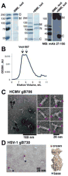
Properties of the HCMV gB706-CHis6. (A). Coomassie-stained gels and a Western blot of purified HCMV gB706-CHis6. Under non-reducing mildly denaturing conditions (-BME, unboiled), the protein migrates as a mixture of monomer and trimer. Under reducing denaturing conditions (+BME, boiled), some protein migrates as an intact monomer while a fraction separates into the N-terminal and C-terminal fragments. O – oligomer, M – monomer, N – N-terminal fragment, C – C-terminal fragment. Molecular size standards are labeled. (B). Size-exclusion chromatogram of purified gB706-CHis6. gB elutes as a broad peak near the void volume. The elution volumes of the void and the 667 kDa standard are shown (arrows). (C). Electron micrograph of purified gB706-CHis6. Arrows indicate long rods (D, dimer of trimers) and three-armed particles (T, trimer of trimers). Close-up view of selected rods and three-armed particles is shown below. Pink asterisks mark crown ends. (D). Electron micrograph of HSV-1 gB730 along with its crystal structure, in surface representation. Pink asterisks mark crown ends, and blue asterisks mark fusion-loop-containing base ends.
Fig. 3
Fig. 3
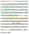
Sequence alignment of gB proteins from HSV-1 (strain KOS), EBV (strain B958), and HCMV (strain AD169), generated using ClustalW2 (Larkin et al., 2007). Residue numbers for HSV-1 gB and HCMV gB are shown above and below their respective sequences. Secondary structure of HSV-1 gB is shown above the sequence alignment, and its coloring matches that in reference (Heldwein et al., 2006). Signal sequences, absent from mature glycoproteins, are shown in red. Identical residues are highlighted in yellow; similar residues, in grey; cysteines, in green; and putatively glycosylated asparagines, in blue. Furin cleavage sites in HCMV and EBV are highlighted in purple. Residues in two fusion loop regions are shown in teal. Figure was generated in ALSCRIPT (Barton, 1993).
Fig. 4
Fig. 4
Fusion loop regions in HCMV gB and HSV-1 gB ectodomains. (A). Crystal structure of HSV-1 gB ectodomain, gB730, is shown side-by-side with a homology model of HCMV gB ectodomain, gB706, in surface representations. Fusion loop regions are shown in side and face-on views. Hydrophobic residues (yellow), positively charged residues (blue), negatively charged residues (red) and histidines (cyan) are shown. Side chains of mutated residues in HCMV gB706-4M mutant and their HSV-1 counterparts are labeled (in one protomer only, for clarity). Figures were generated using PyMol (www.pymol.org). (B). Sequence alignment of the fusion loop regions in HCMV gB, HCMV gB706-4M mutant, HSV-1 gB, and EBV gB. Hydrophobic residues (orange), positively charged residues (blue), negatively charged residues (red), and histidines (cyan) are colored. Mutated residues in HCMV gB and their HSV-1 gB counterparts are underlined.
Fig. 5
Fig. 5
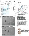
Properties of the quadruple mutant gB706-4M. (A). Coomassie-stained gel of purified gB706-4M. Under non-reducing mildly denaturing conditions (-BME, unboiled), the protein migrates as a mixture of monomer and trimer. Under non-reducing denaturing conditions (-BME, boiled), the protein migrates as a monomer. Under reducing denaturing conditions (+BME, boiled), some protein migrates as an intact monomer while a fraction separates into the N-terminal and C-terminal fragments. O – oligomer, M – monomer, N – N-terminal fragment, C – C-terminal fragment. Molecular size standards are labeled. (B). Overlay of size-exclusion chromatograms of wt HCMV gB706-CHis6, HCMV gB706-4M mutant, and HSV-1 gB730. The elution volumes of the void and the 667 kDa and 232 kDa standards are marked with arrows. (C). Electron micrographs of peaks 1 and 2 with different particles marked with arrows: long rods (D, dimer of trimers), three-armed particles (T, trimer of trimers), and a single short rod (M, monodisperse trimer). (D) Close-up views of individual particles in HCMV gB706-4M, peak 2 and HSV-1 gB730. The crystal structure of HSV-1 gB730 in surface representation is shown for comparison. Pink asterisks mark crown ends, and blue asterisks mark fusion-loop containing base ends.
Fig. 6
Fig. 6
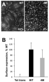
gB-4M mutant does not mediate cell fusion despite adequate surface expression. (A) SYTO 13 green fluorescent nucleic acid staining was used to quantify fusion of ARPE-19 cells mediated by either WT gB or gB-4M mutant, in the presence of gH/gL. The level of fusion was quantified by counting the total number of cell nuclei in syncytia divided by the total number of nuclei in the same field and expressed as the percentage of cells fused. A syncytium was defined as such when membrane enclosed 10 or more nuclei. Reported values represent an average of three experiments. (B) Surface expression in HDFn cells of WT gB and gB-4M mutant was assessed by cell-based ELISA with the mAb 27–156. Non-specific binding of the antibody to cells was measured with cells infected with recombinant adenovirus encoding Tet transactivator instead of gB. In each experiment, the Tet transactivator control was subtracted from the sample. Six replicate samples were averaged, and the signal was expressed as a percentage of WT (100%). Error bars represent standard deviations. Relatively large standard deviations are likely due to the fact that at any time, only a small fraction of total HCMV gB is present on the cell surface, especially when compared to HSV gB or HCMV gB lacking the endocytic motif within the cytoplasmic domain (data not shown).
Fig. 7
Fig. 7
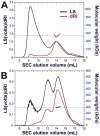
HCMVgB706-4M is a trimer in solution. SEC-coupled multi-angle light scattering (MALS) analysis of HSV-1 gB730 (A) and HCMV gB706-4M (B) is shown. The signal from the 90°-scattering detector is shown in black and the signal from the refractive index detector is shown in red; both are plotted against the left Y-axis. Average molecular weights are plotted in blue against the right Y-axis. Molecular weight corresponding to that of a trimer is shown as a horizontal dashed black line.

Similar articles

Cited by

References

    1. Atanasiu D, Saw WT, Cohen GH, Eisenberg RJ. Cascade of events governing cell-cell fusion induced by herpes simplex virus glycoproteins gD, gH/gL, and gB. Journal of virology. 2010;84:12292–12299. - PMC - PubMed
    1. Backovic M, Jardetzky TS, Longnecker R. Hydrophobic residues that form putative fusion loops of Epstein-Barr virus glycoprotein B are critical for fusion activity. J Virol. 2007a;81:9596–9600. - PMC - PubMed
    1. Backovic M, Leser GP, Lamb RA, Longnecker R, Jardetzky TS. Characterization of EBV gB indicates properties of both class I and class II viral fusion proteins. Virology. 2007b;368:102–113. - PMC - PubMed
    1. Backovic M, Longnecker R, Jardetzky TS. Structure of a trimeric variant of the Epstein-Barr virus glycoprotein B. Proc Natl Acad Sci U S A. 2009;106:2880–2885. - PMC - PubMed
    1. Barton GJ. Alscript: a tool to format multiple sequence alignments. Protein Eng. 1993;6:37–40. - PubMed

Publication types

MeSH terms

Substances