. 2012 Nov 28;31(23):4466-80.
doi: 10.1038/emboj.2012.283. Epub 2012 Oct 19.

Molecular basis for SNX-BAR-mediated assembly of distinct endosomal sorting tubules

Affiliations

Molecular basis for SNX-BAR-mediated assembly of distinct endosomal sorting tubules

Jan R T van Weering et al. EMBO J. .

Abstract

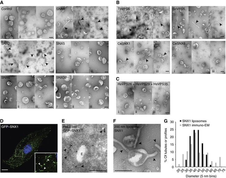
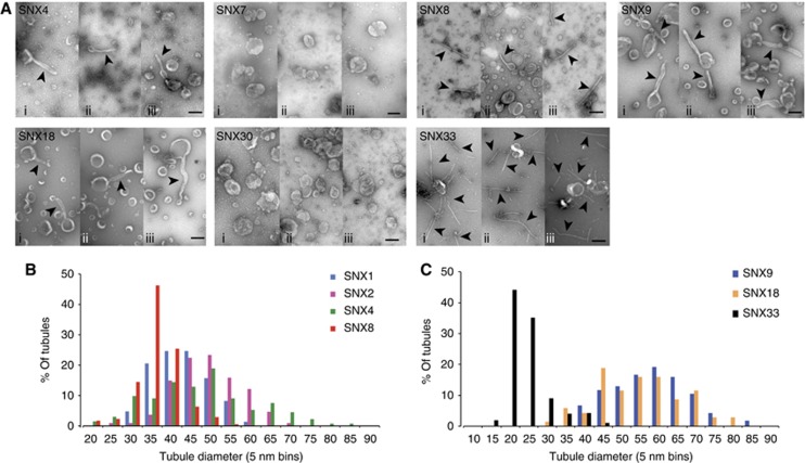
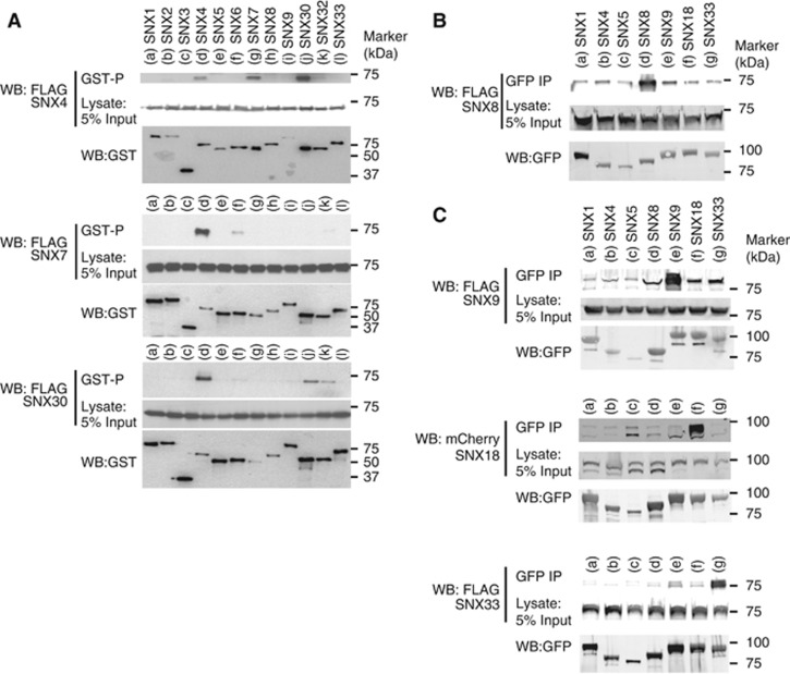
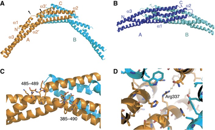
Sorting nexins (SNXs) are regulators of endosomal sorting. For the SNX-BAR subgroup, a Bin/Amphiphysin/Rvs (BAR) domain is vital for formation/stabilization of tubular subdomains that mediate cargo recycling. Here, by analysing the in vitro membrane remodelling properties of all 12 human SNX-BARs, we report that some, but not all, can elicit the formation of tubules with diameters that resemble sorting tubules observed in cells. We reveal that SNX-BARs display a restricted pattern of BAR domain-mediated dimerization, and by resolving a 2.8 Å structure of a SNX1-BAR domain homodimer, establish that dimerization is achieved in part through neutralization of charged residues in the hydrophobic BAR-dimerization interface. Membrane remodelling also requires functional amphipathic helices, predicted to be present in all SNX-BARs, and the formation of high order SNX-BAR oligomers through selective 'tip-loop' interactions. Overall, the restricted and selective nature of these interactions provide a molecular explanation for how distinct SNX-BAR-decorated tubules are nucleated from the same endosomal vacuole, as observed in living cells. Our data provide insight into the molecular mechanism that generates and organizes the tubular endosomal network.


Conflict of interest statement

The authors declare that they have no conflict of interest.

Figures

Figure 1
Figure 1
VPS5 homologues of the SNX-BAR-retromer complex remodel liposomes into tubular membrane structures. (A) Example micrographs of negative stained liposomes, extruded to 200 nm diameter and incubated with buffer control or the individual SNX-BAR proteins of the mammalian SNX-BAR-retromer complex at 10 μM final concentration (i–iii show three different example views). (B) Example micrographs of liposomes incubated with Trypanosoma brucei VPS5 (TbVPS5), Saccharomyces cerevisiae VPS5 (ScVPS5) or Caenorhabditis elegans SNX1 (CeSNX1) and SNX6 (CeSNX6) at 10 μM final concentration (i–iii show three different example views). (C) Example micrographs of liposomes incubated with the trimeric complex of Homo sapiens VPS26–VPS29–VPS36 (i–iii show three different example views). (D) Confocal image of a HeLa cell expressing GFP–SNX1 (green) and stained for nucleus (DAPI, blue). The boxed area indicates the region that is displayed in the insert. Arrowheads indicate GFP–SNX1-positive tubular structures. Scale bar represents 10 μm. (E) Electron micrograph of an endosome (marked by ‘E’) in a HeLa cell expressing GFP–SNX1, which is processed according to the Tokuyasu cryosection method and immunolabelled for GFP-10 nm gold. Arrowheads indicate the tubular/vesicular profiles positive for GFP–SNX1. (F) Electron micrograph of liposomes incubated with 10 μM SNX1 at similar magnification as (E). (G) Histogram of the minimal diameter of SNX1-positive tubular/vesicular profiles in Tokuyasu-processed HeLa cells (n=105) and SNX1-formed tubules on 200 nm liposomes in vitro (n=49), plotted as percentage of all tubules in 5 nm bins. All arrowheads indicate the membrane tubules. All scale bars represent 200 nm unless otherwise indicated.
Figure 2
Figure 2
Most SNX-BAR proteins can remodel membranes in vitro. (A) Example micrographs of negative stained liposomes incubated with the individual SNX-BAR proteins at 10 μM final concentration (i–iii show three different example views), scale bar represents 200 nm. Tubules are indicated by arrowheads. (B) Histogram of the diameter of tubules generated by SNX1 (n=49 tubules; see also Figure 1G), SNX2 (n=36), SNX4 (n=44) and SNX8 (n=57), plotted as percentage of all tubules in 5 nm bins. (C) Histogram of the diameter of tubule generated by the SH3-SNX-BAR proteins SNX9 (n=54 tubules), SNX18 (n=28) and SNX33 (n=114), plotted as percentage of all tubules in 5 nm bins.
Figure 3
Figure 3
SNX-BAR proteins show a specific pattern of homo- and hetero-dimerization. (A) Western blots (WBs) of expressed Flag–SNX4, Flag–SNX7 or Flag–SNX30 co-expressing GST–SNX1 to GST–SNX9, GST–SNX30, GST–SNX32 or GST–SNX33 in HEK-293T cells using glutathione-sepharose precipitation (GST-P). (B) WBs of expressed Flag–SNX8 co-expressing GFP–SNX1, GFP–SNX4, GFP–SNX5, GFP–SNX8, GFP–SNX9, GFP–SNX18 or GFP–SNX33 in HEK-293T cells using GFP-nanotrap immunoprecipitation (IP). (C) WBs of expressed Flag–SNX9, mCherry–SNX18 or Flag–SNX33 co-expressing GFP–SNX1, GFP–SNX4, GFP–SNX5, GFP–SNX8, GFP–SNX9, GFP–SNX18 or GFP–SNX33 in HEK-293T cells using GFP-nanotrap IP. Figure source data can be found with the Supplementary data.
Figure 4
Figure 4
Crystallization of the SNX1 homodimer. (A) Ribbon model of the SNX1-BAR homodimer. Chain A is shown in orange and chain B in cyan. Black arrowheads mark the non-standard breaks in BAR domain helices 2 and 3. (B) Ribbon model of the SNX9-BAR homodimer (PDB 2RAK; Pylypenko et al, 2007) in a similar view as (A). SNX9 molecule A is shown in dark blue and SNX9 molecule B in cyan. (C) Close-up of the breaks in helices 2 and 3 of the SNX1 structure. (D) Close-up of the environment of Arg337 in the SNX1 homodimer, showing that there is no complementary acidic partner residue close enough to neutralize the basic Arg side chain.
Figure 5
Figure 5
Charged residues in the hydrophobic BAR interface determine specific dimerization of SNX-BAR proteins. (A) The homology model of the SNX5:SNX5 homodimer (monomers shown in blue and cyan ribbons). Clashing negatively charged Glu280 and Glu383 are shown as red space filling spheres. (B) Close-up of the environment of Glu280 and Glu383 in the SNX5:SNX5 dimerization interface. (C) Western blots (WB) of expressed Flag–SNX5 wild type (WT) or Flag–SNX5-E280A/E383A (AA) co-expressing GFP control, GFP–SNX1, GFP–SNX5-WT or GFP–SNX5-AA in HEK-293T cells using GFP-nanotrap IP. (D) Example micrographs of liposomes incubated with 10 μM SNX5-AA (i–iii show three different example views), scale bar represents 200 nm. (E) Coomassie-stained gel of SNX5-AA in the pellet (P) and supernatant (S) fractions after sedimentation in the presence or absence of liposomes. Figure source data can be found with the Supplementary data.
Figure 6
Figure 6
All SNX-BAR proteins contain an amphipathic helix, which is essential for membrane remodelling. (A) Table of all predicted amphipathic helices (AHs) in SNX-BAR proteins. The numbers in the region column refer to the amino acids in the full-length protein. (B) Cartoon of the arrangement of the 18-residue AHs of SNX1, SNX5 and SNX8. Arrowheads indicate the hydrophobic residues mutated to acidic residues (dAH). (C) Example micrographs of liposomes incubated with 10 μM SNX1-WT, SNX5-WT, SNX1-dAH, SNX5-dAH or combinations of SNX1-dAH with SNX5-WT or SNX5-dAH at 5 μM final concentration for each protein (i–iii show three different example views), scale bar represents 200 nm. Tubules are indicated by arrowheads. (D) Example micrographs of liposomes incubated with 10 μM SNX8-WT or SNX8-dAH (i–iii show three different example views), scale bar represents 200 nm. Tubules are indicated by arrowheads. (E) Coomassie-stained gel of SNX1-WT, SNX1-dAH, SNX5-WT, SNX5-dAH, SNX8-WT and SNX8-dAH in the pellet (P) and supernatant (S) fractions after sedimentation in the presence or absence of liposomes. Figure source data can be found with the Supplementary data.
Figure 7
Figure 7
Tip connections between SNX-BAR dimers regulate SNX-BAR oligomerization. (A) Ribbon model of the SNX1:SNX1 homodimer (green) indicating Lys442 and Lys445 in blue space-filling spheres and the SNX5-AA-1TIP:SNX5-AA-1TIP homodimer (cyan) indicating the tip–loop region of SNX1 in green and the E280A/E383A substitutions in orange space-filling spheres. (B) Example micrographs of liposomes incubated with 10 μM SNX1-WT, SNX1-K442A, SNX1-K445A and SNX1-K442/445A (i–iii show three different example views). (C) Example micrographs of liposomes incubated with 10 μM SNX5-WT, SNX5-AA, SNX5-WT-1TIP or SNX5-AA-1TIP (i–iii show three different example views). (D) Coomassie-stained gel of SNX1-K442A, SNX1-K445A, SNX1-K442/445A, SNX5-WT-1TIP and SNX5-AA-1TIP in the pellet (P) and supernatant (S) fractions after sedimentation in the presence or absence of liposomes. (E) Example micrographs of liposomes incubated with 10 μM SNX9, SNX33 or preincubated mixture of SNX9 and SNX33 (i–iii show three different example views). (F) Histogram of the diameter of tubule generated by the preincubated mixture of SNX9 and SNX33 (n=141 tubules) plotted as percentage of all tubules in 5 nm bins, in comparison to SNX9 (n=54) or SNX33 (n=114) alone (see also Figure 2C). (G) Cartoon of the required interactions by SNX-BAR proteins to remodel membrane vesicles into tubules: (I) formation the SNX-BAR dimer, (II) association of the SNX-BAR dimer with the membrane with insertion of an amphipathic helix and (III) oligomerization of SNX-BAR dimers on the membrane to stabilize and expand local membrane curvature. (H) Cartoon of an endosomal vacuole (E) with two SNX-BAR-decorated sorting tubules attached. Each of the sorting tubules will extend selectively by recruitment self similar SNX-BAR molecules, due to the specific dimerization and oligomerization through the BAR domain and tip–loop interactions. All scale bars represent 200 nm. Tubules are indicated by arrowheads. Figure source data can be found with the Supplementary data.

Similar articles

Cited by

References

    1. Bhatia VK, Madsen KL, Bolinger PY, Kunding A, Hedegård P, Gether U, Stamou D (2009) Amphipathic motifs in BAR domains are essential for membrane curvature sensing. EMBO J 28: 3303–3314 - PMC - PubMed
    1. Blanc E, Roversi P, Vonrhein C, Flensburg C, Lea SM, Bricogne G (2004) Refinement of severely incomplete structures with maximum likelihood in BUSTER-TNT. Acta Crystallogr D Biol Crystallogr 60: 2210–2221 - PubMed
    1. Boucrot E, Pick A, Çamdere G, Liska N, Evergren E, McMahon HT, Kozlov MM (2012) Membrane fission is promoted by insertion of amphipathic helices and is restricted by crescent BAR domains. Cell 149: 124–136 - PMC - PubMed
    1. Bujny MV, Popoff V, Johannes L, Cullen PJ (2007) The retromer component sorting nexin-1 is required for efficient retrograde transport of Shiga toxin from early endosome to the trans Golgi network. J Cell Sci 120: 2010–2021 - PubMed
    1. Carlton J, Bujny M, Peter B, Oorschot V, Rutherford A, Mellor H, Klumperman J, McMahon HT, Cullen PJ (2004) Sorting nexin-1 mediates tubular endosome-to-TGN transport through coincidence sensing of high- curvature membranes and 3-phosphoinositides. Curr Biol 14: 1791–1800 - PubMed

Publication types