. 2012 Nov 30;48(4):532-46.
doi: 10.1016/j.molcel.2012.09.011. Epub 2012 Oct 18.

Epigenetic regulation of condensin-mediated genome organization during the cell cycle and upon DNA damage through histone H3 lysine 56 acetylation

Affiliations

Epigenetic regulation of condensin-mediated genome organization during the cell cycle and upon DNA damage through histone H3 lysine 56 acetylation

Atsunari Tanaka et al. Mol Cell. .

Abstract

Complex genome organizations participate in various nuclear processes including transcription, DNA replication, and repair. However, the mechanisms that generate and regulate these functional genome structures remain largely unknown. Here, we describe how the Ku heterodimer complex, which functions in nonhomologous end joining, mediates clustering of long terminal repeat retrotransposons at centromeres in fission yeast. We demonstrate that the CENP-B subunit, Abp1, functions as a recruiter of the Ku complex, which in turn loads the genome-organizing machinery condensin to retrotransposons. Intriguingly, histone H3 lysine 56 (H3K56) acetylation, which functions in DNA replication and repair, interferes with Ku localization at retrotransposons without disrupting Abp1 localization and, as a consequence, dissociates condensin from retrotransposons. This dissociation releases condensin-mediated genomic associations during S phase and upon DNA damage. ATR (ATM- and Rad3-related) kinase mediates the DNA damage response of condensin-mediated genome organization. Our study describes a function of H3K56 acetylation that neutralizes condensin-mediated genome organization.


Figures

Figure 1
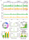
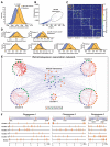
Figure 1. Genome-wide distribution of Ku binding
(A) Chromosomal distributions of Pku70 (green) and Pku80 (yellow) were determined based on ChIP-seq analyses. ChIP was performed using strains carrying Pku70-Myc and Pku80-Myc proteins. (B) Summary of PKu distributions across the fission yeast genome. Pie-chart shows the composition of Pku70/Pku80 common peaks (top). Coverages of LTR, tRNA, and 5S rRNA genes estimated by the presence of significant Pku binding peaks within 300 bp from the genes are shown in the bar graph (bottom). Each number in parenthesis indicates the total gene number. (C) Average binding patterns of Pku proteins at LTR, tRNA, and 5S rRNA genes. (D and E) Pku distributions at the right telomere (D) and centromere (E) of chromosome 2.
Figure 2
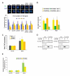
Figure 2. Ku mediates retrotransposon clustering
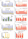
(A) FISH analysis visualizing Tf2 elements (green). Typical microscopic images in the indicated strains are shown on top. Black bar indicates 1 μm. Number of Tf2 dots in the nucleus is summarized at the bottom. (B) ChIP results showing Pku enrichment at Tf elements in wt and abp1Δ cells. (C) ChIP results showing enrichment of Abp1-Pk at Tf elements in wt and pkuΔ cells. (D) Co-immunoprecipitation results showing the interaction between Pku-Myc and Abp1-Flag. (E) RT-PCR results indicating the transcript levels of Tf2 LTR and ORF regions in pkuΔ and abp1Δ cells. The transcript levels in mutants were normalized by those in wt cells. In panels A, B, C, and E, data are represented as mean +/− SD.
Figure 3
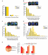
Figure 3. Clustering of retrotransposons at centromeres
(A) Centromere clustering is not affected by pkuΔ. Centromeres were visualized by FISH in the indicated strains. Number of centromeric foci in the nucleus is summarized in a graph. (B) Centromere anchoring to the nuclear periphery is not compromised in pkuΔ cells. Centromeres (red) and nuclear pore complex (NPC; green) were visualized by FISH and IF using anti-Nup189 antibody, respectively (top). Distance between centromeric signal and its nearby NPC focus was measured and summarized in a graph. (C) FISH/IF analysis visualizing Tf2 elements (red) and the NPC subunit, Nup189 (green). Distance between Tf2 dot and its nearest NPC was measured. (D) FISH analysis visualizing centromeres (red) and Tf2 elements (green). Distance between centromeric and Tf2 foci was measured and summarized in a graph. (E) Distance from centromeres was divided into three zones based on the criteria depicted on the left. Distance between centromeres and Tf2 dot from FISH result shown in (D) was binned into one of the assigned zones. n.s. indicates P > 0.05. Black bar indicates 1 μm.
Figure 4
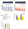
Figure 4. Tf clustering at centromeres is compromised in condensin mutant
(A) The condensin mutation, cut14-208, impairs Tf clustering. FISH analysis was performed as described in Figure 2A. Black bar indicates 1 μm. (B) Centromeric localization of Tf cluster is compromised in condensin mutant cells. The experiment was carried out as described in Figures 3D and 3E. (C) Co-IP analysis investigating an interaction between Pku80-Flag and Cut3-Myc. (D) ChIP result showing enrichment of the condensin subunit Cut14-Pk at Tf elements in wt and pkuΔ cells. (E) ChIP result showing enrichment of Pku70-Pk and Pku80-Pk at Tf1-10 in wt and cut14-208 condensin mutant cells. Data are represented as mean +/− SD. n.s. indicates P > 0.05.
Figure 5
Figure 5. Genomic analyses on LTR clustering
(A) LTR-containing genomic sections significantly associate with centromeres. Distributions for average centromeric association scores for both LTR-containing sections and random control were generated by repeated sampling. We carried out Student’s t-test and observed a significant difference between the two distributions. (B) LTR-containing genomic sections with both high local and global connectivity measures, degree and betweenness, respectively, were designated as potential global interactors and excluded from subsequent clustering analyses. (C) Heat map representation of the final clustering results. Total of five significant clusters were identified at a modularity level of 0.4. Normalized physical proximity values reflect association strength between genomic sections (Tanizawa et al. 2010). (D) Significance of associations between centromeres and genomic sections in each cluster or global interactors were calculated using the same procedure exploited in (A). (E) Graphical representation of the final clustering results. Each colored dot represents an LTR-containing genomic section. Each edge indicates significant association, and its thickness correlates with association strength. (F) Genomic locations of LTR-containing genomic sections in the different clusters. Method details are described in Supplemental Experimental Procedures.
Figure 6
Figure 6. Regulation of Tf clustering at centromeres by H3K56 acetylation
(A) The histone deacetylase mutations, clr6-1 and hst4Δ, impair Tf clustering. FISH analysis was performed as described in Figure 2A. The five HDAC mutants, clr6-1, hst4Δ, clr3Δ, sir2Δ, and hos2Δ, were subjected to the experiment. (B) The clr6-1 and hst4Δ HDAC mutations affect centromeric localization of Tf elements. The analyses were carried out as described in Figures 3D and 3E using the indicated strains. (C) The histone acetyltransferase mutations do not affect Tf clustering. The five HAT mutants, gcn5Δ, hat1Δ, mst2Δ, elp3Δ, and rtt109Δ, were subjected to the experiment. (D) The HAT mutation, rtt109Δ, enhances centromeric localization of Tf elements. (E) ChIP results showing enrichment of PKu-Pk, Abp1-Pk, and Cut14-Pk at Tf1-10 in wt, hst4Δ and rtt109Δ cells. Data are represented as mean +/− SD. (F) Centromeric localization of Tf elements during the cell cycle. The cdc25-22 mutation was used for the cell-cycle synchronization. The septation index is shown at the bottom and the septation peak roughly coincides with S phase. FISH experiment was performed using cells in different stages of the cell cycle. (G and H) Tf clustering at centromeres is compromised upon DNA damage. Wild-type and rtt109Δ cells were cultured in a nutrient-rich (YEA) medium containing 0.5 mM H2O2, 0.08% methyl methanesulfonate (MMS), and 200 mM hydroxyurea (HU). Tf clustering (G) and its centromeric localization (H) were investigated by FISH. (I and J) Histone H3K56Q mutation impairs Tf clustering at centromeres. Tf clustering (I) and its centromeric localization (J) were investigated by FISH. n.s. indicates P > 0.05.
Figure 7
Figure 7. Disruption of Tf clustering at centromeres through ATR
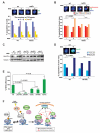
(A) Tf clustering is not impaired upon DNA damage in rad3Δ cells. The MMS treatment was performed as described in Figure 6G. FISH analysis was performed as described in Figure 2A. (B) Centromeric localization of Tf elements is not affected by the MMS treatment in rad3Δ cells. The analyses were carried out as described in Figures 3D and 3E. (C) The MMS treatment reduces the amount of Hst4 in wt cells, but not in rad3Δ, pku70Δ, pku80Δ, and abp1Δ cells. Western blot analysis was performed to detect Hst4-Pk and tubulin using anti-Pk and TAT-1 antibodies, respectively. (D) IF result showing Pku70-Pk localization (green) with and without MMS treatment in wt and rtt109Δ cells. Pku70-Pk staining area on a focal point, which reflects Pku70 occupancy in the nucleus, was measured and the derived data were classified into two groups, below 0.6 μm2 and more than 0.6 μm2. Typical images are shown on top. (E) Nonhomologous end joining assay. Efficiencies of NHEJ in wt, pku70Δ, pku80Δ, rad3Δ, rtt109Δ, and hst4Δ cells were measured by a plasmid-based assay. Data are represented as mean +/− SD. (F) A schematic model for retrotransposon-mediated genome organization and its regulatory mechanism through histone H3K56 acetylation. The CENP-B subunit Abp1 binds to Tf elements and recruits Ku, which in turn loads the genome-organizing machinery condensin onto chromatin. Condensin associating with Tf elements and centromeres mediates Tf clustering at centromeres. H3K56 acetylation by Rtt109 releases Ku and condensin from Tf elements without disrupting Abp1 binding, thereby disassembling this genome organization during S phase of the cell cycle and upon DNA damage. Once H3K56 is deacetylated by Hst4, stable interaction of the recruiter Abp1 with Tf elements would help efficiently reestablish condensin-mediated genome organization. In panels B, D, and E, n.s. indicates P > 0.05.

Comment in

Similar articles

Cited by

References

    1. Bähler J, Wu J, Longtine MS, Shah NG, McKenzie A, 3rd, Steever AB, Wach A, Philippsen P, Pringle JR. Heterologous modules for efficient and versatile PCR-based gene targeting in Schizosaccharomyces pombe. Yeast. 1998;14:943–951. - PubMed
    1. Baumann P, Cech TR. Protection of telomeres by the Ku protein in fission yeast. Mol. Biol. Cell. 2000;11:3265–3275. - PMC - PubMed
    1. Bowen NJ, Jordan IK, Epstein JA, Wood V, Levin HL. Retrotransposons and their recognition of pol II promoters: a comprehensive survey of the transposable elements from the complete genome sequence of Schizosaccharomyces pombe. Genome Res. 2003;13:1984–1997. - PMC - PubMed
    1. Bupp JM, Martin AE, Stensrud ES, Jaspersen SL. Telomere anchoring at the nuclear periphery requires the budding yeast Sad1-UNC-84 domain protein Mps3. J. Cell Biol. 2007;179:845–854. - PMC - PubMed
    1. Cam HP, Noma K, Ebina H, Levin HL, Grewal SI. Host genome surveillance for retrotransposons by transposon-derived proteins. Nature. 2008;451:431–436. - PubMed

Publication types

MeSH terms