Methods for robust in vivo strain estimation in the carotid artery
- PMID: 23079725
- PMCID: PMC3740562
- DOI: 10.1088/0031-9155/57/22/7329
Methods for robust in vivo strain estimation in the carotid artery
Abstract
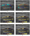
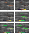
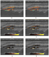
A hierarchical block-matching motion tracking algorithm for strain imaging is presented. Displacements are estimated with improved robustness and precision by utilizing a Bayesian regularization algorithm and an unbiased subsample interpolation technique. A modified least-squares strain estimator is proposed to estimate strain images from a noisy displacement input while addressing the motion discontinuity at the wall-lumen boundary. Methods to track deformation over the cardiac cycle incorporate a dynamic frame skip criterion to process data frames with sufficient deformation to produce high signal-to-noise displacement and strain images. Algorithms to accumulate displacement and/or strain on particles in a region of interest over the cardiac cycle are described. New methods to visualize and characterize the deformation measured with the full 2D strain tensor are presented. Initial results from patients imaged prior to carotid endarterectomy suggest that strain imaging detects conditions that are traditionally considered high risk including soft plaque composition, unstable morphology, abnormal hemodynamics and shear of plaque against tethering tissue can be exacerbated by neoangiogenesis. For example, a maximum absolute principal strain exceeding 0.2 is observed near calcified regions adjacent to turbulent flow, protrusion of the plaque into the arterial lumen and regions of low echogenicity associated with soft plaques. Non-invasive carotid strain imaging is therefore a potentially useful tool for detecting unstable carotid plaque.
Figures


Similar articles
-
MRI-based biomechanical parameters for carotid artery plaque vulnerability assessment.Thromb Haemost. 2016 Mar;115(3):493-500. doi: 10.1160/TH15-09-0712. Epub 2016 Jan 21. Thromb Haemost. 2016. PMID: 26791734 Review.
-
Arterial wall mechanical inhomogeneity detection and atherosclerotic plaque characterization using high frame rate pulse wave imaging in carotid artery disease patients in vivo.Phys Med Biol. 2020 Jan 17;65(2):025010. doi: 10.1088/1361-6560/ab58fa. Phys Med Biol. 2020. PMID: 31746784 Free PMC article.
-
Locally optimized correlation-guided Bayesian adaptive regularization for ultrasound strain imaging.Phys Med Biol. 2020 Mar 19;65(6):065008. doi: 10.1088/1361-6560/ab735f. Phys Med Biol. 2020. PMID: 32028272 Free PMC article.
-
Sites of rupture in human atherosclerotic carotid plaques are associated with high structural stresses: an in vivo MRI-based 3D fluid-structure interaction study.Stroke. 2009 Oct;40(10):3258-63. doi: 10.1161/STROKEAHA.109.558676. Epub 2009 Jul 23. Stroke. 2009. PMID: 19628799 Free PMC article.
-
Contemporary carotid imaging: from degree of stenosis to plaque vulnerability.J Neurosurg. 2016 Jan;124(1):27-42. doi: 10.3171/2015.1.JNS142452. Epub 2015 Jul 31. J Neurosurg. 2016. PMID: 26230478 Review.
Cited by
-
Estimation of ultrasound strain indices in carotid plaque and correlation to cognitive dysfunction.Annu Int Conf IEEE Eng Med Biol Soc. 2014;2014:5627-30. doi: 10.1109/EMBC.2014.6944903. Annu Int Conf IEEE Eng Med Biol Soc. 2014. PMID: 25571271 Free PMC article.
-
Study of the Relationship Between Ultrasound Strain Indices and Cognitive Decline for Vulnerable Carotid Plaque.Annu Int Conf IEEE Eng Med Biol Soc. 2020 Jul;2020:2088-2091. doi: 10.1109/EMBC44109.2020.9175911. Annu Int Conf IEEE Eng Med Biol Soc. 2020. PMID: 33018417 Free PMC article.
-
Noninvasive characterization of carotid plaque strain.J Vasc Surg. 2017 Jun;65(6):1653-1663. doi: 10.1016/j.jvs.2016.12.105. Epub 2017 Mar 6. J Vasc Surg. 2017. PMID: 28274754 Free PMC article.
-
Strain estimation in aortic roots from 4D echocardiographic images using medial modeling and deformable registration.Med Image Anal. 2023 Jul;87:102804. doi: 10.1016/j.media.2023.102804. Epub 2023 Apr 1. Med Image Anal. 2023. PMID: 37060701 Free PMC article.
-
Hierarchical Motion Estimation With Bayesian Regularization in Cardiac Elastography: Simulation and In Vivo Validation.IEEE Trans Ultrason Ferroelectr Freq Control. 2019 Nov;66(11):1708-1722. doi: 10.1109/TUFFC.2019.2928546. Epub 2019 Jul 12. IEEE Trans Ultrason Ferroelectr Freq Control. 2019. PMID: 31329553 Free PMC article.
References
-
- Ajduk M, Pavic L, Bulimbasic S, Sarlija M, Pavic P, Patrlj L, Brkljacic B. Multidetector-row computed tomography in evaluation of atherosclerotic carotid plaques complicated with intraplaque hemorrhage. Ann Vasc Surg. 2009;23:186–93. - PubMed
-
- Boersma HH, Kietselaer BL, Stolk LM, Bennaghmouch A, Hofstra L, Narula J, Heidendal GA, Reutelingsperger CP. Past, present, and future of annexin A5: from protein discovery to clinical applications. J Nucl Med. 2005;46:2035–50. - PubMed
MeSH terms
Grants and funding
LinkOut - more resources
Full Text Sources
