. 2013 Jan;87(1):403-14.
doi: 10.1128/JVI.02465-12. Epub 2012 Oct 17.

Herpes simplex virus membrane proteins gE/gI and US9 act cooperatively to promote transport of capsids and glycoproteins from neuron cell bodies into initial axon segments

Affiliations

Herpes simplex virus membrane proteins gE/gI and US9 act cooperatively to promote transport of capsids and glycoproteins from neuron cell bodies into initial axon segments

Paul W Howard et al. J Virol. 2013 Jan.

Abstract

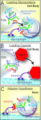
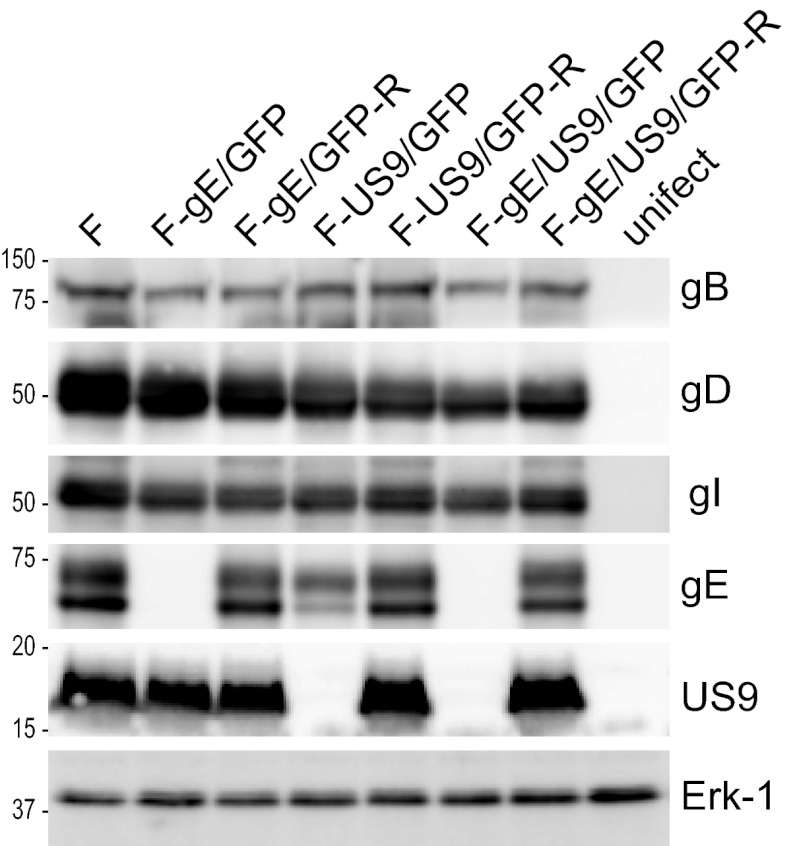
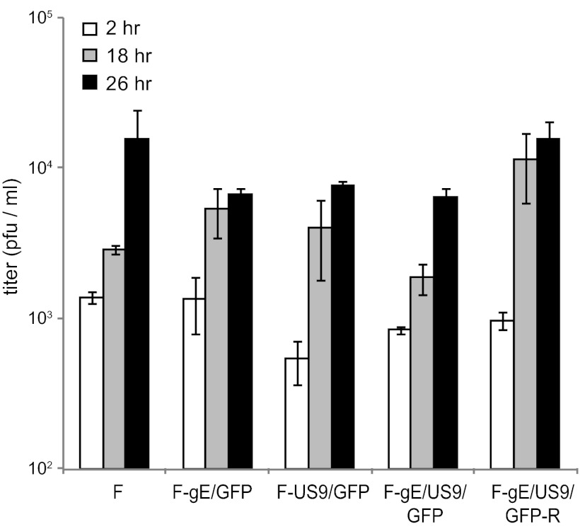
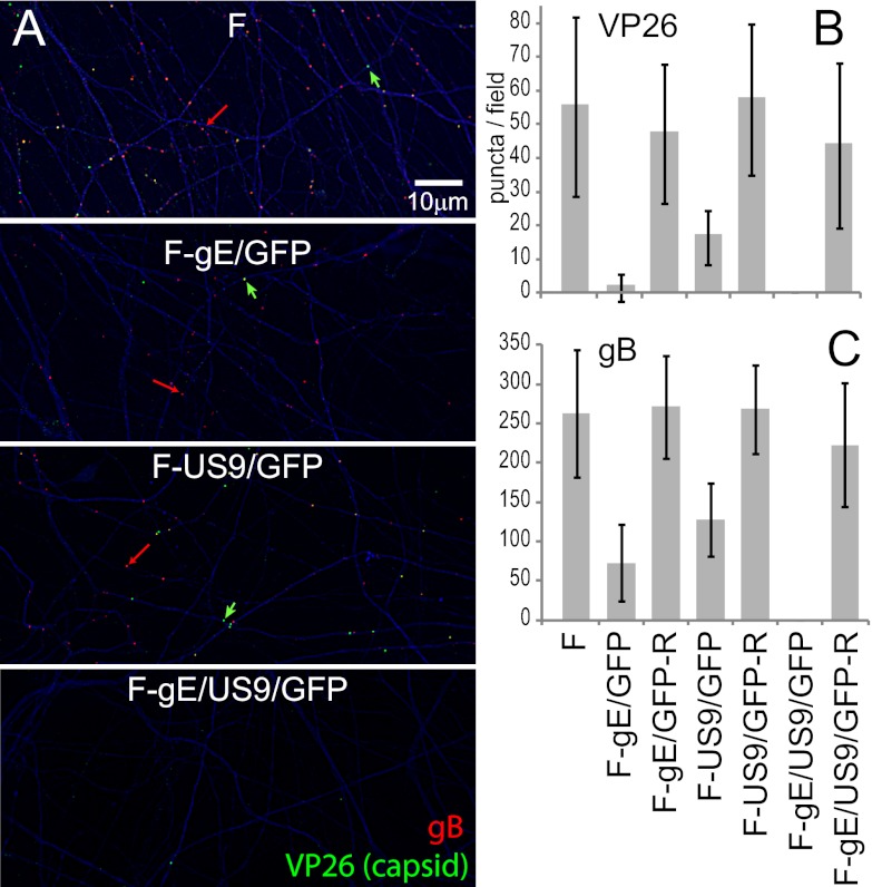
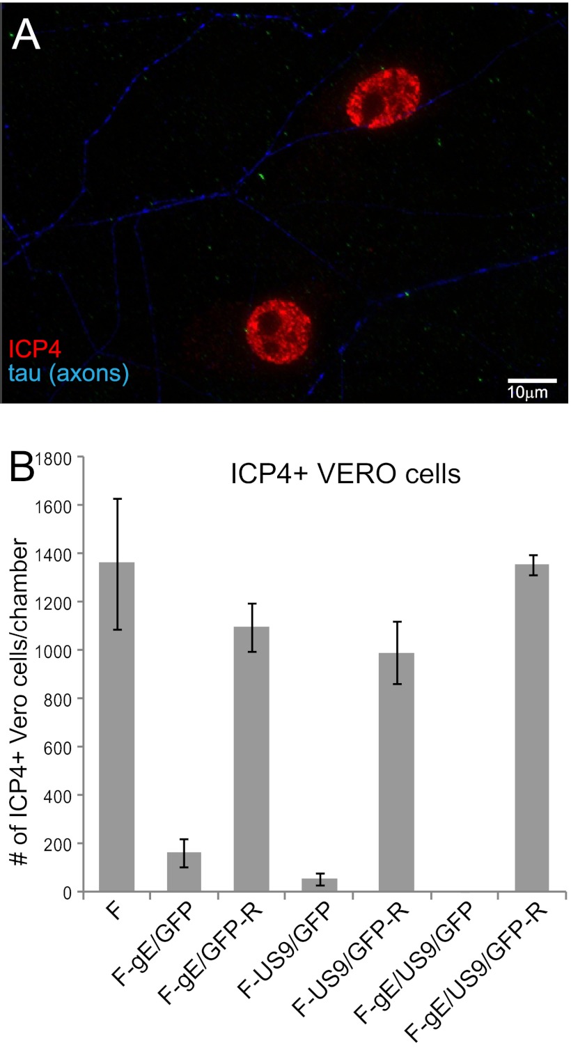
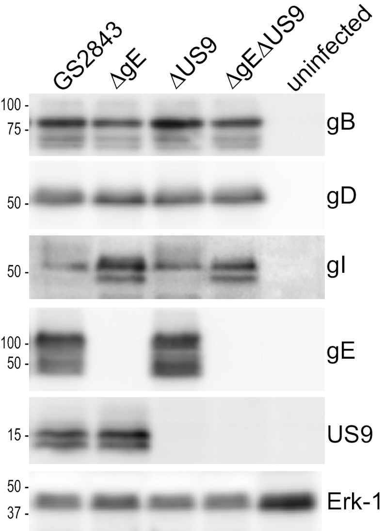
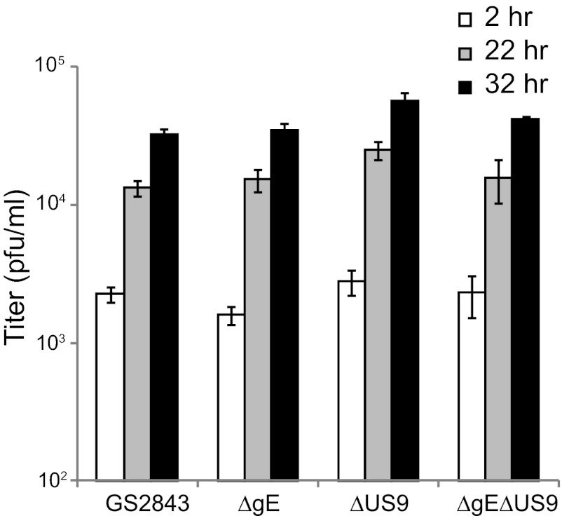
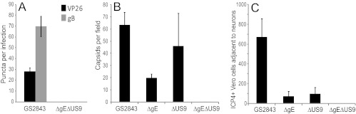
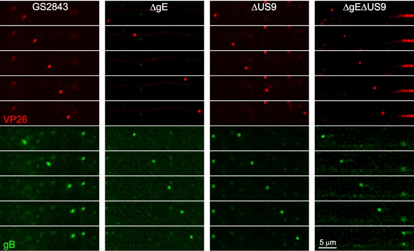
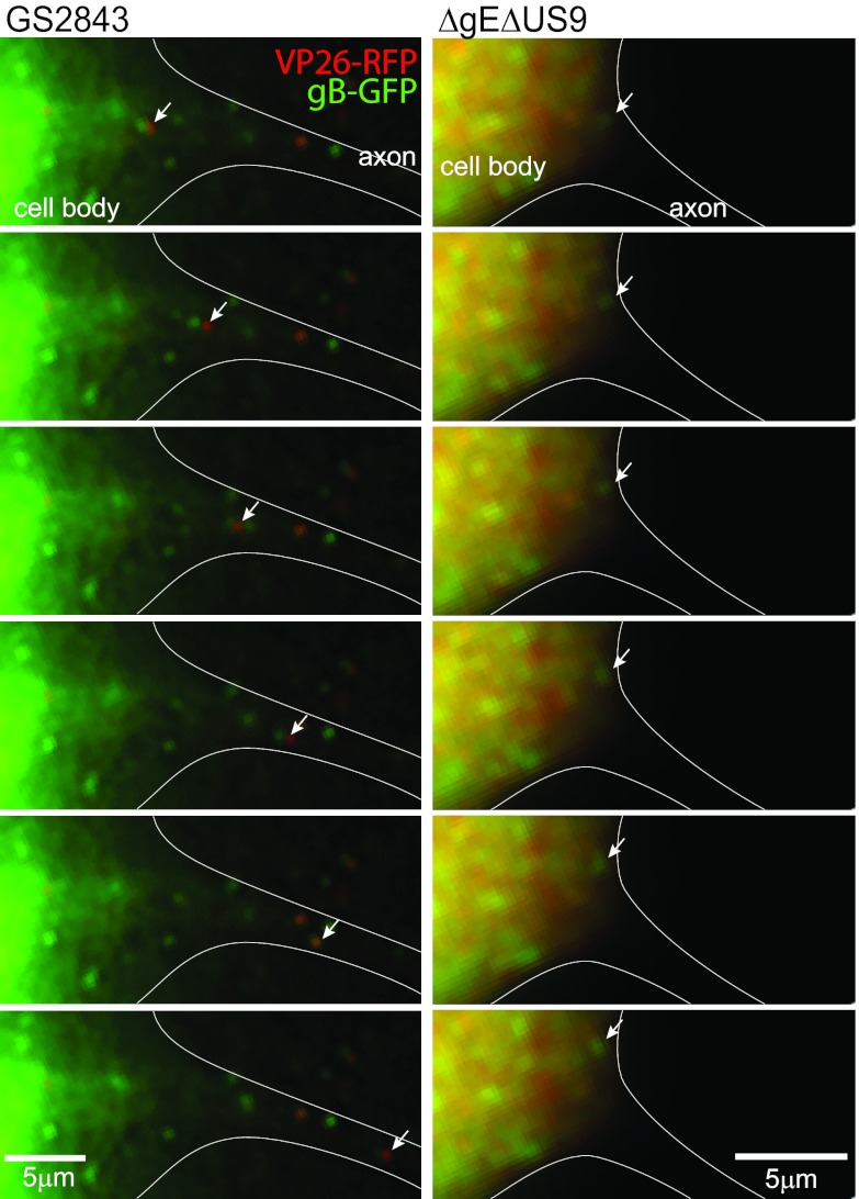
Herpes simplex virus (HSV) and other alphaherpesviruses must move from sites of latency in ganglia to peripheral epithelial cells. How HSV navigates in neuronal axons is not well understood. Two HSV membrane proteins, gE/gI and US9, are key to understanding the processes by which viral glycoproteins, unenveloped capsids, and enveloped virions are transported toward axon tips. Whether gE/gI and US9 function to promote the loading of viral proteins onto microtubule motors in neuron cell bodies or to tether viral proteins onto microtubule motors within axons is not clear. One impediment to understanding how HSV gE/gI and US9 function in axonal transport relates to observations that gE(-), gI(-), or US9(-) mutants are not absolutely blocked in axonal transport. Mutants are significantly reduced in numbers of capsids and glycoproteins in distal axons, but there are less extensive effects in proximal axons. We constructed HSV recombinants lacking both gE and US9 that transported no detectable capsids and glycoproteins to distal axons and failed to spread from axon tips to adjacent cells. Live-cell imaging of a gE(-)/US9(-) double mutant that expressed fluorescent capsids and gB demonstrated >90% diminished capsids and gB in medial axons and no evidence for decreased rates of transport, stalling, or increased retrograde transport. Instead, capsids, gB, and enveloped virions failed to enter proximal axons. We concluded that gE/gI and US9 function in neuron cell bodies, in a cooperative fashion, to promote the loading of HSV capsids and vesicles containing glycoproteins and enveloped virions onto microtubule motors or their transport into proximal axons.


Figures

Fig 1
Fig 1
Models for how HSV gE/gI and US9 might promote anterograde axonal transport of capsids and glycoproteins. (A) The loading hypothesis suggests that gE/gI and US9 accumulate in the TGN of neuron cell bodies and cause other HSV membrane proteins to accumulate there. Transport vesicles that bud from these specific TGN membranes are loaded onto kinesins for transport into proximal axons. Similar vesicles containing enveloped HSV (Married) particles might also be loaded in this way. (B) The loading of capsids onto kinesin motors may similarly be affected by gE/gI and US9 accumulation in the TGN. gE/gI is known to extensively interact with tegument proteins that, in turn, interact with capsids. Thus, by causing the accumulation of capsids in TGN loading compartments, gE/gI and US9 may promote the axonal transport of unenveloped capsids. (C) The adaptor hypothesis functions more extensively in axons rather than cell bodies. In this model, the cytoplasmic domains of gE/gI and US9 interact with kinesin adaptors or directly with kinesins to tether vesicles containing other HSV glycoproteins (gD and gB) and cellular cargo (synaptophysin) onto motors during transport.
Fig 2
Fig 2
Expression of gE and US9 in cells infected with HSV mutant viruses. Vero cells were infected with wild-type HSV strain F; the gE, US9, or gE/US9 mutant; or repaired versions of these viruses for 16 h, and SDS-containing cell extracts were then produced. These extracts were resolved on polyacrylamide gels, proteins were transferred onto PVDF membranes, and the membranes were incubated with antibodies specific for gB, gD, gI, gE, US9, or the cellular protein ERK-1. Membranes were washed and incubated with horseradish peroxidase-conjugated secondary antibodies and a chemiluminescent reagent. Numbers indicate molecular mass markers in kilodaltons.
Fig 3
Fig 3
Production of infectious progeny in neurons infected with gE, US9, and gE/US9 mutants. SCG neurons in 24-well dishes (∼40,000 neurons per well) were infected with wild-type HSV strain F; the gE, US9, or gE/US9 mutant; or repaired viruses at 5 PFU/cell for 2 h, and the cells were then washed once with medium. At this time (2 h) and after 18 and 26 h, the cells were scraped into the medium and then frozen and sonicated, and viral titers were determined using plaque assays involving Vero cells. Each time point was assayed in triplicate, and the data are presented with standard deviations.
Fig 4
Fig 4
Numbers of capsids and gB puncta in distal axons of SCG neurons infected with the gE, US9, or gE/US9 mutant. SCG neurons were plated in the somal compartment of microfluidic chambers, and axons were allowed to grow into the axonal side. HSV-1 (8 PFU/cell) was introduced into somal chambers, and 18 h later, the devices were disassembled, and axons in the axonal chambers were fixed with paraformaldehyde and simultaneously immunostained with antibodies specific for VP26 (capsids) (one per panel marked with a green arrow), gB (red) (one per panel marked with a red arrow), and the microtubule-associated protein tau (blue) and then with secondary fluorescent antibodies. (A) Representative images of axons in the axonal compartment. VP26 is stained in green, gB puncta are red, and tau is blue. The puncta were small, and one gB punctum (red) and one VP26 punctum are indicated by arrows. Puncta containing both VP26 and gB (Married particles) appear yellow. (B and C) The ImageJ software program was used to count capsid and gB puncta in 10 distinct 10,551-μm2 fields of the axonal compartments from three separate infections. Puncta with an intensity of >1,500 and a size of 7 square pixels or greater were counted. The total area of tau staining was also measured and was used to correct the data for the quantities of axons present.
Fig 5
Fig 5
Spread of gE, US9, and gE/US9 mutants from distal axons to adjacent nonneuronal cells. SCG neurons were plated into microfluidic chambers, and axons were allowed to grow into the axonal compartment. Vero cells were plated in the axonal compartment 24 h before the neurons were infected with HSV-1 (8 PFU/cell) added to the somal compartments. Two to four hours after infection, 0.1% human gamma globulin was added to the axonal chambers, and 18 h after infection, the devices were disassembled. Cells in the axonal chambers were fixed with 4% paraformaldehyde, permeabilized with 0.1% Triton X-100, and immunostained with antibodies specific for the immediate-early gene product ICP4 (red) and simultaneously with antibodies specific for the axon protein tau (blue). (A) Representative image of ICP4+ Vero cells adjacent to two axons stained with tau. (B) ICP4+ Vero cells were manually counted in 10 axonal compartments involving 3 separate experiments. Total numbers of ICP4+ Vero cells per chamber are shown with standard deviations.
Fig 6
Fig 6
Immunoblot analyses of viral proteins produced by fluorescent HSV recombinants with deletions in the gE, US9, or gE and US9 genes. Vero cells were left uninfected or were infected with HSV recombinant strain GS2843, GS2843ΔgE, GS2843ΔUS9, or GS2843ΔgEΔUS9 for 16 h, and SDS cell extracts were then made, and proteins were resolved on polyacrylamide gels and transferred onto PVDF membranes. The membranes were immunoblotted with antibodies specific for gB, gD, gI, gE, US9, or the cellular protein ERK-1. Numbers represent molecular mass markers in kilodaltons.
Fig 7
Fig 7
Replication of fluorescent HSV recombinants with deletions in the gE, US9, or gE and US9 genes in neurons. SCG neurons in 24-well plates were infected with GS2843, GS2843ΔgE, GS2843ΔUS9, or GS2843ΔgEΔUS9 at 5 PFU/cell. After 2 h, cells were washed once with medium. Cells and medium were harvested immediately (2 h) or after 22 or 32 h and then frozen and subsequently sonicated, and the viral titers were then determined using Vero cells. Each time point represents three separate wells, and the data are presented as the averages with standard deviations.
Fig 8
Fig 8
Capsids and gB in distal axons following infection with fluorescent HSV recombinants lacking gE, US9, or both, and virus spread to nonneuronal cells. SCG neurons were infected with GS2843, GS2843ΔgE, GS2843ΔUS9, or GS2843ΔgEΔUS9 at 5 PFU/cell in somal compartments and then incubated for 22 h. (A) Quantification of mRFP-VP26 and gB puncta in axons found in axonal compartments. (B) Methanol-acetone (1:1) at −20°C was added to somal compartments for 10 min, the devices were then disassembled, and axons were stained with anti-VP5 polyclonal antibodies and secondary fluorescent antibodies. (C) Vero cells were plated in axonal compartments, neurons were infected in somal compartments as described above, and after 22 h, the Vero cells were then stained with anti-ICP4 antibodies, as described in the legend of Fig. 5.
Fig 9
Fig 9
Sequential still images of medial axons derived from live-cell analyses. Images of mRFP-VP26 capsid puncta (red) or GFP-gB puncta (green) moving in neuronal axons were derived from live-cell imaging of axons in microchannels. Neurons were infected with GS2843, GS2843ΔgE, GS2843ΔUS9, or GS2843ΔgEΔUS9, and after 22 h, live-cell imaging was then performed on axons in microchannels. Still images taken 2 s apart of capsids or gB puncta are shown.
Fig 10
Fig 10
Sequential images of neuron cell bodies. Neurons were infected with HSV recombinant strain GS2843 or GS2843ΔgEΔUS9. Live-cell imaging of neuron cell bodies in somal compartments was performed, and still images (separated by 2 s) were derived from these movies. The left panels represent a neuron infected with GS2843 in an earlier stage of infection, with the majority of RFP-VP26 remaining in the nucleus, which was intentionally cropped out of these images, related to intense fluorescence. A Married HSV particle is shown moving from the neuron cell body into an axon initial segment. Note that live-cell imaging requires switching filters, which creates a small separation of red and green signals with Married particles. A Separate capsid was observed moving from this neuron cell body into the initial axon segment in Fig. S1 in the supplemental material. The right panels show a neuron infected with GS2843ΔgEΔUS9. This neuron was in a later stage of infection, when RFP-VP26 and GFP-gB were present at higher levels in the cytoplasm. With this neuron, there were no capsids or gB puncta that moved into axons.

Similar articles

Cited by

References

    1. Diefenbach RJ, Miranda-Saksena M, Douglas MW, Cunningham AL. 2008. Transport and egress of herpes simplex virus in neurons. Rev. Med. Virol. 18:35–51 - PubMed
    1. Johnson DC, Baines JD. 2011. Herpesviruses remodel host membranes for virus egress. Nat. Rev. Microbiol. 9:382–394 - PubMed
    1. Kratchmarov R, Taylor MP, Enquist LW. 16 July 2012. Making the case: married versus separate models of alphaherpes virus anterograde transport in axons. Rev. Med. Virol. [Epub ahead of print.] - PMC - PubMed
    1. Snyder A, Polcicova K, Johnson DC. 2008. Herpes simplex virus gE/gI and US9 proteins promote transport of both capsids and virion glycoproteins in neuronal axons. J. Virol. 82:10613–10624 - PMC - PubMed
    1. Miranda-Saksena M, Armati P, Boadle RA, Holland DJ, Cunningham AL. 2000. Anterograde transport of herpes simplex virus type 1 in cultured, dissociated human and rat dorsal root ganglion neurons. J. Virol. 74:1827–1839 - PMC - PubMed

Publication types

MeSH terms

Substances

LinkOut - more resources