. 2012;7(10):e47638.
doi: 10.1371/journal.pone.0047638. Epub 2012 Oct 11.

Staphylococcus aureus proteins Sbi and Efb recruit human plasmin to degrade complement C3 and C3b

Affiliations

Staphylococcus aureus proteins Sbi and Efb recruit human plasmin to degrade complement C3 and C3b

Tina K Koch et al. PLoS One. 2012.

Abstract

Upon host infection, the human pathogenic microbe Staphylococcus aureus (S. aureus) immediately faces innate immune reactions such as the activated complement system. Here, a novel innate immune evasion strategy of S. aureus is described. The staphylococcal proteins surface immunoglobulin-binding protein (Sbi) and extracellular fibrinogen-binding protein (Efb) bind C3/C3b simultaneously with plasminogen. Bound plasminogen is converted by bacterial activator staphylokinase or by host-specific urokinase-type plasminogen activator to plasmin, which in turn leads to degradation of complement C3 and C3b. Efb and to a lesser extend Sbi enhance plasmin cleavage of C3/C3b, an effect which is explained by a conformational change in C3/C3b induced by Sbi and Efb. Furthermore, bound plasmin also degrades C3a, which exerts anaphylatoxic and antimicrobial activities. Thus, S. aureus Sbi and Efb comprise platforms to recruit plasmin(ogen) together with C3 and its activation product C3b for efficient degradation of these complement components in the local microbial environment and to protect S. aureus from host innate immune reactions.


Conflict of interest statement

Competing Interests: The authors have declared that no competing interests exist.

Figures

Figure 1
Figure 1. Plasminogen is recruited by Sbi and Efb.
(A) Plasminogen bound to Sbi and Efb (lane 1 and 2). The accuracy of the recombinant staphylococcal proteins was confirmed as both, Sbi and Efb, bound to C3 (lane 3 and 4) and Sbi also bound IgG. Recombinant Sbi or Efb was separated by SDS-PAGE and either blotted to a membrane (lane 1–6) or silver stained (lane 7–8). Membranes were incubated with biotinylated PLG (PLGb), C3, or HRP- coupled IgG and bound proteins were detected as indicated. (B) Binding of plasminogen to Sbi or Efb was confirmed by ELISA. Bacterial proteins (equimolar) were immobilized and PLGb was applied and detected using streptavidin-HRP. Plasminogen binding to Sbi and Efb was comparable to plasminogen binding to the borrelial protein CRASP-5. Plasminogen did not bind to HSA or the plate (buffer). Data represent mean values ± standard deviations from three independent experiments. (C) Plasminogen was recruited to Sbi and Efb. Non-labeled plasminogen was applied to immobilized bacterial proteins. Bound proteins were eluted, separated by SDS-PAGE and plasminogen (92 kDa) was detected by Western blot analysis. CRASP-5 bound plasminogen in contrast to HSA and buffer. A representative experiment out of three is shown.
Figure 2
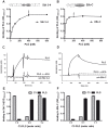
Figure 2. Characterization of the Plasminogen interactions with Sbi and Efb-C.
(A+B) Plasminogen (PLG) bound to the fragments 3–4 of Sbi (A) and the C-terminus of Efb (B). Sbi 3–4 or Efb-C was immobilized and increasing amounts of plasminogen were added. Bound plasminogen was detected with specific antisera. Plasminogen concentrations showing saturated binding are marked by arrows. (C+D) Plasminogen (solid lines) associated with Sbi 3–4 (C) or Efb-C (D) was determined by surface plasmon resonance. Sbi 3–4 or Efb-C was immobilized and plasminogen was applied in fluid phase. Binding of plasminogen to Sbi 3–4 or Efb-C was inhibited by εACA (dashed line). (E+F) Plasminogen (black columns) and C3 (grey columns) bound simultaneously to Sbi 3–4 (E) and Efb-C (F). Sbi 3–4 or Efb-C was immobilized and C3 and plasminogen were added either alone or together (200 nM C3:200 nM PLG  = 1∶1 for EfbC, 200 nM C3:400 nM PLG = 1∶2 for Sbi3-4). Representative experiments out of three independent experiments are shown.
Figure 3
Figure 3. Plasminogen bound to Sbi or Efb is converted to plasmin.
(A) Plasmin (PL) bound to Sbi, Efb, and to the borrelial CRASP-5 converted a chromogenic substrate as followed by measuring absorbance at 405 nm after 24 h. Equimolar amounts of bacterial proteins were immobilized and incubated with plasminogen (PLG). After washing, the activator uPA was applied together with the chromogenic substrate S-2251. Human serum albumin (HSA) had no effect. (B+C) Plasmin generation of plasminogen bound to Sbi (B) and Efb (C) by uPa or staphylokinase (SAK). The plasmin activators uPa and SAK or no activator (w/o) were applied to plasminogen bound to Sbi or Efb together with the chromogenic substrate. Conversion of the chromogenic substrate was determined at various time points. Data represent mean values ± standard deviations from three independent experiments.
Figure 4
Figure 4. Plasmin complexed with C3(b) and Sbi or Efb degrades C3 and C3b within the complex.
Plasmin degraded C3/C3b within the Sbi:plasmin:C3/C3b or Efb:plasmin:C3/C3b complexes (lane 1 and 4) and C3/C3b cleavage products appeared (marked with arrows). Bacterial proteins were immobilized (equimolar) and plasminogen was added together with C3 (A) or C3b (B). The activator SAK was applied and C3/C3b cleavage was followed by Western blot analysis using anti-C3-HRP (Fab). The mobility of the α (α ´) and β chains of C3/C3b and the cleavage products are indicated by arrows. CRASP-5 and HSA had no effect on cleavage (lane 7 and 8). Data shown are representative of three independent experiments. (C) Proposed schematic model of the C3 cleavage products generated by plasmin modified after , . The C3 dg cleavage product is not recognized by the C3-antibodies used in this study.
Figure 5
Figure 5. Plasmin mediated cleavage of C3 is enhanced by Sbi and Efb.
(A) In the presence of Sbi and Efb (lane 1 and 2) the C3 α-chain decreased and C3 cleavage products (indicated by arrows) appeared. CRASP-5 did not show this effect. C3 was incubated with SAK-activated plasmin (PLSAK) in the presence of Sbi, Efb, CRASP-5, or HSA. Samples were separated by SDS-PAGE and C3 cleavage products were visualized using Western blot analysis. (B) A similar assay was performed with uPa as activator and this time the fragments Sbi 1–2, Sbi 3–4, and Efb-C were assayed. Sbi 3–4 (lane 5 and 6) and Efb-C (lane 8 and 9) enhanced the plasmin mediated C3 cleavage as seen by the appearance of small cleavage products. The IgG-binding Sbi 1–2 (mobility of 17 kDa) did not affect the C3 cleavage pattern (lane 2 and 3). Polyclonal C3 antiserum was used for detection. (C) In the presence of Sbi 3–4 and Efb-C the plasmin mediated C3 proteolysis was enhanced as measured by ELISA. C3 was immobilized and plasmin (activated by uPa or SAK) together with Sbi 3–4 or Efb-C were added. C3 deposition was detected after 3 h with C3a antiserum. CRASP-5 and HSA did not influence plasmin cleavage of C3. (D) Sbi 3–4 and Efb-C significantly enhanced C3 degradation by plasmin in a dose dependent manner. The C3 amount after incubation with plasmin was set to 100%. Data in C and D are mean values of three independent experiments; error bars indicate standard deviations. *p<0.05; **p<0.01 and ***p<0.001.
Figure 6
Figure 6. Plasmin cleavage of C3b is enhanced by Sbi and Efb.
(A) Sbi 3–4 (lane 5–6) and Efb-C (lane 8–9) enhanced cleavage of C3b by plasmin, as seen by the accumulation of C3b cleavage products. Sbi 1–2 did not influence C3b degradation (lane 2–3). C3b was cleaved by plasmin in the presence of Sbi 1–2, Sbi 3–4, and Efb-C. (B+C) Sbi 3–4 and Efb-C enhanced the anti-opsonic activity of plasmin. C3b was deposited on S. aureus and Sbi 1–2, Sbi 3–4, Efb-C, or HSA was incubated in the absence or presence of plasmin. Remaining C3b fragments on S. aureus were measured by flow-cytometry. The C3b amount in the absence of plasmin was set to 100%. Data represent mean values ± standard deviations of five independent experiments. *p<0.05. (C) In parallel the supernatants were separated by SDS-PAGE and analyzed by Western blotting using anti-C3-HRP (Fab). Sbi 1–2 did not influence the amount of C3b-fragments after plasmin degradation (lane 1). Sbi 3–4 (lane 2) and Efb-C (lane 4) increased the plasmin degradation and more C3b cleavage products were detected.
Figure 7
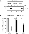
Figure 7. Plasmin degrades C3a.
(A) C3a was completely degraded by plasmin; plasminogen had a weak degrading effect. S. aureus was incubated with C3a in the presence of plasmin (PLG+SAK) (lane 4) or plasminogen alone (lane 6). The supernatants were analyzed by SDS-PAGE and Western blot analysis. (B) Plasmin increased the survival of S. aureus to 85% and plasminogen alone to 50%. In parallel S. aureus treated with C3a and or plasmin(ogen) was cultivated overnight on LB agar plates. CFUs were counted and the survival without C3a was set to 100% (white columns). Data represent mean values ± standard deviations from three independent experiments. ***p<0.001.

Similar articles

Cited by

References

    1. Lowy FD (1998) Staphylococcus aureus infections. N Engl J Med 339: 520–532. - PubMed
    1. Klevens RM, Morrison MA, Nadle J, Petit S, Gershman K, et al. (2007) Invasive methicillin-resistant Staphylococcus aureus infections in the United States. JAMA 298: 1763–1771. - PubMed
    1. Walport MJ (2001) Complement. First of two parts. N Engl J Med 344: 1058–1066. - PubMed
    1. Zipfel PF, Skerka C (2009) Complement regulators and inhibitory proteins. Nature Reviews Immunology 9: 729–740. - PubMed
    1. Walport MJ (2001) Complement. Second of two parts. N Engl J Med 344: 1140–1144. - PubMed

Publication types

Grants and funding

Tina Koch is a doctoral researcher at the International Leibniz Research School (ILRS), part of the Jena school of Microbial Communication (JSMC), Jena, Germany. The funders had no role in study design, data collection and analysis, decision to publish, or preparation of the manuscript.

LinkOut - more resources