A novel anticancer therapy that simultaneously targets aberrant p53 and Notch activities in tumors
- PMID: 23071601
- PMCID: PMC3468572
- DOI: 10.1371/journal.pone.0046627
A novel anticancer therapy that simultaneously targets aberrant p53 and Notch activities in tumors
Abstract
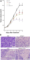
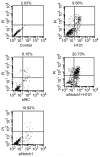
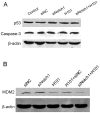
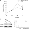
Notch signaling pathway plays an important role in tumorigenesis by maintaining the activity of self-renewal of cancer stem cells, and therefore, it is hypothesized that interference of Notch signaling may inhibit tumor formation and progression. H101 is a recombinant oncolytic adenovirus that is cytolytic in cells lacking intact p53, but it is unable to eradicate caner stem cells. In this study, we tested a new strategy of tumor gene therapy by combining a Notch1-siRNA with H101 oncolytic adenovirus. In HeLa-S3 tumor cells, the combined therapy blocked the Notch pathway and induced apoptosis in tumors that are p53-inactive. In nude mice bearing xenograft tumors derived from HeLa-S3 cells, the combination of H101/Notch1-siRNA therapies inhibited tumor growth. Moreover, Notch1-siRNA increased Hexon gene expression at both the transcriptional and the translational levels, and promoted H101 replication in tumors, thereby enhancing the oncolytic activity of H101. These data demonstrate the feasibility to combine H101 p53-targted oncolysis and anti-Notch siRNA activities as a novel anti-cancer therapy.
Conflict of interest statement
Figures


Similar articles
-
A novel apoptotic mechanism of genetically engineered adenovirus-mediated tumour-specific p53 overexpression through E1A-dependent p21 and MDM2 suppression.Eur J Cancer. 2012 Sep;48(14):2282-91. doi: 10.1016/j.ejca.2011.12.020. Epub 2012 Jan 13. Eur J Cancer. 2012. PMID: 22244827
-
Enhanced therapeutic efficacy by simultaneously targeting two genetic defects in tumors.Mol Ther. 2009 Jan;17(1):57-64. doi: 10.1038/mt.2008.236. Epub 2008 Nov 18. Mol Ther. 2009. PMID: 19018252 Free PMC article.
-
The oncolytic virus H101 combined with GNAQ siRNA-mediated knockdown reduces uveal melanoma cell viability.J Cell Biochem. 2019 Apr;120(4):5766-5776. doi: 10.1002/jcb.27863. Epub 2018 Oct 15. J Cell Biochem. 2019. PMID: 30320917
-
Recombinant oncolytic adenovirus H101 combined with siBCL2: cytotoxic effect on uveal melanoma cell lines.Br J Ophthalmol. 2012 Oct;96(10):1331-8. doi: 10.1136/bjophthalmol-2011-301470. Epub 2012 Jul 27. Br J Ophthalmol. 2012. PMID: 22843987
-
Functional genomic screening to enhance oncolytic virotherapy.Br J Cancer. 2013 Feb 5;108(2):245-9. doi: 10.1038/bjc.2012.467. Epub 2012 Nov 20. Br J Cancer. 2013. PMID: 23169279 Free PMC article. Review.
Cited by
-
Effects of dexamethasone on angiotensin II-induced changes of monolayer permeability and F-actin distribution in glomerular endothelial cells.Exp Ther Med. 2013 Nov;6(5):1131-1136. doi: 10.3892/etm.2013.1278. Epub 2013 Aug 30. Exp Ther Med. 2013. PMID: 24223634 Free PMC article.
-
An Extensive Review on Preclinical and Clinical Trials of Oncolytic Viruses Therapy for Pancreatic Cancer.Front Oncol. 2022 May 24;12:875188. doi: 10.3389/fonc.2022.875188. eCollection 2022. Front Oncol. 2022. PMID: 35686109 Free PMC article. Review.
-
Effects of inhibition of hedgehog signaling on cell growth and migration of uveal melanoma cells.Cancer Biol Ther. 2014 May;15(5):544-59. doi: 10.4161/cbt.28157. Epub 2014 Mar 11. Cancer Biol Ther. 2014. PMID: 24553082 Free PMC article.
-
Detection and validation of circulating endothelial cells, a blood-based diagnostic marker of acute myocardial infarction.PLoS One. 2013;8(3):e58478. doi: 10.1371/journal.pone.0058478. Epub 2013 Mar 4. PLoS One. 2013. PMID: 23484031 Free PMC article.
-
Oncolytic Virotherapy: From Bench to Bedside.Front Cell Dev Biol. 2021 Nov 26;9:790150. doi: 10.3389/fcell.2021.790150. eCollection 2021. Front Cell Dev Biol. 2021. PMID: 34901031 Free PMC article. Review.
References
-
- Bischoff JR, Kirn DH, Williams A, Heise C, Horn S, et al. (1996) An adenovirus mutant that replicates selectively in p53-deficient human tumor cells. Science 274: 373–376. - PubMed
-
- Wiman KG (2006) Strategies for therapeutic targeting of the p53 pathway in cancer. Cell Death Differ 13: 921–926. - PubMed
-
- Post LE (2002) Selectively replicating adenoviruses for cancer therapy: an update on clinical development. Curr Opin Investig Drugs 3: 1768–1772. - PubMed
-
- Yu W, Fang H (2007) Clinical trials with oncolytic adenovirus in China. Curr Cancer Drug Targets 7: 141–148. - PubMed
Publication types
MeSH terms
Substances
Grants and funding
LinkOut - more resources
Full Text Sources
Research Materials
Miscellaneous

