

Early life stress differentially modulates distinct forms of brain plasticity in young and adult mice
- PMID: 23071534
- PMCID: PMC3465301
- DOI: 10.1371/journal.pone.0046004
Early life stress differentially modulates distinct forms of brain plasticity in young and adult mice
Abstract
Background: Early life trauma is an important risk factor for many psychiatric and somatic disorders in adulthood. As a growing body of evidence suggests that brain plasticity is disturbed in affective disorders, we examined the short-term and remote effects of early life stress on different forms of brain plasticity.
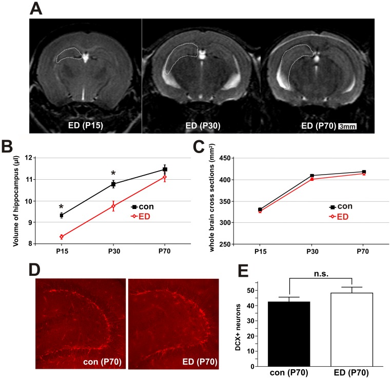
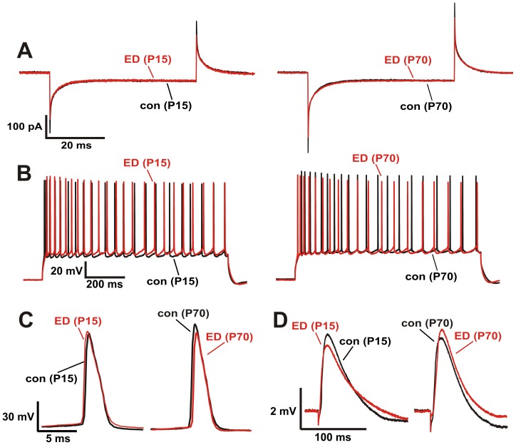
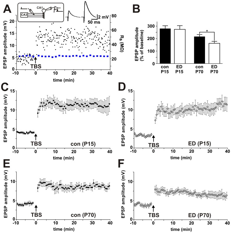
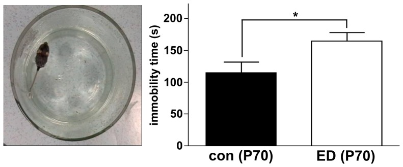
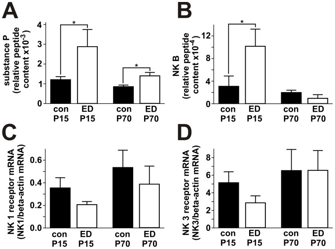
Methodology/principal findings: Mice were subjected to early deprivation by individually separating pups from their dam in the first two weeks after birth. Distinct forms of brain plasticity were assessed in the hippocampus by longitudinal MR volumetry, immunohistochemistry of neurogenesis, and whole-cell patch-clamp measurements of synaptic plasticity. Depression-related behavior was assessed by the forced swimming test in adult animals. Neuropeptides and their receptors were determined by real-time PCR and immunoassay. Early maternal deprivation caused a loss of hippocampal volume, which returned to normal in adulthood. Adult neurogenesis was unaffected by early life stress. Long-term synaptic potentiation, however, was normal immediately after the end of the stress protocol but was impaired in adult animals. In the forced swimming test, adult animals that had been subjected to early life stress showed increased immobility time. Levels of substance P were increased both in young and adult animals after early deprivation.
Conclusion: Hippocampal volume was affected by early life stress but recovered in adulthood which corresponded to normal adult neurogenesis. Synaptic plasticity, however, exhibited a delayed impairment. The modulation of synaptic plasticity by early life stress might contribute to affective dysfunction in adulthood.
Conflict of interest statement
Figures
Similar articles
-
Severe early life stress hampers spatial learning and neurogenesis, but improves hippocampal synaptic plasticity and emotional learning under high-stress conditions in adulthood.J Neurosci. 2010 May 12;30(19):6635-45. doi: 10.1523/JNEUROSCI.0247-10.2010. J Neurosci. 2010. PMID: 20463226 Free PMC article.
-
Enhanced long-term synaptic depression in an animal model of depression.Biol Psychiatry. 2007 Jul 1;62(1):92-100. doi: 10.1016/j.biopsych.2006.07.007. Epub 2006 Dec 4. Biol Psychiatry. 2007. PMID: 17141742
-
Maternal separation prior to neonatal hypoxia-ischemia: Impact on emotional aspects of behavior and markers of synaptic plasticity in hippocampus.Int J Dev Neurosci. 2016 Aug;52:1-12. doi: 10.1016/j.ijdevneu.2016.04.002. Epub 2016 May 7. Int J Dev Neurosci. 2016. PMID: 27165447
-
Early-life stress mediated modulation of adult neurogenesis and behavior.Behav Brain Res. 2012 Feb 14;227(2):400-9. doi: 10.1016/j.bbr.2011.07.037. Epub 2011 Jul 28. Behav Brain Res. 2012. PMID: 21821065 Review.
-
Enhancement of Hippocampal Plasticity by Physical Exercise as a Polypill for Stress and Depression: A Review.CNS Neurol Disord Drug Targets. 2019;18(4):294-306. doi: 10.2174/1871527318666190308102804. CNS Neurol Disord Drug Targets. 2019. PMID: 30848219 Review.
Cited by
-
A preclinical perspective on the enhanced vulnerability to Alzheimer's disease after early-life stress.Neurobiol Stress. 2018 Feb 23;8:172-185. doi: 10.1016/j.ynstr.2018.02.003. eCollection 2018 Feb. Neurobiol Stress. 2018. PMID: 29888312 Free PMC article. Review.
-
Neonatal Maternal Separation Induces Sexual Dimorphism in Brain Development: The Influence on Amino Acid Levels and Cognitive Disorders.Biomolecules. 2023 Sep 26;13(10):1449. doi: 10.3390/biom13101449. Biomolecules. 2023. PMID: 37892131 Free PMC article.
-
Early-Life Social Isolation Impairs the Gonadotropin-Inhibitory Hormone Neuronal Activity and Serotonergic System in Male Rats.Front Endocrinol (Lausanne). 2015 Nov 10;6:172. doi: 10.3389/fendo.2015.00172. eCollection 2015. Front Endocrinol (Lausanne). 2015. PMID: 26617573 Free PMC article.
-
Maternal Forced Swimming Reduces Cell Proliferation in the Postnatal Dentate Gyrus of Mouse Offspring.Front Neurosci. 2016 Aug 29;10:402. doi: 10.3389/fnins.2016.00402. eCollection 2016. Front Neurosci. 2016. PMID: 27621701 Free PMC article.
-
Early life stress-induced alterations in rat brain structures measured with high resolution MRI.PLoS One. 2017 Sep 25;12(9):e0185061. doi: 10.1371/journal.pone.0185061. eCollection 2017. PLoS One. 2017. PMID: 28945761 Free PMC article.
References
-
- Gage FH (2000) Mammalian neural stem cells. Science 287: 1433–1438. - PubMed
-
- Holderbach R, Clark K, Moreau JL, Bischofberger J, Normann C (2007) Enhanced long-term synaptic depression in an animal model of depression. Biol Psychiatry 62: 92–100. - PubMed
-
- Leventopoulos M, Ruedi-Bettschen D, Knuesel I, Feldon J, Pryce CR, et al. (2007) Long-term effects of early life deprivation on brain glia in Fischer rats. Brain Res 1142: 119–126. - PubMed
Publication types
MeSH terms
Substances
Grants and funding
LinkOut - more resources
Full Text Sources
Medical
Research Materials
