The biochemistry of hematopoietic stem cell development
- PMID: 23069720
- PMCID: PMC3587313
- DOI: 10.1016/j.bbagen.2012.10.004
The biochemistry of hematopoietic stem cell development
Abstract
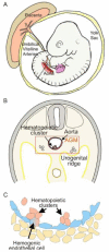
Background: The cornerstone of the adult hematopoietic system and clinical treatments for blood-related disease is the cohort of hematopoietic stem cells (HSC) that is harbored in the adult bone marrow microenvironment. Interestingly, this cohort of HSCs is generated only during a short window of developmental time. In mammalian embryos, hematopoietic progenitor and HSC generation occurs within several extra- and intraembryonic microenvironments, most notably from 'hemogenic' endothelial cells lining the major vasculature. HSCs are made through a remarkable transdifferentiation of endothelial cells to a hematopoietic fate that is long-lived and self-renewable. Recent studies are beginning to provide an understanding of the biochemical signaling pathways and transcription factors/complexes that promote their generation.
Scope of review: The focus of this review is on the biochemistry behind the generation of these potent long-lived self-renewing stem cells of the blood system. Both the intrinsic (master transcription factors) and extrinsic regulators (morphogens and growth factors) that affect the generation, maintenance and expansion of HSCs in the embryo will be discussed.
Major conclusions: The generation of HSCs is a stepwise process involving many developmental signaling pathways, morphogens and cytokines. Pivotal hematopoietic transcription factors are required for their generation. Interestingly, whereas these factors are necessary for HSC generation, their expression in adult bone marrow HSCs is oftentimes not required. Thus, the biochemistry and molecular regulation of HSC development in the embryo are overlapping, but differ significantly from the regulation of HSCs in the adult.
General significance: HSC numbers for clinical use are limiting, and despite much research into the molecular basis of HSC regulation in the adult bone marrow, no panel of growth factors, interleukins and/or morphogens has been found to sufficiently increase the number of these important stem cells. An understanding of the biochemistry of HSC generation in the developing embryo provides important new knowledge on how these complex stem cells are made, sustained and expanded in the embryo to give rise to the complete adult hematopoietic system, thus stimulating novel strategies for producing increased numbers of clinically useful HSCs. This article is part of a Special Issue entitled Biochemistry of Stem Cells.
Copyright © 2012 Elsevier B.V. All rights reserved.
Figures


Similar articles
-
Hematopoietic stem cell development and regulatory signaling in zebrafish.Biochim Biophys Acta. 2013 Feb;1830(2):2370-4. doi: 10.1016/j.bbagen.2012.06.008. Epub 2012 Jun 15. Biochim Biophys Acta. 2013. PMID: 22705943 Review.
-
Current approaches in biomaterial-based hematopoietic stem cell niches.Acta Biomater. 2018 May;72:1-15. doi: 10.1016/j.actbio.2018.03.028. Epub 2018 Mar 22. Acta Biomater. 2018. PMID: 29578087 Review.
-
The emergence of definitive hematopoietic stem cells in the mammal.Curr Opin Hematol. 2005 May;12(3):197-202. doi: 10.1097/01.moh.0000160736.44726.0e. Curr Opin Hematol. 2005. PMID: 15867575 Review.
-
Role of key regulators of the cell cycle in maintenance of hematopoietic stem cells.Biochim Biophys Acta. 2013 Feb;1830(2):2335-44. doi: 10.1016/j.bbagen.2012.07.004. Epub 2012 Jul 20. Biochim Biophys Acta. 2013. PMID: 22820018 Review.
-
Hematopoietic (stem) cell development - how divergent are the roads taken?FEBS Lett. 2016 Nov;590(22):3975-3986. doi: 10.1002/1873-3468.12372. Epub 2016 Sep 1. FEBS Lett. 2016. PMID: 27543859 Free PMC article. Review.
Cited by
-
Defining the path to hematopoietic stem cells.Nat Biotechnol. 2013 May;31(5):416-8. doi: 10.1038/nbt.2571. Nat Biotechnol. 2013. PMID: 23657396 No abstract available.
-
Odd couple: The unexpected partnership of glucocorticoid hormones and cysteinyl-leukotrienes in the extrinsic regulation of murine bone-marrow eosinopoiesis.World J Exp Med. 2017 Feb 20;7(1):11-24. doi: 10.5493/wjem.v7.i1.11. eCollection 2017 Feb 20. World J Exp Med. 2017. PMID: 28261551 Free PMC article. Review.
-
Transition of mesenchymal stem/stromal cells to endothelial cells.Stem Cell Res Ther. 2013 Aug 14;4(4):95. doi: 10.1186/scrt306. Stem Cell Res Ther. 2013. PMID: 23953698 Free PMC article.
-
ATF4 plays a pivotal role in the development of functional hematopoietic stem cells in mouse fetal liver.Blood. 2015 Nov 19;126(21):2383-91. doi: 10.1182/blood-2015-03-633354. Epub 2015 Sep 17. Blood. 2015. PMID: 26384355 Free PMC article.
-
Let-7 microRNA-dependent control of leukotriene signaling regulates the transition of hematopoietic niche in mice.Nat Commun. 2017 Jul 25;8(1):128. doi: 10.1038/s41467-017-00137-y. Nat Commun. 2017. PMID: 28743859 Free PMC article.
References
-
- Murray P. The development in vitro of the blood of the early chick embryo. Proc Roy Soc London. 1932;11:497–521.
-
- Sabin F. Studies on the origin of blood vessels and of red blood corpuscles as seen in the living blastoderm of chicks during the second day of incubation, Carnegie Inst. Wash. Pub. # 272. Contrib. Embryol. 1920;9:214.
-
- Park C, Ma YD, Choi K. Evidence for the hemangioblast. Exp Hematol. 2005;33:965–970. - PubMed
-
- Shalaby F, Rossant J, Yamaguchi TP, Gertsenstein M, Wu XF, Breitman ML, Schuh AC. Failure of blood-island formation and vasculogenesis in Flk-1-deficient mice. Nature. 1995;376:62–66. - PubMed
Publication types
MeSH terms
Grants and funding
LinkOut - more resources
Full Text Sources
Other Literature Sources
Medical
Research Materials

