. 2012 Nov 14;31(22):4318-33.
doi: 10.1038/emboj.2012.275. Epub 2012 Oct 12.

RUNX1 reshapes the epigenetic landscape at the onset of haematopoiesis

Affiliations

RUNX1 reshapes the epigenetic landscape at the onset of haematopoiesis

Monika Lichtinger et al. EMBO J. .

Abstract

Cell fate decisions during haematopoiesis are governed by lineage-specific transcription factors, such as RUNX1, SCL/TAL1, FLI1 and C/EBP family members. To gain insight into how these transcription factors regulate the activation of haematopoietic genes during embryonic development, we measured the genome-wide dynamics of transcription factor assembly on their target genes during the RUNX1-dependent transition from haemogenic endothelium (HE) to haematopoietic progenitors. Using a Runx1-/- embryonic stem cell differentiation model expressing an inducible Runx1 gene, we show that in the absence of RUNX1, haematopoietic genes bind SCL/TAL1, FLI1 and C/EBPβ and that this early priming is required for correct temporal expression of the myeloid master regulator PU.1 and its downstream targets. After induction, RUNX1 binds to numerous de novo sites, initiating a local increase in histone acetylation and rapid global alterations in the binding patterns of SCL/TAL1 and FLI1. The acquisition of haematopoietic fate controlled by Runx1 therefore does not represent the establishment of a new regulatory layer on top of a pre-existing HE program but instead entails global reorganization of lineage-specific transcription factor assemblies.


Conflict of interest statement

The authors declare that they have no conflict of interest.

Figures

Figure 1
Figure 1
Time course of expression of haematopoietic regulator genes during ES cell differentiation. (A) Schematic outline of the haematopoietic differentiation model. Haemangioblast (Flk1+, Bry+) cells are sorted from embryoid bodies on day 0 and during a 4-day blast culture through the stage of the haemogenic endothelium give rise to haematopoietic progenitors, which can be differentiated into macrophages (CD11b+, F4/80+). Relevant surface markers of sorted cell populations are indicated. (B) Relative expression analysis of haematopoietic regulator genes in purified cells from all stages of differentiation. Flk1− cells served as control and experiments were carried out at least in triplicates where STDEV was applied, otherwise the average of two biological duplicates and the respective values are shown.
Figure 2
Figure 2
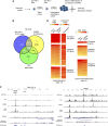
Genome-wide SCL/TAL1, FLI1 and C/EBPβ binding in the haemogenic endothelium. (A) Schematic overview of iRUNX1 differentiation. In RUNX1-deficient iRUNX1 cells a two-step protocol was used, including a second Flk1 sort during blast culture to isolate haemogenic endothelium cells, which are arrested at that stage. (B) Venn diagram demonstrating the intersection of SCL/TAL1, C/EBPβ and FLI1 peaks. (C) UCSC genome browser screenshots showing the binding pattern of transcription factors, RNA-Pol II and H3K9Ac at Sfpi1 (Pu.1) and Notch1 and depicting unmanipulated aligned reads at each chromosomal location. (D) Heat maps showing the distribution of RNA-Pol II and H3K9 acetylation around SCL/TAL1 (left), FLI1 (middle) and C/EBPβ (right) binding sites within promoter/intragenic and intergenic regions. RNA Pol II and H3K9Ac levels for each promoter/intragenic and intergenic division were counted using a 1-kb window flanking the peak summits.
Figure 3
Figure 3
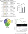
Sequence motifs and genes associated with SCL/TAL1, C/EBPβ and FLI1 binding. (A) Unbiased binding motif search underlying unique SCL/TAL1, C/EBPβ or FLI1 peaks and those overlapping between SCL/TAL1 and FLI1. (B) Frequency of the corresponding binding site motifs within ChIP-sequencing peaks. (C) Venn diagram showing the overlap of genes bound by the three factors (2400) and the sections analysed by GSEA (red numbers, Supplementary Figure 3A). (D) Gene set enrichment analysis (GSEA) of genes bound by all three factors, SCL/TAL1, FLI1 and C/EBPβ comparing these genes with genes expressed in haematopoietic stem cells (HSCs) and differentiated mature blood cells.
Figure 4
Figure 4
The FLI1 binding site in the URE is required for the correct timing of Pu.1 upregulation. (A) Upper panel: Map of regulatory elements of the Pu.1 gene. Transcription factor binding sites of the −14 kb upstream regulatory element (URE) and promoter are highlighted in the enlarged detailed map. DNase I hypersensitive sites are marked by vertical arrows and the transcription start site by a horizontal arrow. Lower panel: Section of the −14 kb URE containing a mutation in the FLI1 binding site. (B) Relative expression analysis during the ES cell to macrophage differentiation in heterozygous Pu.1+/ki cells (control: black bars) and the homozygous Pu.1ki/ki cells (white bars). Experiments represent the mean values of three biological replicates.
Figure 5
Figure 5
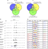
FLI1 and SCL/TAL1 binding profiles in haematopoietic c-kit+ precursor cells. (A) Venn diagram showing the intersection of ChIP-sequencing data of FLI1 and SCL/TAL1 in c-kit+/CD41+/Tie2− (c-kit+) wild-type progenitor cells prepared on day 4 of blast differentiation. (B) UCSC genome browser screenshots providing examples of genes where the binding pattern of FLI1 and SCL/TAL1 changes during differentiation from the uninduced haemogenic endothelium (HE) to c-kit+ progenitors. (C) Venn diagrams showing the overlap of peaks of SCL/TAL1 (left) and FLI1 (right) between the uninduced HE and wild-type c-kit+ progenitors. (D) Hierarchical clustering of transcription factor binding sequences in different cell types (data sets from this study are highlighted in blue). Based on similar binding patterns of the different ChIP-seq data, a correlation matrix was generated and sequence similarities are displayed after hierarchical clustering and using a colour code.
Figure 6
Figure 6
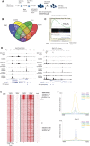
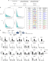
Identification of genome-wide RUNX1 binding sites in the HE. (A) Experimental strategy for iRUNX ES cell differentiation. (B) Four-way Venn diagram of genes bound by RUNX1, SCL/TAL1, FLI1 and C/EBPβ. (C) GSEA of this group of genes. (D) Screenshots from the UCSC genome browser showing genes binding SCL/TAL1, FLI1 C/EBPβ and RNA Pol II in the uninduced HE. RUNX1 binding and H3K9Ac are shown before and after induction of RUNX1. (E) Heat map showing two groups of RUNX1 bound sequences and corresponding H3K9Ac levels before and after RUNX1 induction. ChIP enrichment is shown 5 kb upstream and downstream of the peak centre. (F) Integration of the sequence enrichment of each group into a density plot clearly demonstrating RUNX1 binding to pre-existing sites of histone acetylation and de novo sites.
Figure 7
Figure 7
Redistribution of SCL/TAL1 and FLI1 binding after RUNX1 induction. (A) Venn diagrams intersecting SCL/TAL1 peaks and FLI1 peaks in the uninduced HE, the induced HE and in c-kit+ cells. (B) UCSC genome browser screenshot showing the Sox17 locus exemplifying the movement of different transcription factors after RUNX1 induction in the different cell types. (C) Induction of RUNX1 leads to a shift in the binding of SCL/TAL1 and FLI1 into genomic regions containing RUNX1 motifs. Unbiased motif analysis of genomic regions uniquely bound by the two transcription factors in uninduced or induced iRUNX1 cells.
Figure 8
Figure 8
SCL/TAL1 and FLI1 move towards RUNX1 peaks after induction. (A) Total number and the percentage of peaks that disappear or emerge upon RUNX1 induction. (B) Frequency of distances between SCL/TAL1 or FLI1 peaks and the corresponding nearest RUNX1 binding sites for peaks that were lost (left panel) and peaks that were gained (right panel) after DOX induction. Both SCL/TAL1 and FLI1 gained peak overlaps with RUNX1 peaks were found to be significant (indicated by two asterisks), with Z scores of 107.6 and 34.9, respectively. We found no evidence that RUNX displaced SCL/TAL (Z=0.54), however, it had a weak propensity to replace FLI-1 near its binding sites (Z=24.7, one asterisk). (C) Unbiased motif analysis of SCL/TAL1 and FLI1 bound regions near RUNX1 sites that were either gained or lost after RUNX1 binding, demonstrating the presence of RUNX1 motifs in each analysed peak population. The analysed peak populations are depicted in Supplementary Figure 8. (D) Experimental strategy for withdrawal experiments. (E) Expression (upper panels) and factor binding (lower panels) before and after RUNX1 induction and withdrawal. Amplicons containing a RUNX1 binding site are marked by an asterisk. Error bars indicate the standard deviation between three independent differentiation and ChIP experiments.

Similar articles

Cited by

References

    1. Begay V, Smink J, Leutz A (2004) Essential requirement of CCAAT/enhancer binding proteins in embryogenesis. Mol Cell Biol 24: 9744–9751 - PMC - PubMed
    1. Bereshchenko O, Mancini E, Moore S, Bilbao D, Mansson R, Luc S, Grover A, Jacobsen SE, Bryder D, Nerlov C (2009) Hematopoietic stem cell expansion precedes the generation of committed myeloid leukemia-initiating cells in C/EBPalpha mutant AML. Cancer Cell 16: 390–400 - PubMed
    1. Chambers SM, Boles NC, Lin KY, Tierney MP, Bowman TV, Bradfute SB, Chen AJ, Merchant AA, Sirin O, Weksberg DC, Merchant MG, Fisk CJ, Shaw CA, Goodell MA (2007) Hematopoietic fingerprints: an expression database of stem cells and their progeny. Cell Stem Cell 1: 578–591 - PMC - PubMed
    1. Chen MJ, Li Y, De Obaldia ME, Yang Q, Yzaguirre AD, Yamada-Inagawa T, Vink CS, Bhandoola A, Dzierzak E, Speck NA (2011) Erythroid/Myeloid progenitors and hematopoietic stem cells originate from distinct populations of endothelial cells. Cell Stem Cell 9: 541–552 - PMC - PubMed
    1. Chen MJ, Yokomizo T, Zeigler BM, Dzierzak E, Speck NA (2009) Runx1 is required for the endothelial to haematopoietic cell transition but not thereafter. Nature 457: 887–891 - PMC - PubMed

Publication types