. 2012;7(10):e46459.
doi: 10.1371/journal.pone.0046459. Epub 2012 Oct 8.

Angiogenesis inhibitor vasohibin-1 enhances stress resistance of endothelial cells via induction of SOD2 and SIRT1

Affiliations

Angiogenesis inhibitor vasohibin-1 enhances stress resistance of endothelial cells via induction of SOD2 and SIRT1

Hiroki Miyashita et al. PLoS One. 2012.

Abstract

Vasohibin-1 (VASH1) is isolated as an endothelial cell (EC)-produced angiogenesis inhibitor. We questioned whether VASH1 plays any role besides angiogenesis inhibition, knocked-down or overexpressed VASH1 in ECs, and examined the changes of EC property. Knock-down of VASH1 induced premature senescence of ECs, and those ECs were easily killed by cellular stresses. In contrast, overexpression of VASH1 made ECs resistant to premature senescence and cell death caused by cellular stresses. The synthesis of VASH1 was regulated by HuR-mediated post-transcriptional regulation. We sought to define the underlying mechanism. VASH1 increased the expression of (superoxide dismutase 2) SOD2, an enzyme known to quench reactive oxygen species (ROS). Simultaneously, VASH1 augmented the synthesis of sirtuin 1 (SIRT1), an anti-aging protein, which improved stress tolerance. Paraquat generates ROS and causes organ damage when administered in vivo. More VASH1 (+/-) mice died due to acute lung injury caused by paraquat. Intratracheal administration of an adenovirus vector encoding human VASH1 augmented SOD2 and SIRT1 expression in the lungs and prevented acute lung injury caused by paraquat. Thus, VASH1 is a critical factor that improves the stress tolerance of ECs via the induction of SOD2 and SIRT1.


Conflict of interest statement

Competing Interests: The authors have declared that no competing interests exist.

Figures

Figure 1
Figure 1. Knockdown of VASH1 induces premature senescence and enhances stress-induced cell death of HUVECs.
(A) HUVECs were transfected with VASH1 siRNA or control siRNA. After a 24-hour incubation, RT-PCR and Western blotting for VASH1 were performed. (B) Phase-contrast photomicrographs (on the left: 48 hours after siRNA transfection) and SA beta-gal staining (on the right: 5 days after siRNA transfection) are shown. Scale bars are 250 microm. SA beta-gal-positive HUVECs were quantified, and the % senescent cells was calculated. Values are the ratio of SA beta-gal-positive cells to total cells, and are means and SDs of 3 wells. (*P<0.01, N = 3). (C) HUVECs were transfected with VASH1 or control siRNA. After a 24-hour incubation, Western blotting for VASH1, ATM and p-ATM was performed. (D) HUVECs were transfected with VASH1 siRNA or control siRNA. After a 24-hour incubation, LC3 (red) was immunostained. Scale bars are 25 microm. (E) HUVECs were cultured in growth medium with 100 microM H2O2 including mouse IgG (control) or 10 microg/ml VASH-1 antibody (4E12) for 48 h. Trypan blue exclusion assay was performed. Blue-stained cells quantified, and the % of dead cells was calculated (*P<0.01, N = 3). All the studies were repeated at least 3 times to confirm the reproducibility.
Figure 2
Figure 2. Overexpression of VASH1 inhibits premature senescence and cell death of HUVECs induced by cellular stresses.
(A) HUVECs were infected with AdVASH1 or AdLacZ. After a 24-hour incubation, RT-PCR and Western blotting for VASH1 were performed. (B) HUVECs infected with AdVASH1 or AdLacZ were exposed to 100 µmol/L H2O2 for 1 hour, followed culture for 24 hours (on the left) or to 0% FCS/αMEM 24 hours (on the right). After a 6-day culture, SA β-gal staining was performed. Scale bars are 250 microm. SA β-gal-positive HUVECs were quantified, and the % of senescent cells was calculated (*P<0.01, N = 3). (C) HUVECs were transfected with VASH1 siRNA or control siRNA. After a 24-hour incubation, HUVECs were exposed to 100 µmol/L H2O2 or to 0% FCS/αMEM for 24 hours, and then the trypan blue exclusion assay was performed. Blue-stained cells were quantified, and the % of dead cells was calculated (*P<0.01, N = 3). (D) HUVECs infected with AdVASH1 or AdLacZ were exposed to 100 µmol/L H2O2 for 48 hours or to 0% FCS/αMEM for 24 hours for 24 hours; and then the trypan blue exclusion assay was performed (*P<0.01, N = 3). (E) HUVECs were transfected with VASH1 siRNA or control siRNA. After a 24-hour incubation, the growth medium was replaced with 50% conditioned medium derived from mock or VASH1 over-expressing MS1 clone 4. After a 24-hour incubation, the trypan blue exclusion assay was performed (*P<0.01, N = 3). All the studies were repeated at least 3 times to confirm the reproducibility.
Figure 3
Figure 3. VASH1 inhibits premature senescence and cell death of HAECs.
(A) HAECs were transfected with VASH1 siRNA or control siRNA. After a 24-hour incubation, RT-PCR and Western blotting for VASH1 were performed. (B) After a 6-day incubation, SA beta-gal staining was performed on HAECs that had been transfected with VASH1 siRNA or control siRNA. SA beta-gal-positive HAECs were quantified, and the % of senescent cells was calculated (*P<0.01, N = 3). (C) HAECs were infected with AdVASH1 or control AdLacZ. After a 24-hour incubation, RT-PCR and Western blotting for VASH1 were then performed. (D) HAECs infected with AdVASH1 or control AdLacZ were exposed to 100 µmol/L H2O2 or to 0% FCS/DMEM for 24 hours. The trypan blue exclusion assay was performed to judge cell death (*P<0.01, N = 3). All the studies were repeated at least 3 times to confirm the reproducibility.
Figure 4
Figure 4. HuR increases VASH1 protein level in HUVECs.
(A) HUVECs were incubated in 0% FCS/αMEM, and total RNA and protein were extracted at the indicated time points. Thereafter, RT-PCR and Western blotting for VASH1 were performed. Values below each band represent the mean fold change in RNA or protein expression level compared with the cognate zero time. (B) The AU-rich element (ARE) in the 3′ non coding region of the VASH1 gene is shown. (C) Immunoprecipitation and reverse transcription-polymerase chain reaction were performed as described in Materials and Methods. (D) HUVECs were transfected with HuR siRNA or control siRNA. After a 24- hour incubation, total RNA and protein were extracted; and then RT-PCR for HuR and Western blotting for VASH1 were performed. All the studies were repeated at least 3 times to confirm the reproducibility.
Figure 5
Figure 5. VASH1 controls SOD2 level and decreases ROS level in HUVECs.
(A) HUVECs were transfected with VASH1 siRNA or control siRNA. After a 24-hour incubation, cellular ROS was determined as described in Materials and Methods (*P<0.01, N = 3). (B) HUVECs were transfected with VASH1 siRNA or control siRNA. Twenty-four hours later, RT-PCR for the indicated genes was performed. (C) HUVECs were transfected with VASH1 siRNA or control siRNA in the presence or absence of 50 µmol/L NAC. After a 12-hour incubation, the culture medium was replaced with growth medium containing vehicle or 50 µmol/L NAC. After a 6-day culture, SA β-gal staining was performed. Scale bars are 250 µm. SA β-gal-positive HUVECs were quantified, and the % of senescent cells was calculated (*P<0.01, N = 3). (D) HUVECs were infected with AdVASH1 or AdLacZ. After a 24-hour incubation, the cells were exposed to 100 µmol/L H2O2 for 1 hour or to 0% FCS/αMEM for 6 hours. Thereafter, the cellular ROS level was determined (*P<0.01, N = 3). (E) HUVECs were infected with AdVASH1 or AdLacZ. After a 24-hour incubation, RT-PCR for VASH1 and SOD2 was performed. (F) HUVECs were infected with AdVASH1 or AdLacZ. After a 24-hour incubation, HUVECs were then transfected with SOD2 siRNA or control siRNA. After a subsequent 24-hour incubation, the cells were exposed to 100 µ mol/L H2O2 for 1 hour followed by a 48-hour incubation in growth medium. Scale bars are 250 µm. SA β-gal staining and Western blotting for VASH1 and SOD2 were then performed. SA β-gal-positive HUVECs were quantified, and the % of senescent cells was calculated (*P<0.01, N = 3). All the studies were repeated at least 3 times to confirm the reproducibility.
Figure 6
Figure 6. VASH1 controls SIRT1 level and increases stress resistance of HUVECs.
(A) HUVECs were transfected with VASH1 siRNA or control siRNA. After a 72-hour incubation, Western blotting for VASH1 and SIRT1 was performed. (B) HUVECs were transfected with VASH1 siRNA or control siRNA. After a 24-hour incubation, SIRT1 activity was determined (*P<0.01, N = 3) (C) HUVECs were preteated with vehicle or 5 µmol/L SIRT1 activator 3 for 12 hours, and then transfected with VASH1 siRNA or control siRNA. After a 6-day incubation, SA β-gal staining was performed. Scale bars are 100 µm. SA β-gal-positive HUVECs were quantified, and the % of senescent cells was calculated (*P<0.01, N = 4). (D) HUVECs were transfected with SIRT1 siRNA or control siRNA. After a 72-hour incubation, Western blotting for VASH1 and SIRT1 was performed. (E) HUVECs were infected with AdVASH1 or AdLacZ. After a 72-hour incubation, Western blotting for VASH1 and SIRT1. (F) HUVECs were infected with AdVASH1. After a 24-hour incubation, HUVECs were then transfected with SIRT1 siRNA or control siRNA. After a subsequent 24-hour incubation, the cells were exposed to 100 µmol/L H2O2 for 1 hour followed by a 48-hour incubation in growth medium. Scale bars are 250 µm. SA β-gal staining and Western blotting for VASH1 and SIRT1 were then performed. β-gal-positive HUVECs were quantified, and the % of senescent cells was calculated (*P<0.01, N = 3). All the studies were repeated at least 3 times to confirm the reproducibility.
Figure 7
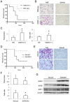
Figure 7. VASH1 protects mice from death with acute lung injury induced by paraquat treatment.
(A) Paraquat was administered to WT (N = 8, dotted line) or VASH1 (+/−) mice (N = 8, solid line), and the survival was observed over 10 days. Kaplan-Meier survival analysis showed significant difference. (B) Two days after paraquat administion, histological analyses of the lungs were performed. H&E staining is shown on the left, and immunostaining for 8-OHdG on the right. (C) Two days after paraquat administration, total protein and number of cells in the BALF were determined. *Significant difference (N = 7). (D) Paraquat was administered to VASH1 (+/−) mice intratracheally infected with AdVASH1 (N = 10, dotted line) or AdLacZ (N = 11, solid line), and the survival was observed over 10 days. Kaplan-Meier survival analysis showed significant difference. (E) Two days after paraquat administion to AdLacZ or AdVASH1 mice, histological analyses of lungs were performed. H&E staining is shown on the left; and immunostaining fo 8-OHdG, on the right. (F) Two days after paraquat administration, BALF was collected; and total protein and number of cells in the BALF were determined. *Significant difference (N = 10). (G) Three days after the intratracheal infection of mice with AdVASH1 or AdLacZ, their lungs were removed and tissue extracts prepared. Western blotting for VASH1, SOD2, and SIRT1 in the extracts was performed.

Similar articles

Cited by

References

    1. Phng LK, Gerhardt H (2009) Angiogenesis: a team effort coordinated by notch. Dev Cell 16: 196–208. - PubMed
    1. Watanabe K, Hasegawa Y, Yamashita H, Shimizu K, Ding Y, et al. (2004) Vasohibin as an endothelium-derived negative feedback regulator of angiogenesis. J Clin Invest 114: 898–907. - PMC - PubMed
    1. Kimura H, Miyashita H, Suzuki Y, Kobayashi M, Watanabe K, et al. (2009) Distinctive localization and opposed roles of vasohibin-1 and vasohibin-2 in the regulation of angiogenesis. Blood 113: 4810–4818. - PubMed
    1. Andreassi MG (2009) Metabolic syndrome, diabetes and atherosclerosis: influence of gene-environment interaction. Mutat Res 667: 35–43. - PubMed
    1. Erusalimsky JD, Kurz DJ (2005) Cellular senescence in vivo: its relevance in ageing and cardiovascular disease. Exp Gerontol 40: 634–642. - PubMed

Publication types

Grants and funding

This work was supported by grants from the programs Grant-in-Aid for Scientific Research on Innovative Areas “Integrative Research on Cancer Microenvironment Network” (22112006) and Grants-in-Aid for Scientific Research (C) [22590821] from the Ministry of Education, Culture, Sports, Science, and Technology of Japan, and by the 38th Research Grants in the Natural Sciences from the Mitsubishi Foundation. The funders had no role in study design, data collection and analysis, decision to publish, or preparation of the manuscript.