. 2012;7(10):e45366.
doi: 10.1371/journal.pone.0045366. Epub 2012 Oct 9.

Increased synapse formation obtained by T cell epitopes containing a CxxC motif in flanking residues convert CD4+ T cells into cytolytic effectors

Affiliations

Increased synapse formation obtained by T cell epitopes containing a CxxC motif in flanking residues convert CD4+ T cells into cytolytic effectors

Vincent A Carlier et al. PLoS One. 2012.

Abstract

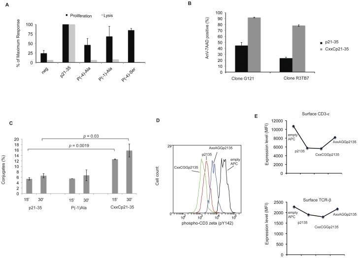
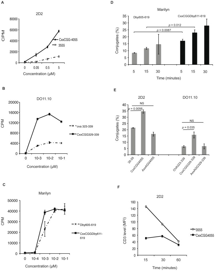
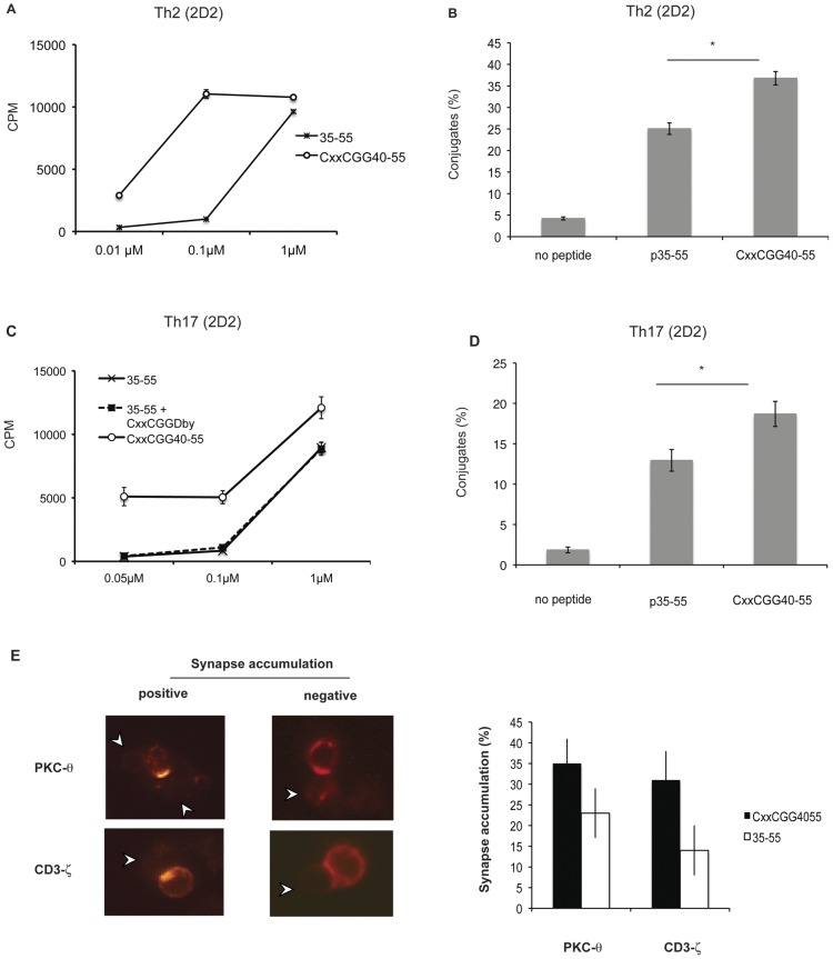
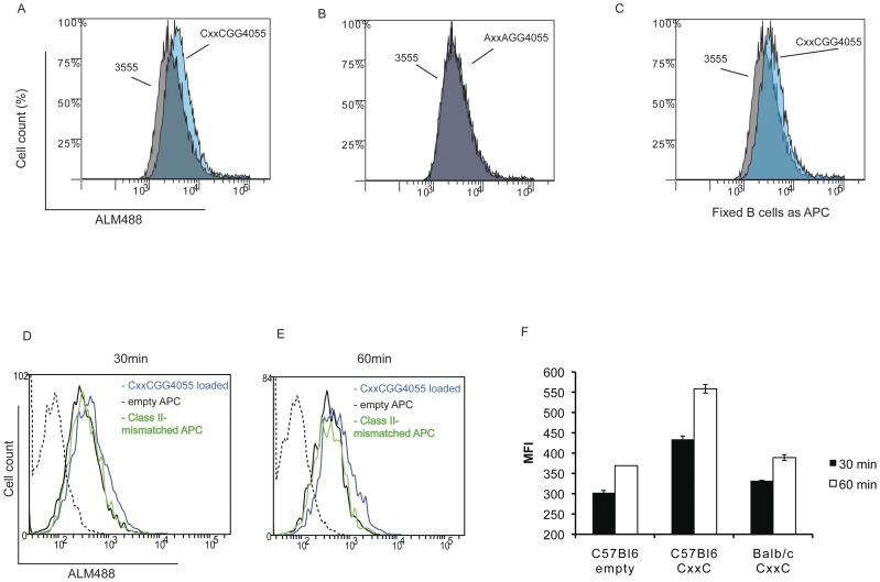
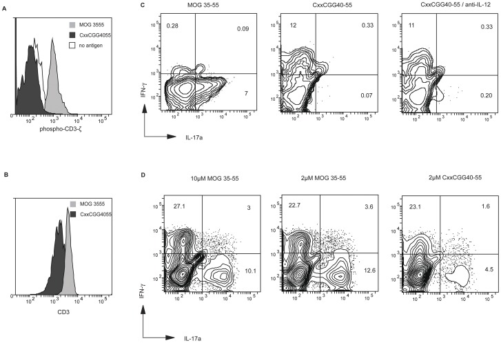
The nature of MHC class II-binding epitopes not only determines the specificity of T cell responses, but may also alter effector cell functions. Cytolytic CD4+ T cells have been observed primarily in anti-viral responses, but very little is known about the conditions under which they can be elicited. Their potential as regulators of immune responses, however, deserves investigations. We describe here that inclusion of a thiol-disulfide oxidoreductase motif within flanking residues of class II-restricted epitopes results, both in vitro and in vivo, in elicitation of antigen-specific cytolytic CD4+ T cells through increased synapse formation. We show that both naïve and polarized CD4+ T cells, including Th17 cells, can be converted by cognate recognition of such modified epitopes. Cytolytic CD4+ T cells induce apoptosis on APCs by Fas-FasL interaction. These findings potentially open the way towards a novel form of antigen-specific immunosuppression.


Conflict of interest statement

Competing Interests: The authors have declared that no competing interests exist.

Figures

Figure 1
Figure 1. Effects of aminoacid residue mutation in the p21–35 flanking sequence.
(A) Proliferation response and cytolytic properties of a p21–35 specific CD4+ T cell clone (G121) obtained from peptide-immunized BALB/c mice. Various mutant peptides (0.1 µM; sequence as indicated) were cultured with mitomycin C-treated splenocytes (T cell depleted). After 48 h, 3H-thymidine was added and its incorporation measured after 18 h of culture. Results are shown as percentage of incorporation obtained by comparison with p21–35 wildtype sequence (containing a CxxS motif). G121-induced cytolysis was measured on Dioc18-stained WEHI 231 B cells loaded with 0.1 µM of each peptide. After 24 h of incubation, cells were stained with Annexin V and 7-AAD and analysed by flow-cytometry. WEHI cell lysis (Dioc18+) is represented as percentage of lysis obtained by comparison with p21–35 wildtype sequence. Error bars represent 1 standard deviation (SD). Data are representative of three independent experiments. (B) Cytolytic assay of WEHI 231 B cells carried out as in (A) using two different CD4+ clones (G121 and R3TB7) exposed to peptides in either p21–35 wildtype sequence or after introduction of a cysteine in P(−1) thereby creating a thio-oxidoreductase motif. Error bars represent 1 SD. Data representative of two experiments. (C) Conjugate formation. DioC18 stained WEHI 231 B cells were loaded for 1 h with 1 µM of either Der p 2 p21–35, mutant peptide P(−1)Ala or with peptide containing the sequence CxxCGG. A CellTrace DDAO stained p21–35 specific clone (R3TB7) was added to each WEHI cell culture (ratio 1/1) and cell mixtures were briefly centrifuged. After 15 or 30 minutes, cells were softly resuspended, fixed for 10 minutes with 4% paraformaldhyde and analyzed by flow cytometry. Percentages of conjugates stand for the proportion of T cells staining positive for both labels over the total T cell population. Error bars represent 1 SD. Two tailed p values are derived from unpaired t tests. Data representative of two experiments. (D) Catabolism of CD3-ζ. On day 2 after restimulation, a p21–35 specific clone R3TB6 was washed and cultured for 15 minutes with WEHI 231 B cells either unloaded or preloaded with p21–35, CxxCGGp21–35 or loss of function peptide AxxAGGp21–35 (50 µM each). After fixation and permeation, expression of phosphorylated CD3-ζ chain was determined by flow cytometry. Data representative of three experiments and different p21–35 specific cell lines. (E) The same cells as in (D) were stained for surface CD3-ε or TCR-β after 1 h of culture with WEHI 231 B cells either unloaded or preloaded with p21–35, CxxCGGp21–35 or loss of function peptide AxxAGGp21–35 (50 µM each). Data representative of two experiments.
Figure 2
Figure 2. CD4+ T cell stimulatory capacity of modified peptides.
CD4+CD62L+ splenic T cells (naïve T cells) were purified from 2D2 TCR transgenic mice (A), DO11.10 transgenic mice (B) or Marylin TCR transgenic mice (C). (A,B,C) The proliferative response of naive cells was assayed after 72 h in cocultures with mitomycin C-treated T cell-depleted splenocytes loaded with indicated peptide concentrations. 3H-thymidine was added for the last 12 h of culture. Error bars represent 1 SD. Data representative of three experiments. (D) Conjugate formation. C57BL/6 T cell-depleted splenocytes were stained with DIOC18, preloaded with 1 µM of either natural sequence or peptide modified as indicated, and cultured with CellTrace DDAO-SE far red stained CD4+CD62L+ T cells from Marylin transgenic mice (1/1 ratio). At indicated time-points, cells were gently resuspended, fixed for 10 minutes and analysed by flow cytometry. The percentage of cells forming conjugates was calculated as described in Figure 1C. Error bars represent 1 SD. Two tailed p values are derived from unpaired t tests. Data representative of two experiments. (E) Same experiment as in (D), with CD4+CD62L+ T cells from either 2D2 TCR or DO11.10 mice. Conjugates formation was analysed after 30 minutes of culture. Data representative of two experiments. Two tailed p values are derived from unpaired t tests. Error bars represent 1 SD. (F) Kinetics of CD3 or TCR downregulation. CD4+CD62L+ T cells purified from 2D2 transgenic mice were stimulated with T cell depleted splenocytes loaded with 50 µM natural sequence or modified peptide containing a CxxC motif. At indicated time points, cells were resuspended, stained with anti-CD4 and anti-CD3ε antibodies followed by analysis by flow-cytometry. Data set is representative of two experiments.
Figure 3
Figure 3. Polarized CD4+ T cells show significantly higher proliferation and conjugate formation when stimulated with peptides containing a CxxC motif.
(A) CD4+CD62L+ T cells obtained from 2D2 transgenic mice were polarized by 3 cycles of stimulation under Th2 conditions by addition of IL-4, anti-IFN-γ and anti-IL-12 antibodies in the presence of 1 µM MOG35–55. On day 14 after the last stimulation, cells were cultured for 72 h with mitomycin C-treated T cell-depleted splenocytes from naive C56BL/6 mice in the presence of indicated concentrations of either MOG35–55 or MOG CxxCGG40–55. 3H-thymidine was added for the final 12 h culture. Error bars represent 1 SD. Data are representative of two experiments. (B) Conjugate formation. CD4+ T cells from 2D2 transgenic mice were polarized under Th2 conditions and MOG35–55 as described in (A). Cells were then cultured with DiOC18-stained T cell-depleted splenocytes from naive C57BL/6 mice, either unloaded, loaded with 1 µM MOG35–55 or MOG CxxCGG40–55. After 30 minutes, cells were gently resuspended, fixed for 10 minutes and analysed by flow-cytometry. Percentage of cells forming conjugates was calculated as described in Figure 1C. Error bars represent 1 SD. (*: p<0.05; unpaired t test). Data representative of two experiments. (C) CD4+CD62L+ T cells obtained from 2D2 transgenic mice were polarized by 3 cycles of stimulation under Th17 conditions by addition of TGF-β, IL-6, anti-IL-4, anti-IFN-γ, anti-IL-12 and anti-IL-10 antibodies in the presence of 1 µM MOG35–55. Proliferative response of resting cells (day14) was measured as in (A). Dotted line represents 3H-thymidine incorporation of cells incubated with indicated concentrations of MOG 35–55 peptide together with a constant concentration (5 µM) of an irrelevant peptide containing a CxxC motif (CxxC-GG-Dby). (D) The same cells as in (C) were then with DiOC18-stained T cell-depleted splenocytes from naive C57BL/6 mice, either unloaded, loaded with 1 µM MOG35–55 or MOG CxxCGG40–55. After 30 minutes, cells were gently resuspended, fixed for 10 minutes and analysed by flow-cytometry. Percentage of cells forming conjugates was calculated as described in Figure 1C. Error bars represent 1 SD. (*: p<0.05; unpaired t test) Data representative of two experiments; (E) Peptide-induced translocation of CD3-ζ and PKC-θ to the immune synapse. Th17 polarized T cells (as in D) were incubated with B cells preloaded with 30 µM MOG35–55 or MOG CxxCGG40–55. After 20 minutes, cells were gently resuspended and added to poly-L-lysine-coated slides for 25 minutes. After fixation and permeabilization, they were stained for CD3-ζ and PKC-θ. Slides were mounted and analysed by confocal microscopy. Between 25 and 75 conjugates from two experiments were analysed. Left panel shows examples of scoring for CD3-ζ and PKC-θ accumulation in conjugates. The results are shown as the average percentage of CD4+ T cells accumulating CD3-ζ and PKC-θ at the contact zone as percentage of total conjugates. White arrows indicate B cells in conjugates. Data representative of three experiments.
Figure 4
Figure 4. CD4+ T Cell surface thiol level is increased when exposed to CxxC containing peptides.
Splenic B cells were purified from C57BL6 mice by depletion, loaded with 50 µM of peptide MOG35–55, CxxCGG40–55 or loss of function AxxAGG40–55, and added to a Th17 cell line specific for MOG 35–55 (ratio APC/T:1/1). After 1 h, cells were washed and stained with Alexa-maleimide 488 (ALM488) and for CD3 and Ia/Ie molecules and analysed by flow cytometry. Blue histograms show ALM488 staining on T cells (CD3 positive/Ia-Ie negative cells) incubated with CxxCGG40–55 (A) or AxxAGG40–55 (B) loaded B cells in overlay with staining on cells incubated with MOG35–55 loaded B cells (grey histograms). (C), same as in (A), but B cells were fixed with glutaraldehyde after loading with CxxCGG40–55. Data representative of three experiments. (D,E) APC (T cell depleted splenocytes) were loaded with 50 µM peptide CxxCGG40–55 (blue line histogram) or kept unloaded (black line histogram) for 2 h at 37°C. After extensive washes, they were added to the same Th17 cells as described before. Histograms show ALM488 staining of CD3 positive/Ia-Ie negative cells after 30 min (D) or 60 min (E) of co-culture. Class II –mismatched APC (BALB/c) loaded with 50 µM peptide CxxCGG40–55 were used as non-specific binding control (green histogram). Dotted histogram is for background fluorescence of CD4+ T cells. Median fluorescence intensity (MFI) obtained for the two time points (D,E) are shown in (F). Representative of two experiments.
Figure 5
Figure 5. CxxC-containing epitopes prevent and suppress the generation of specific Th17 cells.
(A) CD4+CD62L+ T cells obtained from 2D2 transgenic mice were polarized under Th17 conditions with T cell-depleted splenocytes from naive C57BL/6 mice either unloaded (open histogram), loaded with 2 µM MOG35–55 (light grey histogram) or MOG CxxCGG40–55 (dark grey histogram). After 20 minutes, cells were permeated and stained for intracellular phosphorylated CD3-ζ chain. Data are representative of two experiments. (B) The same cells as in (A) were stained for surface CD3 after 18 h of culture with MOG35–55 (light grey histogram) or MOG CxxCGG40–55 (dark grey histogram). (C) On day 6, cells polarized as in (a) and stimulated with MOG35–55 (left panel), modified CxxCGG40–55 (middle) or CxxCGG40–55 with anti-IL-12 antibody (right) were resuspended and restimulated with PMA/ionomycin for 6 h. After permeation, cells were stained for intracellular IL-17 and IFN-γ and analysed by flow cytometry. Numbers indicate percentages of cells in each quadrant. Data are representative of three experiments. (D) A CD4 T cell line was obtained from C57BL6 mice induced into EAE. After three in-vitro stimulations with peptide MOG35–55 in the presence of TGF-β and IL-23, cells were restimulated for three cycles with peptide MOG35–55 either at high concentration (10 µM) or normal concentration (2 µM), or with peptide CxxCGG40–55 (2 µM) in the presence of polarizing cytokines. On day 8 after the last stimulation, cells were tested for intracellular cytokines after stimulation with PMA/ionomycin as in (c).
Figure 6
Figure 6. Induction of cytolytic factors in CD4 T cells stimulated with CxxC modified peptides.
(A) CD4+CD62L+ cells from 2D2 transgenic mice stimulated for three cycles under Th17 polarizing conditions with wt or modified peptide (as in Figure 5) were added to LPS activated dendritic GFP-transduced JAWS II cells (ratio T/DC: 2/1) loaded with peptide MOG 35–55. After 20 h, apoptosis of JAWS cells was measured by Annexin V staining of GFP positive cells. Error bars respresent 1 SD. (***p = 0.0018; NS p = 0.02). Two tailed p values are from unpaired t test. (B) quantitative PCR for FAS-L and GZB on polyclonal 2D2 CD4 cells differentiated under TH17 conditions with wild type or CxxC peptide (same cells as in Figure 5A). Fold change for CxxC-GG40–55 cells relative to MOG35–55 generated cells was determined after stimulation with APC loaded wild type MOG35–55. Results were normalized to 18S rRNA. Two tailed p values are derived from unpaired t tests. (C) Quantitative PCR on EAE CD4 cells after 3 cycles of in vitro amplification (in the presence of IL-23 and TGF–β) with MOG35–55 or CxxCGG40–55. Fold change for CxxCGG40–55 cells relative to MOG35–55 generated cells was determined after stimulation with T cell depleted splenocytes loaded wild type 35–55. Grey histograms are for CxxCGG40–55 stimulated in the presence of an anti-IL-12 antibody. Results were normalized to 18S rRNA. Results are representative of two independent experiments. Two tailed p values are derived from unpaired t tests. (D) CD4+CD62L+ cells from 2D2 transgenic mice were expanded for three cycles with CxxCGG40–55 or loss of function SxxSGG40–55. Ten days after the third stimulation, cells were stimulated with MOG 35–55 for 18 h and stained for intracellular GZB. Grey histogram is for cells expanded with CxxCGG40–55 and black histogram for SxxSGG40–55 expanded cells. Open histogram is for isotype control staining (iso). Data representative of two experiments. (E) C57BL/6 mice were immunized by 3 footpad injections of 10 µM MOG3555 or CxxCGG40–55 peptides in Incomplete Freund’s adjuvant at 11 days intervals. Ten days after the second (SC2) and third (SC3) injections, mice (n = 5 in each group) were bled and the presence of anti-MOG35–55 IgG antibodies was detected in a quantitative Elisa assay.

Similar articles

Cited by

References

    1. Swain SL, Agrewala JN, Brown DM, Jelley-Gibbs DM, Golech S, et al. (2006) CD4+ T-cell memory: generation and multi-faceted roles for CD4+ T cells in protective immunity to influenza. Immunol Rev 211: 8–22. - PMC - PubMed
    1. Bluestone JA, Mackay CR, O'Shea JJ, Stockinger B (2009) The functional plasticity of T cell subsets. Nat Rev Immunol - PMC - PubMed
    1. Zhou X, Bailey-Bucktrout S, Jeker LT, Bluestone JA (2009) Plasticity of CD4(+) FoxP3(+) T cells. Curr Opin Immunol 21: 281–285. - PMC - PubMed
    1. Araki Y, Wang Z, Zang C, Wood WH 3rd, Schones D, et al. (2009) Genome-wide analysis of histone methylation reveals chromatin state-based regulation of gene transcription and function of memory CD8+ T cells. Immunity 30: 912–925. - PMC - PubMed
    1. Fooksman DR, Vardhana S, Vasiliver-Shamis G, Liese J, Blair DA, et al. (2010) Functional anatomy of T cell activation and synapse formation. Annu Rev Immunol 28: 79–105. - PMC - PubMed

Publication types

MeSH terms

Substances

Grants and funding

The work was supported by University of Leuven/Collen Research foundation funding. The funders had no role in study design, data collection and analysis, decision to publish, or preparation of the manuscript.