. 2012;8(10):e1002934.
doi: 10.1371/journal.ppat.1002934. Epub 2012 Oct 4.

DENV inhibits type I IFN production in infected cells by cleaving human STING

Affiliations

DENV inhibits type I IFN production in infected cells by cleaving human STING

Sebastian Aguirre et al. PLoS Pathog. 2012.

Abstract

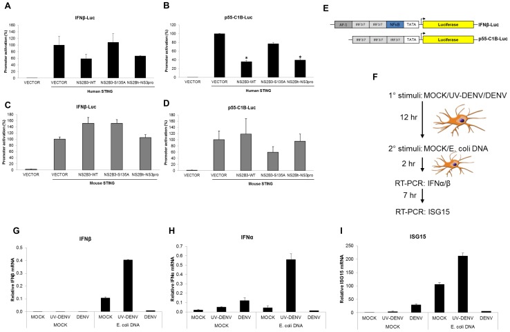
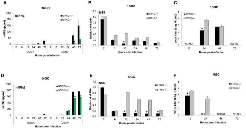
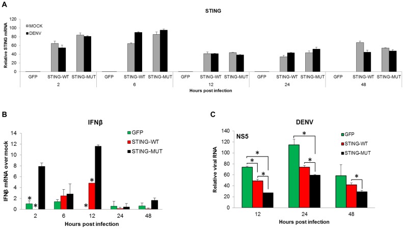
Dengue virus (DENV) is a pathogen with a high impact on human health. It replicates in a wide range of cells involved in the immune response. To efficiently infect humans, DENV must evade or inhibit fundamental elements of the innate immune system, namely the type I interferon response. DENV circumvents the host immune response by expressing proteins that antagonize the cellular innate immunity. We have recently documented the inhibition of type I IFN production by the proteolytic activity of DENV NS2B3 protease complex in human monocyte derived dendritic cells (MDDCs). In the present report we identify the human adaptor molecule STING as a target of the NS2B3 protease complex. We characterize the mechanism of inhibition of type I IFN production in primary human MDDCs by this viral factor. Using different human and mouse primary cells lacking STING, we show enhanced DENV replication. Conversely, mutated versions of STING that cannot be cleaved by the DENV NS2B3 protease induced higher levels of type I IFN after infection with DENV. Additionally, we show that DENV NS2B3 is not able to degrade the mouse version of STING, a phenomenon that severely restricts the replication of DENV in mouse cells, suggesting that STING plays a key role in the inhibition of DENV infection and spread in mice.


Conflict of interest statement

The authors have declared that no competing interests exist.

Figures

Figure 1
Figure 1. DENV NS2B3 protease complex cleaves human STING but not mouse STING.
(A) Schemes of the DENV-NS2B3 protease constructs used [wild type (WT), proteolyticaly inactive mutant (S135A) and proteolytic core (NS2Bh-NS3pro)]. (B) Representative map of the human adaptor STING showing the five transmembrane domains (TM) presented at the N-terminal region. Highlighted aminoacid sequence alignment of human and mouse STING at the N-termini of TM3 (light blue), and cleavage site of DENV NS2B3 (highlighted in red). (asterisk) indicates positions which have a single, fully conserved residue. (C) Co-transfection of DENV-NS2B3 plasmid constructs and human STING expressing plasmid in 293T cells. The samples were analyzed by SDS-PAGE and detected by Western blotting (WB). (D) Same co-transfection experiment described in (C) but using mouse STING expression plasmid. (E) Analysis of the interaction between DENV-NS2B3 protease and STING by co-immunoprecipitation assay. HA-tagged NS2B3 and FLAG-tagged human and mouse STING plasmids were transfected in 293T cells. After 48 h, cells lysates were prepared and immunoprecipitation was carried out using anti-FLAG antibody. (F) Evaluation of cleavage ability of DENV-NS2B3 on WT human STING and mutant version of human STING harboring aminoacids sequence from mouse STING (RRG/HCM) by WB using anti-HA and anti-FLAG antibodies. (G) Evaluation of cleavage ability of DENV-NS2B3 on WT mouse STING and mutant version of mouse STING harboring aminoacids sequence from human STING (IHCM/LRRG) by WB using anti-HA antibodies. (H) Degradation of endogenous human STING by DENV in primary human DCs analyzed by WB of endogenous levels of human STING and DENV NS3 protein in human DCs infected with MOCK, DENV and UV-DENV at different times post infection using anti-STING and anti-NS3 antibodies described in materials and methods.
Figure 2
Figure 2. DENV NS2B3 inhibits induction of IFNβ and p55-C1B promoters by human STING.
(A) 293T cells were co-transfected with a vector encoding for an IFNβ promoter driving the luciferase gene, a plasmid encoding for human STING, and plasmids for different versions of DENV protease (see Fig. 1D). Luciferase activity was measured 24 hours after transfection. (B) Same as described in (A) but using a plasmid encoding for the mouse version of STING. (C) 293T cells were co-transfected with a vector encoding for p55-C1B promoter driving the luciferase gene, a plasmid encoding for the human STING and plasmids for three different version of the DENV protease (see Fig. 1D). (D) Same as described in (C) but using a plasmid encoding for the mouse version of STING. (E) Scheme of the luciferase expressing vectors used, harboring the promoters for IFNβ and p55-CIB (F) Scheme of experimental design. MDDCs were treated with MOCK, DENV and UV inactivated DENV. After 12 hours, the three sets of cells were stimulated with MOCK or purified E. coli DNA and total RNA was purified at 2 and 7 hours post treatment. Relative expressions of type I IFN and ISGs, normalized against rsp11 and α-tubulin, were measured by qRT-PCR at specific time points. (G) INFβ mRNA. (H) IFNα mRNA. (I) ISG15 mRNA. Data shown represents one of two independent experiments. Error bars are the standard deviation of two individual replicates. *, p<0.05.
Figure 3
Figure 3. DENV NS2B3 inhibits type I IFN production in human but not in mouse dendritic cells.
Human dendritic cells were infected with MOCK, DENV-2 (16681), SFV-NS2B3-WT and SFV- NS2B3-S135A using a MOI = 1, and total RNA was extracted at 2, 6, 12, 24, 48 and 72 hpi (A to F). Relative expressions of different proteins and viral RNAs were measured by qRT-PCR at specific time points normalized against rsp11 and α-tubulin. (A) INFβ mRNA. (B) IFNα mRNA. (C) TNFα mRNA. (D) STING mRNA and (E) DENV RNA, (F) SFV RNA. (G to L) Mouse dendritic cells were infected with MOCK, DENV, SFV-NS2B3-WT and SFV- NS2B3-S135A using an MOI = 1, and total RNA was extracted at 2, 6, 12, 24, 48 and 72 hpi. Relative amounts of mRNA were measured by qRT-PCR using 18S and β-actin as housekeeping genes. (G) IFNβ mRNA. (H) IFNα mRNA. (I) TNFα mRNA. (J) STING mRNA. (K) DENV RNA (L) SFV RNA. Data re representative of three independent experiments from three different donors (A to F) and two individual experiments (G to L). Error bars indicate standard deviations of the mean from duplicate samples.*, p<0.05.
Figure 4
Figure 4. STING restricts replication of DENV in mouse cells.
Sting +/+ and Sting −/− MEFs were infected with DENV-2 (16681 strain) (A, B and C) and DENV-2 (NGC strain) (D, E and F) at an MOI of 5 and total RNA was extracted at the indicated times post infection. Levels of IFNβ were measured by ELISA(A and D) and DENV RNA levels were measured by qRT-PCR (B and E) using 18S and β-actin for normalization.. Infectious particles released to the supernatants of infected MEFs were measured by plaque assay on BHK cells (C and F) as described in materials and methods. Data are representative of two independent experiments. Error bars represent standard deviations of the mean of two replicate samples. *, p<0.05.
Figure 5
Figure 5. Expression of mutant STING in Sting −/− MEFs rescues their ability to produce type I IFN and to control DENV infection.
Two versions of recombinant human STING (WT and MUT) and GFP control were expressed in Sting −/− MEFs using lentiviral vectors (described in materials and methods). After 24 hours, infection by lentivirus was confirmed by GFP expression and MEFs were subsequently infected with DENV-2 16681 (A–C) or DENV-2 NGC (D–F). Total RNA was extracted at the specified times post infection and the levels of IFNβ mRNA (A and D), IFNα mRNA (B and E), and DENV RNA (C and F) were measured by qRT-PCR. (G) Schematic representation of lentiviral vectors used in these experiments. Error bars represent standard deviations of the mean of two replicate samples.*, p<0.05.
Figure 6
Figure 6. Over expression of STING in MDDCs potentiates IFNβ induction and attenuates DENV replication.
Primary human monocytes were transduced with lentiviruses expressing either STING-WT, STING-MUT or GFP as a control (Figure 5G and materials and methods). After five days of culture in the presence of GMCSF and IL-4 (see materials and methods) the differentiated MDDCs, were infected with DENV-2, 16681 using an MOI of 5 and total RNA was extracted at the specified times post infection. (A) Levels of STING mRNA were measured for mock and DENV infected cells at 2, 6, 12, 24 and 48 hpi. (B) Levels of mRNA for IFNβ were measured as indicated for (A). (C) Replication of DENV as measured by qRT-PCR (materials and methods) using specific primers for DENV-2 at the specified times post infection. Data shown are representative of three individual experiments from three independent donors. Error bars represent the standard deviation of the mean of two replicate samples. *, p<0.05.
Figure 7
Figure 7. Silencing of STING in human MDDCs results in enhanced DENV replication.
Primary human MDDCs were transfected with two scramble siRNAs and three STING specific siRNAs as described in the materials and methods section. 48 hours post transfection, cells were mock or DENV-2 infected (16681)at an MOI of 1. (A) Levels of mRNA for STING were quantified by qRT-PCR at the specified times post infection. (B) Levels of DENV RNA were measured at 24 hpi by qRT-PCR (data from donor 4 is shown). (C) DENV titers were determined by plaque assay of the supernatants from infected MDDCs treated with scramble and STING siRNA from six independent donors at 24 hpi (ND: not detected). Under these experimental conditions the limit of detection for the plaque assay was 101 PFU (indicated with a dashed line). Data for A and B is representative of six independent donors tested in 3 separate experiments. Error bars represent the standard deviation of the mean of two replicate samples.
Figure 8
Figure 8. Human primary immune cells from blood fail to produce type I IFN after DENV infection.
Several cell populations (pDCs, B cells, cDCs, Monocytes and MDDCs) were isolated from human blood as described in material and methods and subsequently infected with DENV-2 (166810 at MOI of 1. (A) viral RNA levels were measured in each cell population 12 h after infection by qRT-PCR using specific primers for DENV-2. (B) Cell supernatants from samples described above were collected to measure cytokine and chemokine levels by multiplex ELISA. (C) Whole PBMCs were infected with DENV-2 at MOI of 1 (black bars) or uninfected (grey bars) after ficoll centrifugation. Cytokine and chemokine levels in the PBMC supernatants were measured at 18 h post infection by multiplex ELISA. Monocyte derived macrophages (MDMs) (materials and methods) were infected with DENV-2 (16681) or SFV-NS2B3-WT and SFV- NS2B3-S135A at an MOI = 1 or mock infected for the indicated times. (D) DENV RNA levels were measured by qRT-PCR at the indicated times. Cytokines present in the supernatants from those infected MDMs were measured by multiplex ELISA: (E) TNFα levels, (F) IL-6 levels and (G) IFNα levels. (H) MDDC were infected with DENV as described for D to G and IFNα levels were measured in the supernatants by multiplex ELISA at the indicated times. Data shown for each graph are representative of three individual experiments from three independent donors. Error bars represent the standard deviation of the mean of three replicate samples.

Similar articles

Cited by

References

    1. Ho LJ, Wang JJ, Shaio MF, Kao CL, Chang DM, et al. (2001) Infection of human dendritic cells by dengue virus causes cell maturation and cytokine production. J Immunol 166: 1499–1506. - PubMed
    1. Wu SJ, Grouard-Vogel G, Sun W, Mascola JR, Brachtel E, et al. (2000) Human skin Langerhans cells are targets of dengue virus infection. Nat Med 6: 816–820. - PubMed
    1. Cologna R, Rico-Hesse R (2003) American genotype structures decrease dengue virus output from human monocytes and dendritic cells. J Virol 77: 3929–3938. - PMC - PubMed
    1. Jessie K, Fong MY, Devi S, Lam SK, Wong KT (2004) Localization of dengue virus in naturally infected human tissues, by immunohistochemistry and in situ hybridization. J Infect Dis 189: 1411–1418. - PubMed
    1. Kou Z, Quinn M, Chen H, Rodrigo WW, Rose RC, et al. (2008) Monocytes, but not T or B cells, are the principal target cells for dengue virus (DV) infection among human peripheral blood mononuclear cells. J Med Virol 80: 134–146. - PubMed

Publication types

MeSH terms