. 2012 Nov 23;287(48):40535-46.
doi: 10.1074/jbc.M112.385641. Epub 2012 Oct 8.

Src homology 2 domain-containing phosphatase 2 (Shp2) is a component of the A-kinase-anchoring protein (AKAP)-Lbc complex and is inhibited by protein kinase A (PKA) under pathological hypertrophic conditions in the heart

Affiliations

Src homology 2 domain-containing phosphatase 2 (Shp2) is a component of the A-kinase-anchoring protein (AKAP)-Lbc complex and is inhibited by protein kinase A (PKA) under pathological hypertrophic conditions in the heart

Brian T Burmeister et al. J Biol Chem. .

Abstract

Background: AKAP-Lbc is a scaffold protein that coordinates cardiac hypertrophic signaling.

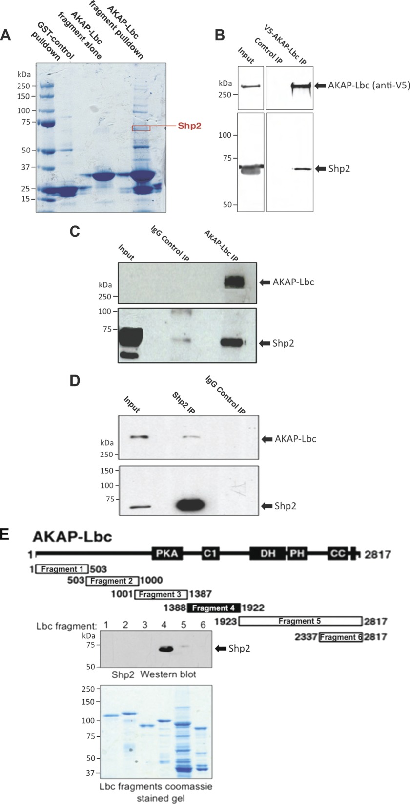
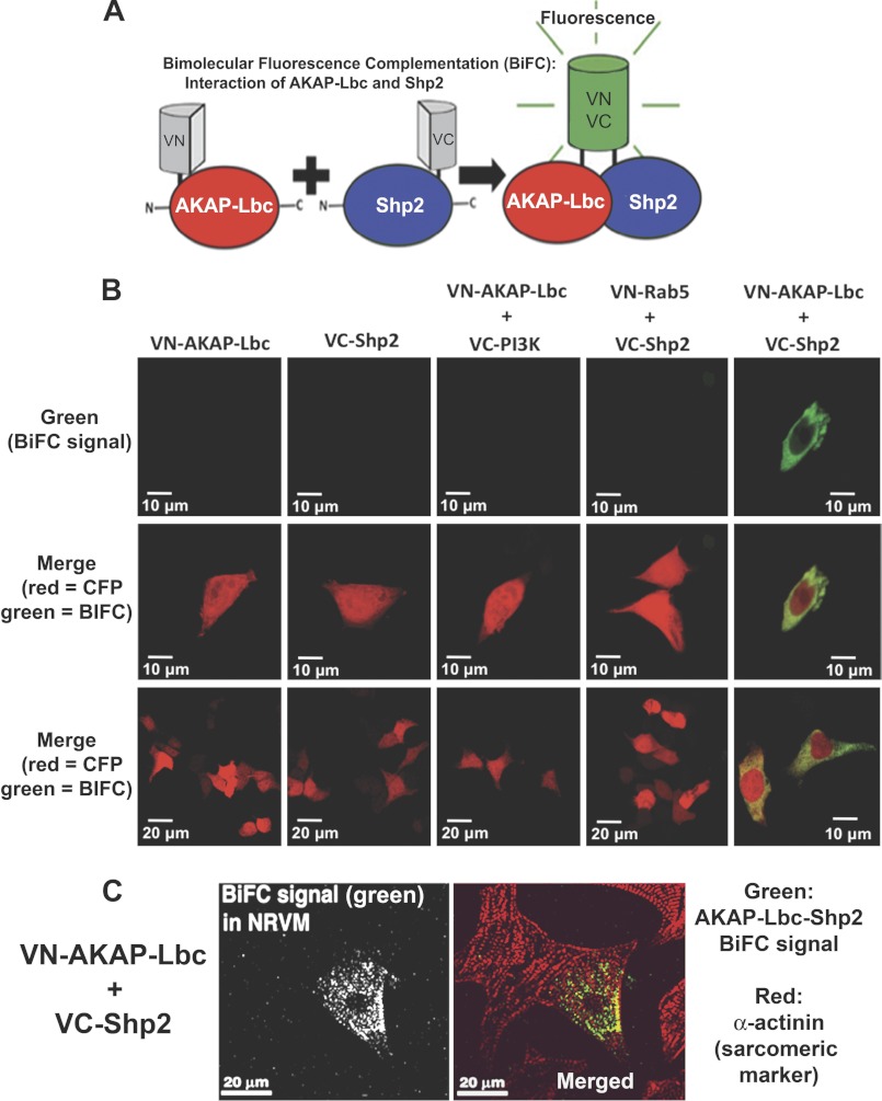
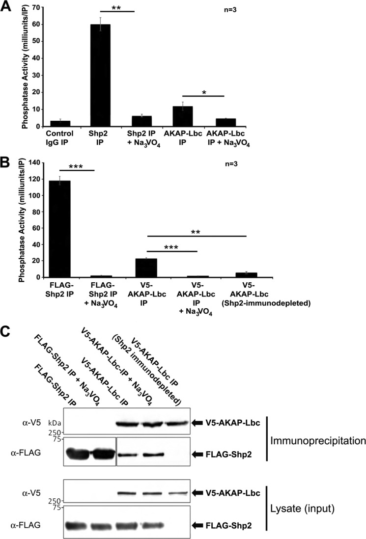
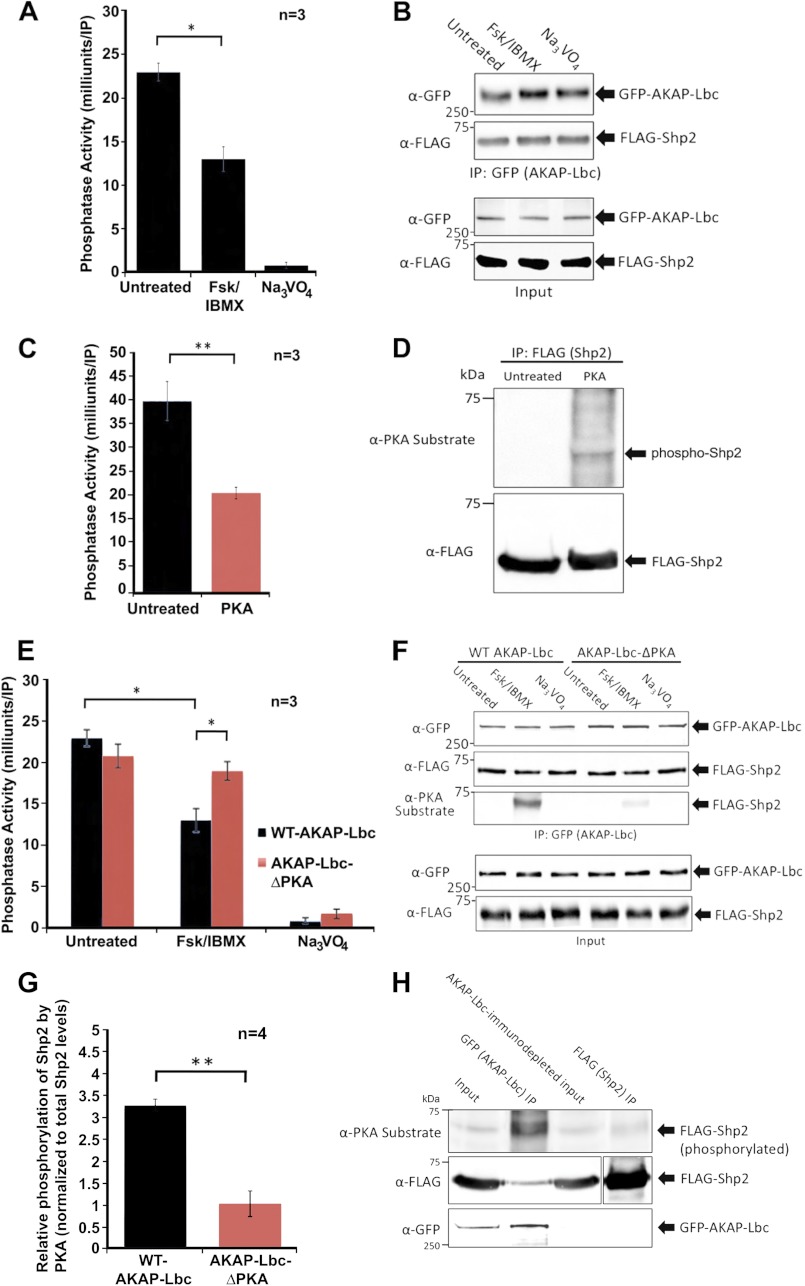
Results: AKAP-Lbc interacts with Shp2, facilitating its regulation by PKA.

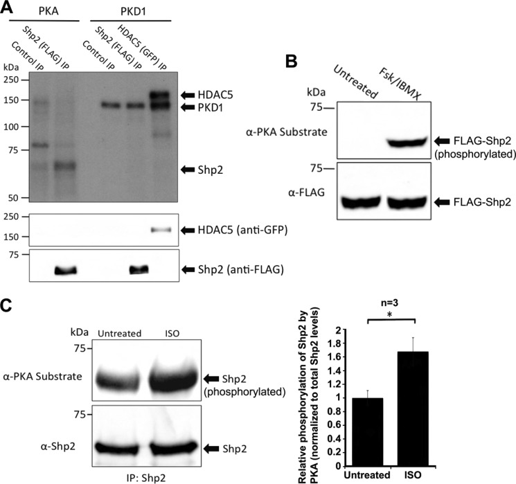
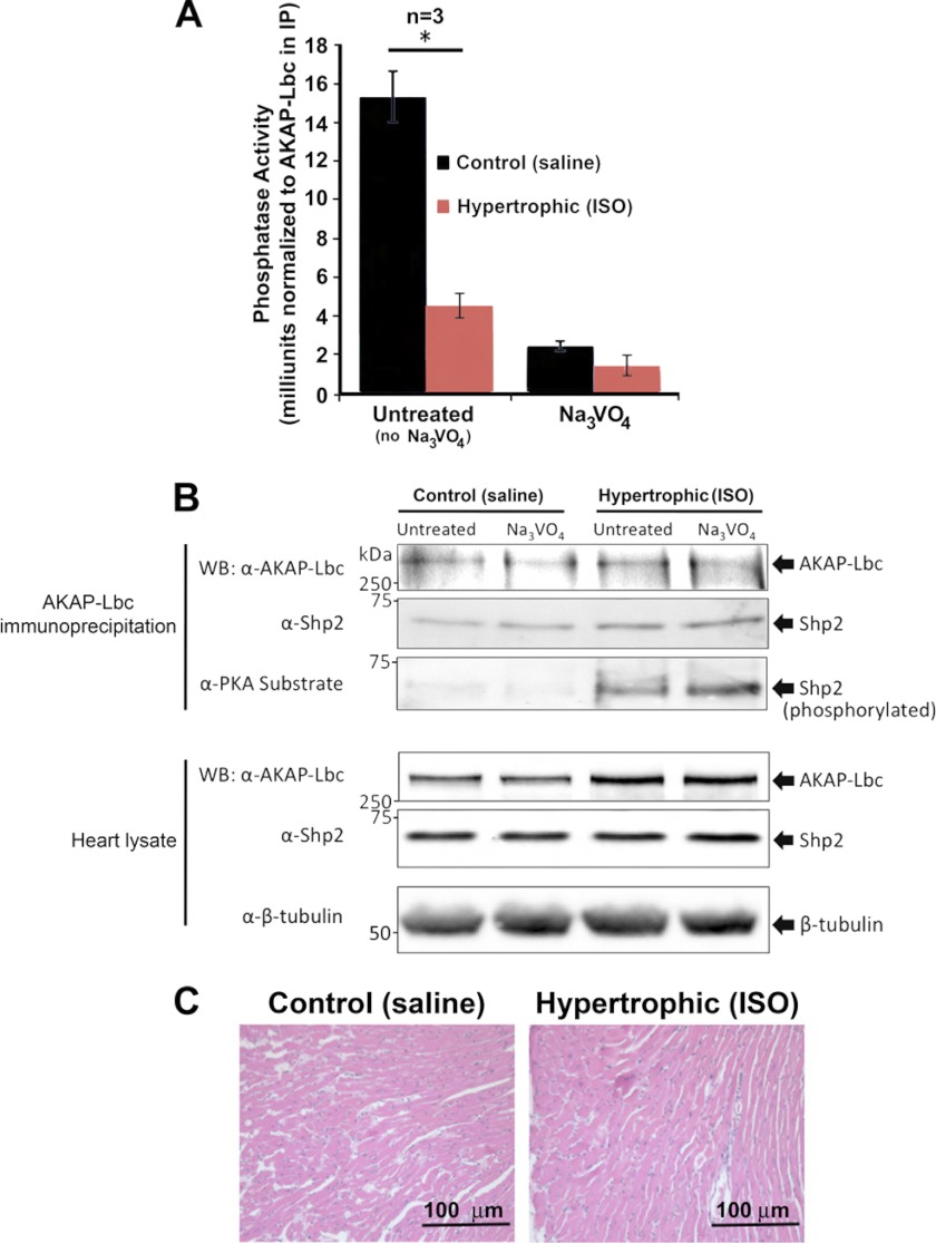
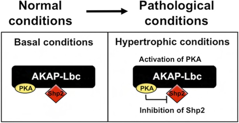
Conclusion: AKAP-Lbc integrates PKA and Shp2 signaling in the heart. Under pathological hypertrophic conditions Shp2 is phosphorylated by PKA, and phosphatase activity is inhibited.

Significance: Inhibition of Shp2 activity through AKAP-Lbc-anchored PKA is a previously unrecognized mechanism that may promote pathological cardiac hypertrophy. Pathological cardiac hypertrophy (an increase in cardiac mass resulting from stress-induced cardiac myocyte growth) is a major factor underlying heart failure. Our results identify a novel mechanism of Shp2 inhibition that may promote cardiac hypertrophy. We demonstrate that the tyrosine phosphatase, Shp2, is a component of the A-kinase-anchoring protein (AKAP)-Lbc complex. AKAP-Lbc facilitates PKA phosphorylation of Shp2, which inhibits its protein-tyrosine phosphatase activity. Given the important cardiac roles of both AKAP-Lbc and Shp2, we investigated the AKAP-Lbc-Shp2 interaction in the heart. AKAP-Lbc-tethered PKA is implicated in cardiac hypertrophic signaling; however, mechanism of PKA action is unknown. Mutations resulting in loss of Shp2 catalytic activity are also associated with cardiac hypertrophy and congenital heart defects. Our data indicate that AKAP-Lbc integrates PKA and Shp2 signaling in the heart and that AKAP-Lbc-associated Shp2 activity is reduced in hypertrophic hearts in response to chronic β-adrenergic stimulation and PKA activation. Thus, while induction of cardiac hypertrophy is a multifaceted process, inhibition of Shp2 activity through AKAP-Lbc-anchored PKA is a previously unrecognized mechanism that may promote compensatory cardiac hypertrophy.


Figures

FIGURE 1.
FIGURE 1.
Shp2 interacts with AKAP-Lbc. A, identification of Shp2 co-purifying with AKAP-Lbc by mass spectrometry. Heart extract was used for GST-AKAP-Lbc fragment (amino acid residues 1756–1801) pulldowns (2 mg of heart extract per pulldown). The resulting material was eluted from the GST-agarose and concentrated. Proteins were resolved by SDS-PAGE and Coomassie staining and identified by tandem MS. B, co-IP of endogenous Shp2 with V5-AKAP-Lbc. HEK293 cells were transfected for expression of V5-tagged AKAP-Lbc. AKAP-Lbc was immunoprecipitated with anti-V5-agarose from cell lysates. Parallel control IPs were performed using an equal amount of lysate where V5-AKAP-Lbc was not expressed (Control IP). IPs were washed, and the bound proteins were separated by SDS-PAGE and transferred to nitrocellulose. Detection of Shp2 and AKAP-Lbc was carried out by immunoblotting. C, co-IP of endogenous Shp2 with endogenous AKAP-Lbc in heart. AKAP-Lbc was immunoprecipitated from mouse heart extract (5 mg of total protein). Parallel control IgG IPs were carried out using an equal amount of heart extract. IPs were washed, and the bound proteins were separated by SDS-PAGE and transferred to nitrocellulose. Detection of AKAP-Lbc and co-purifying Shp2 were carried out by immunoblotting. D, co-IP of endogenous AKAP-Lbc with endogenous Shp2 in heart. Shp2 was immunoprecipitated from mouse heart extract, and detection of AKAP-Lbc and co-purifying Shp2 were carried out by immunoblotting (as described for C). E, mapping of Shp2 binding to AKAP-Lbc indicating that Shp2 binds to amino acid residues 1388–1922 of AKAP-Lbc. Diagram shows GST-AKAP-Lbc fragments used for pulldown experiments. Shaded area indicates Shp2 binding fragment as determined by immunoblot detection of Shp2 co-purifying with AKAP-Lbc fragments by GST pulldown from heart extract. Coomassie-stained gel indicates equal expression of the AKAP-Lbc fragments that were used for GST pulldown.
FIGURE 2.
FIGURE 2.
Visualization of AKAP-Lbc-Shp2 interaction inside cells by BiFC. A, schematic diagram illustrates the BiFC assay. The Venus (green) fluorescent protein is split into two nonfluorescent halves which are fused to AKAP-Lbc and Shp2. Specific protein-protein interaction results in a functional Venus (green) fluorescent protein. B, HEK293 cells were co-transfected with VN-AKAP-Lbc and VC-Shp2 and CFP (pseudo-colored red), as a marker for transfected cells. Cells were fixed 20 h after transfection and imaged. No BiFC (green) fluorescence is observed in cells expressing either VN-AKAP-Lbc or VC-Shp2 alone or when VN-AKAP-Lbc is co-expressed with a noninteracting control VC-protein (VC-PI3K). Similarly, no BiFC (green) fluorescence is observed when VC-Shp2 is co-expressed with a control VN-protein (VN-Rab5), whereas specific interaction of VC-Shp2 with VN-AKAP-Lbc results in fluorescence. Equal protein expression was determined by Western blotting (data not shown). C, neonatal rat ventricular cardiac myocytes were electroporated for expression of VN-AKAP-Lbc and VC-Shp2. After 48-h treatment with phenylephrine (10 μm), cells were fixed, permeabilized, and immunostained for the sarcomeric marker protein α-actinin (red in merged image). BiFC signal is shown in green in the merged image.
FIGURE 3.
FIGURE 3.
Measurement of PTP activity co-purifying with AKAP-Lbc. A, measurement of PTP activity co-purifying with AKAP-Lbc from mouse heart. AKAP-Lbc complexes were isolated from mouse heart extract using anti-AKAP-Lbc antibody. Immunoprecipitates were washed, and PTP activity was measured by a fluorometric in vitro assay using fluorescein diphosphate as substrate. Sodium orthovanadate (100 μm working concentration) was added to inhibit PTP activity, confirming that released phosphate was due to phosphatase activity and not proteolytic activity. Parallel negative control IgG IP assays and positive control Shp2 IP assays were also were carried out. Results indicate mean PTP activity per IP ± S.E. (error bars). All assays were performed in triplicate for three independent experiments. B, measurement of PTP activity co-purifying with AKAP-Lbc expressed in HEK293 cells. V5-AKAP-Lbc complexes were isolated from HEK293 cells using anti-V5-agarose. Immunoprecipitates from cell lysates (2 mg of total protein) were washed, and PTP activity was measured as described above. As a control, PTP activity co-purifying with AKAP-Lbc expressed in HEK293 cells previously immunodepleted for Shp2 was measured. Results presented show mean PTP activity per IP ± S.E. after control IgG IP background activity has been subtracted. All assays were performed in triplicate for three independent experiments. Differences in quantitative variables were examined by ANOVA. p = 0.05 is considered significant (*), p = 0.01 is considered very significant (**), and p = 0.001 is considered extremely significant (***). C, Western blots showing corresponding levels of AKAP-Lbc and Shp2 in samples used for PTP activity measurement in Fig. B.
FIGURE 4.
FIGURE 4.
Shp2 is a PKA, but not a PKD1 substrate. A, PKA, but not PKD1, phosphorylates Shp2 in vitro. Immunoprecipitated FLAG-tagged Shp2 from HEK293 cells was phosphorylated in vitro in kinase assay buffer supplemented with [γ-32P]ATP and bacterially purified recombinant PKA catalytic subunit (0.2 mg), or recombinant PKD1 (0.2 mg). Reactions were for 20 min at 30 °C and were terminated by washing twice with fresh kinase buffer prior to resuspension in Laemmli sample buffer. Incorporation of phosphate was determined by autoradiography following SDS-PAGE and transfer to nitrocellulose. GFP-HDAC5 (a well characterized PKD1 substrate) was used as a positive control in this experiment. Additionally, autophosphorylation of PKD1 is observed, indicating that PKD1 was active in this experiment. Immunoblotting confirmed similar levels of Shp2 in both the PKA and PKD1 phosphorylation reactions. A control FLAG-IP was also carried out using cell lysate where FLAG-Shp2 was not expressed. B, activation of PKA promotes Shp2 phosphorylation in HEK293 cells. Cells were treated with either DMSO (untreated) or forskolin (20 μm) and IBMX (75 μm) for 20 min to activate PKA. Western blotting using an anti-PKA-phosphosubstrate antibody was carried out following immunoprecipitation of FLAG-Shp2, SDS-PAGE, and transfer to nitrocellulose. The immunoblot was stripped and then probed for total levels of Shp2 with anti-FLAG antibody. C, activation of PKA promotes Shp2 phosphorylation in cardiac myocytes. Neonatal rat ventricular myocytes were treated with either DMSO (untreated) or isoproterenol (10 μm isoproterenol for 20 min) to activate PKA. Western blotting using an anti-PKA-phosphosubstrate antibody was carried out following immunoprecipitation of endogenous Shp2, SDS-PAGE, and transfer to nitrocellulose. The immunoblot was stripped and then probed for total levels of Shp2 with anti-Shp2 antibody. The relative increase in Shp2 phosphorylation by PKA in response to isoproterenol was quantified over three independent experiments. Error bars, indicate S.E. A p value of 0.05 is considered significant (*).
FIGURE 5.
FIGURE 5.
Shp2 activity in the AKAP-Lbc complex is inhibited by PKA. A, PKA activation promotes a decrease in Shp2 activity in the AKAP-Lbc complex. HEK293 cells transfected for expression of AKAP-Lbc and FLAG-Shp2 were treated for 20 min with either DMSO (untreated), or with forskolin (20 μm) and IBMX (75 μm) to activate PKA. Subsequent AKAP-Lbc immunoprecipitations were used for in vitro measurement of PTP activity. All assays were performed in triplicate for three independent experiments. Sodium orthovanadate was used in all assays to determine that PTP activity was being measured. B, Western blot loading controls demonstrate even expression of AKAP-Lbc and Shp2. To confirm even levels of AKAP-Lbc and co-immunoprecipitating Shp2 under all conditions, each IP was equally divided into four tubes; three tubes were used for PTP assay, and the fourth was used for SDS-PAGE and Western blotting, probed with anti-GFP antibodies for AKAP-Lbc detection and anti-FLAG antibodies for Shp2 detection. C, in vitro phosphorylation of Shp2 by PKA inhibits Shp2 activity. Immunoprecipitated Shp2 was phosphorylated in vitro in kinase assay buffer supplemented with bacterially purified recombinant PKA C-subunit (0.2 mg) for 20 min at 30 °C. Parallel control reactions were performed where no PKA was added. Reactions were terminated by washing twice with fresh kinase buffer prior to PTP activity assay. All assays were performed in triplicate for three independent experiments. D, Western blot loading controls confirm phosphorylation of Shp2 by PKA and that equal amounts of Shp2 were present in all assay conditions. E, AKAP-Lbc-anchored PKA promotes a decrease in Shp2 activity. HEK293 cells were transfected for the expression of wild-type (WT)-AKAP-Lbc and FLAG-Shp2 or AKAP-Lbc-ΔPKA (a mutant that cannot bind PKA) and FLAG-Shp2. Prior to lysis, cells were treated for 20 min with either DMSO (untreated), or with forskolin/IBMX to activate PKA. AKAP-Lbc was immunoprecipitated, and immune complexes were used for in vitro measurement of PTP activity. All assays were performed in triplicate for three independent experiments. Sodium orthovanadate was used in all assays to confirm that PTP activity was being measured. F, Western blot loading controls demonstrate comparable expression of AKAP-Lbc-WT, AKAP-Lbc-ΔPKA, and associated Shp2 for all assays; however, PKA phosphorylation of Shp2 (assessed using the anti-PKA-phosphosubstrate antibody) is reduced in cells expressing AKAP-Lbc-ΔPKA compared with AKAP-Lbc-WT. G, Shp2 phosphorylation by AKAP-Lbc-anchored PKA is quantified using ImageJ software. H, Shp2 not associated with AKAP-Lbc is minimally phosphorylated by PKA, in contrast to Shp2 in the AKAP-Lbc complex. Prior to lysis, HEK293 cells expressing GFP-AKAP-Lbc and FLAG-Shp2 were treated for 20 min with forskolin/IBMX to activate PKA. AKAP-Lbc was immunoprecipitated, and subsequent AKAP-Lbc-immunodepleted lysate was used for immunoprecipitation of Shp2. Immune complexes were subjected to SDS-PAGE and transfer to nitrocellulose. Immunoblotting was performed with anti-PKA-phosphosubstrate antibody to determine the extent of Shp2 phosphorylation. Membrane was stripped and re-probed with anti-FLAG and anti-GFP antibodies to confirm the levels of Shp2 and AKAP-Lbc in each experimental condition. A shorter exposure is shown for total FLAG-Shp2 in the FLAG (Shp2) IP, compared with the other lanes (indicated by separation of the two exposures), due to relative high levels of total FLAG-Shp2 immunoprecipitated. Error bars, indicate S.E. p = 0.05 is considered significant (*), and p = 0.01 is considered very significant (**).
FIGURE 6.
FIGURE 6.
Shp2 activity is decreased in isoproterenol-induced hypertrophic hearts. A, AKAP-Lbc complexes were isolated from healthy (Control; saline-treated) and hypertrophic (ISO-treated) mouse heart extract by immunoprecipitation using anti-AKAP-Lbc antibody. Immunoprecipitates were washed, and PTP activity was measured as described previously. Results presented show PTP activity ± S.E. measured in triplicate from three hypertrophic hearts and three age-matched control healthy hearts. Control IgG IP background activity has been subtracted, and the PTP activity was normalized to AKAP-Lbc expression in the immunoprecipitation. Untreated refers to no Na3VO4. B, Western blotting of AKAP-Lbc and associated Shp2 levels in the IPs used for PTP assay. Bottom panels show levels of AKAP-Lbc and Shp2 in heart lysate used for IP. C, histological analysis of mouse hearts corresponding to samples used for PTP assay. Hematoxylin/Eosin staining of left ventricle sections indicates isoproterenol-induced hypertrophic myocytes (Table 1). Echocardiography characteristics indicate ventricular dilation and hypertrophy.
FIGURE 7.
FIGURE 7.
Model showing the role of AKAP-Lbc in the regulation of Shp2 activity by PKA. AKAP-Lbc assembles a signaling complex composed of PKA and Shp2 in cardiac myocytes. Stress conditions (e.g. chronic activation of the β-adrenergic receptor) lead to PKA activation, thereby promoting inhibition of Shp2 activity, which may contribute to the induction of cardiac hypertrophy.

Similar articles

Cited by

References

    1. Shi F., Lemmon M. A. (2011) Biochemistry: KSR plays CRAF-ty. Science 332, 1043–1044 - PubMed
    1. Carnegie G. K., Means C. K., Scott J. D. (2009) A-kinase anchoring proteins: from protein complexes to physiology and disease. IUBMB Life 61, 394–406 - PMC - PubMed
    1. Wong W., Scott J. D. (2004) AKAP signalling complexes: focal points in space and time. Nat. Rev. Mol. Cell Biol. 5, 959–970 - PubMed
    1. Beene D. L., Scott J. D. (2007) A-kinase anchoring proteins take shape. Curr. Opin. Cell Biol. 19, 192–198 - PMC - PubMed
    1. Newlon M. G., Roy M., Morikis D., Carr D. W., Westphal R., Scott J. D., Jennings P. A. (2001) A novel mechanism of PKA anchoring revealed by solution structures of anchoring complexes. EMBO J. 20, 1651–1662 - PMC - PubMed

Publication types

MeSH terms

Substances

LinkOut - more resources