. 2012 Nov;38(5):532-42.
doi: 10.1097/SHK.0b013e31826c5b19.

Radiation combined with thermal injury induces immature myeloid cells

Affiliations

Radiation combined with thermal injury induces immature myeloid cells

April Elizabeth Mendoza et al. Shock. 2012 Nov.

Abstract

The continued development of nuclear weapons and the potential for thermonuclear injury necessitates the further understanding of the immune consequences after radiation combined with injury (RCI). We hypothesized that sublethal ionization radiation exposure combined with a full-thickness thermal injury would result in the production of immature myeloid cells. Mice underwent either a full-thickness contact burn of 20% total body surface area or sham procedure followed by a single whole-body dose of 5-Gy radiation. Serum, spleen, and peripheral lymph nodes were harvested at 3 and 14 days after injury. Flow cytometry was performed to identify and characterize adaptive and innate cell compartments. Elevated proinflammatory and anti-inflammatory serum cytokines and profound leukopenia were observed after RCI. A population of cells with dual expression of the cell surface markers Gr-1 and CD11b were identified in all experimental groups, but were significantly elevated after burn alone and RCI at 14 days after injury. In contrast to the T-cell-suppressive nature of myeloid-derived suppressor cells found after trauma and sepsis, myeloid cells after RCI augmented T-cell proliferation and were associated with a weak but significant increase in interferon γ and a decrease in interleukin 10. This is consistent with previous work in burn injury indicating that a myeloid-derived suppressor cell-like population increases innate immunity. Radiation combined injury results in the increase in distinct populations of Gr-1CD11b cells within the secondary lymphoid organs, and we propose these immature inflammatory myeloid cells provide innate immunity to the severely injured and immunocompromised host.


Conflict of interest statement

There are no conflicts of interest to disclose.

Figures

Figure 1
Figure 1. Survival is decreased after RCI when radiation doses exceed 5-Gy
A) C57BL/6 mice received either sham injury, burn injury (20% TBSA, full-thickness burn), or radiation exposure (2-, 5-, 9-Gy). B) C57BL/6 mice received a thermal injury (20%TBSA, full-thickness burn) followed by radiation exposure (2-, 5- 9-Gy). Survival was observed up to 4 weeks. Data is expressed as percent survival. Groups include 9–12 mice; *P<0.05 vs. sham group.
Figure 2
Figure 2. Lymphopenia is radiation dose-dependent
A) C57BL/6 mice received either sham injury or radiation exposure (2-, 5-, 9-Gy). Spleens were harvested at day 21 post-injury. Flow cytometry was performed to quantify the total number of splenic CD4+ and CD8+ T-cells. B) C57BL/6 mice received either sham, burn (20%TBSA, full-thickness burn), or thermal injury (20%TBSA, full-thickness burn) followed by radiation exposure (2-, 5- 9-Gy). Spleens were harvested at day 21 post-injury. Flow cytometry was performed to quantify the total number of splenic CD4+ and CD8+ T-cells. Groups include 10–12 mice; *P<0.05.
Figure 3
Figure 3. Gr-1+CD11b+ cells are elevated after RCI at day 21
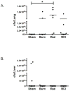
A) C57BL/6 mice received either sham injury or radiation exposure (2-, 5-, 9-Gy). Spleens were harvested at day 21 post-injury. Flow cytometry was performed to quantify the total number of splenic Gr-1+CD11b+ cells. B) C57BL/6 mice received either sham, burn (20%TBSA, full-thickness burn), or thermal injury (20%TBSA, full-thickness burn) followed by radiation exposure (2-, 5- 9-Gy). Spleens were harvested at day 21 post-injury. Flow cytometry was performed to quantify the total number of splenic Gr-1+CD11b+ cells. Groups include 10–12 mice; *P<0.05.
Figure 4
Figure 4. Significant weight loss is observed by three days post-radiation exposure (5-Gy) and RCI (20%TBSA+5-Gy)
C57BL/6 mice received either sham injury, burn injury (20% TBSA, full-thickness burn), radiation exposure (5-Gy) or RCI (20% TBSA + 5-Gy). Body weight (g) of mice was measured at days 0, 3, 7 and 14 post-injury. Experiments were repeated in triplicate. Groups include 3–7 mice. *P<0.05, **P<0.01 vs. sham group.
Figure 5
Figure 5. RCI (20%TBSA+5-Gy) increases pro-inflammatory serum cytokines at day 3 and 14
C57BL/6 mice received either sham injury, burn injury (20% TBSA, full-thickness burn), radiation exposure (5-Gy) or RCI (20% TBSA + 5-Gy). Serum was collected by cardiac puncture on the day of harvest. Serum cytokines of sham, burn, radiation and RCI mice at day 3 (A) and day 14 (B) were analyzed by multiplex bead array. Each experimental time point was repeated. Groups consist of 4–6 mice; *P<0.05.
Figure 6
Figure 6. Gr-1+CD11b+ cells consist of a heterogenous population of Ly6G+Ly6C+ and Ly6C+Ly6G myeloid cells
Gr-1+CD11b+ cells were further defined by their Ly6C and Ly6G expression. A) A heterogenous population of CD11b+ cells were identified and consisted of Ly6G+Ly6C+ and Ly6C+Ly6G cells. B) Side scatter and forward scatter of light suggest that 1.) the Ly6G+Ly6C+ population is granulocytic with high side scatter consistent with increased granularity, and 2.) the Ly6C+Ly6G population have intermediate side scatter consistent with a monocytic population. C) Histograms represent the CD11b expression of the gated cell populations.
Figure 7
Figure 7. Innate and adaptive cell populations are reduced at day 3 after radiation (5-Gy) and RCI (20%TBSA+5Gy)
C57BL/6 mice received either sham injury, burn injury (20% TBSA, full-thickness burn), radiation exposure (5-Gy) or RCI (20% TBSA + 5-Gy). Spleens were harvested from sham, burn, radiation, and RCI mice at day 3. A) Cell populations were identified by cell surface markers using flow cytometric staining. B) Rectangle gates represent the proportion of Gr-1+ CD11b+ cells within each histogram. Experiments were repeated. Each group consists of 4–5 mice; *P<0.05, **P<0.001 vs. sham group.
Figure 8
Figure 8. Gr-1+ CD11b+ cells are elevated at day 14 after burn (20%TBSA) and RCI (20%TBSA+5-Gy)
C57BL/6 mice received either sham injury, burn injury (20% TBSA, full-thickness burn), radiation exposure (5-Gy) or RCI (20% TBSA + 5-Gy). Spleens were harvested from sham, burn, radiation (5-Gy) and RCI (20%TBSA+5-Gy) at day 14. Cell populations were identified by cell surface markers using flow cytometric staining. Experiments were repeated four times. Each group consists of 4–7 mice. *P<0.05, **P<0.01 ***P<0.001.
Figure 9
Figure 9. Gr-1+CD11b+ cells are a major cell type at day 14 after RCI (20%TBSA+5-Gy)
C57BL/6 mice received either sham injury, burn injury (20% TBSA, full-thickness burn), radiation exposure (5-Gy) or RCI (20% TBSA + 5-Gy). Spleens were harvested from sham, burn, radiation (5-Gy) and RCI (20%TBSA+5-Gy) at day 14. A) Percent of splenic Gr-1+ CD11b+ cells in each experimental group were determined by flow cytometry. B) Rectangle gates represent percent of Gr-1+CD11b+ cells within each histogram. Experiments were repeated four times. Each group consists of 4–7 mice. *P<0.05, **P<0.01 ***P<0.001.
Figure 10
Figure 10. Gr-1+CD11b+ cells are increased in the PLN after RCI
C57BL/6 mice received either sham injury, burn injury (20% TBSA, full-thickness burn), radiation (5-Gy) or RCI (20%TBSA + 5-Gy). A) At day 3 and B) day 14 inguinal and axillary lymph nodes (PLN) were harvested and pooled together. Cell populations were identified by cell surface markers using flow cytometric staining. Experiments were repeated. Each group consists of 4–7 mice. *P<0.05.
Figure 11
Figure 11. A heterogeneous population of Gr-1+CD11b+ myeloid cells is observed in the bone marrow at day 14
C57BL/6 mice received either sham injury, burn injury (20% TBSA, full-thickness burn), radiation (5-Gy) or RCI (20%TBSA + 5-Gy). Bone marrow was harvested from bilateral femurs at day 14. A) Populations of Gr-1+CD11b+ cells were identified by cell surface markers using flow cytometric staining. B) Gr-1+CD11b+ cells in the bone marrow consist of a heterogeneous Ly6G+Ly6C+ and Ly6C+Ly6G populations.
Figure 12
Figure 12. Gr-1+CD11b+ cells after RCI express cell surface markers consistent with immature and blast myeloid progenitors
A) Spleens and B) bone marrow were harvested at day 14 from RCI (20%TBSA + 5-Gy) mice. Gr-1+CD11b+ cells were identified by cell surface markers using flow cytometric staining. Rectangle gates represent Gr-1+CD11b+ cells and their CD31 (PECAM) expression.
Figure 13
Figure 13. Thymidine incorporation by T-cells is increased after co-culture with Gr-1+ CD11b+ cells from radiation (5-Gy) and RCI (20%TBSA+5-Gy) mice
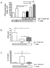
C57BL/6 mice received either sham injury, burn injury (20% TBSA, full-thickness burn), radiation exposure (5-Gy) or RCI (20% TBSA + 5-Gy). Spleens were harvested from sham, burn, radiation (5-Gy) and RCI (20%TBSA+5-Gy) at day 14. Cells were harvested from spleens. Gr-1+CD11b+ cells were sorted and purified by FACS from single cell suspensions. T-cells were purified from untouched C57BL/6 spleens by negative magnetic selection. T-cells were then cultured with plate bound anti-CD3, soluble anti-CD28 and Gr-1+ CD11b+ cells (ratio 1:1; T-cell: Gr-1+ CD11b+ cells) for 72 hours. Controls included stimulated and unstimulated T-cells. [3H]-thymidine was pulsed during the last 18 hours of culture. Proliferation is expressed as cpm [3H]-thymidine incorporation. Experiments were repeated in triplicate. Groups consist of 6–7 mice; *P<0.05, **P<0.01. B, C). Supernatants were collected prior to the addition of [3H]-thymidine, and analyzed by multiplex bead array. IFN-γ (B) and IL-10 (C) were analyzed and compared between RCI, sham and stimulated T-cell controls (±SEM; n=6–7); *P<0.05, **P<0.01.
Figure 14
Figure 14. Gr-1+CD11b+ cells recruit to the lung after pulmonary infection
C57BL/6 mice sustained either sham injury, burn injury (20% TBSA, full-thickness burn), radiation exposure (5-Gy) or RCI (20% TBSA + 5-Gy). Each experimental group was infected at day 14 post-injury with (1×10⁷cfu/inoculum) by intratracheal inoculation; another group remained uninfected. Lung was harvested 24 hours after infection. One lobe was further processed into a single cell suspension. Gr-1+CD11b+ cells were identified by cell surface markers using flow cytometric staining. A) Total Gr-1+CD11b+ cell numbers were enumerated and analyzed. B) Percent of Gr-1+CD11b+ cells in each experimental group were quantified by flow cytometry (±SEM; n=4–5); *P<0.05.
Figure 15
Figure 15. Splenic Gr-1+CD11b+ cells in RCI mice increase after pulmonary infection
C57BL/6 mice received either sham injury, burn injury (20% TBSA, full-thickness burn)radiation exposure (5-Gy) or RCI (20% TBSA + 5-Gy). Each experimental group was infected at day 14 post-injury with (1×10⁷cfu/inoculum) by intratracheal inoculation.; another group remained uninfected. Spleens were harvested 24 hours after infection. Gr-1+CD11b+ cells were identified by cell surface markers using flow cytometric staining. Total Gr-1+CD11b+ cell numbers were enumerated and analyzed (±SEM; n=4–5); *P<0.05.
Figure 16
Figure 16. Pulmonary microbial clearance is similar among sham, burn and RCI mice, but decreased in radiation (5-Gy) mice
C57BL/6 mice received either sham injury, burn injury (20% TBSA, full-thickness burn), radiation exposure (5-Gy) or RCI (20%TBSA + 5-Gy). Each experimental group was infected at day 14 post-injury with (1×10⁷cfu/inoculum) by intratracheal inoculation. A) Lung was harvested 24 hours after infection. One lobe was homogenized and inoculated onto tryptic soy agar containing kanamycin (25mg/mL) (TSA-k). Colony-forming units were enumerated per plate at 24 hours post-harvest (±SEM; n=6–9); *P<0.05. B) Liver was harvested 24 hours after infection. The entire organ was homogenized and inoculated onto TSA-k. Colony-forming units were enumerated per plate at 24 hours post-harvest (±SEM; n=10–11).

Similar articles

Cited by

References

    1. DiCarlo AL, Hatchett RJ, Kaminski JM, Ledney GD, Pellmar TC, Okunieff P, Ramakrishnan N. Medical countermeasures for radiation combined injury: radiation with burn, blast, trauma and/or sepsis. report of an NIAID Workshop; March 26–27, 2007; 2008. pp. 712–721. - PMC - PubMed
    1. DiCarlo AL, Ramakrishnan N, Hatchett RJ. Radiation combined injury: overview of NIAID research. 2010;98:863–867. - PMC - PubMed
    1. DiCarlo AL, Maher C, Hick JL, Hanfling D, Dainiak N, Chao N, Bader JL, Coleman CN, Weinstock DM. Radiation injury after a nuclear detonation: medical consequences and the need for scarce resources allocation. 2011;5 (Suppl 1):S32–44. - PMC - PubMed
    1. Kiang JG, Jiao W, Cary LH, Mog SR, Elliott TB, Pellmar TC, Ledney GD. Wound trauma increases radiation-induced mortality by activation of iNOS pathway and elevation of cytokine concentrations and bacterial infection. 2010;173:319–332. - PMC - PubMed
    1. Buchanan IB, Maile R, Frelinger JA, Fair JH, Meyer AA, Cairns BA. The effect of burn injury on CD8+ and CD4+ T cells in an irradiation model of homeostatic proliferation. 2006;61:1062–1068. - PubMed

Publication types

MeSH terms