

Sea anemone (Cnidaria, Anthozoa, Actiniaria) toxins: an overview
- PMID: 23015776
- PMCID: PMC3447340
- DOI: 10.3390/md10081812
Sea anemone (Cnidaria, Anthozoa, Actiniaria) toxins: an overview
Abstract
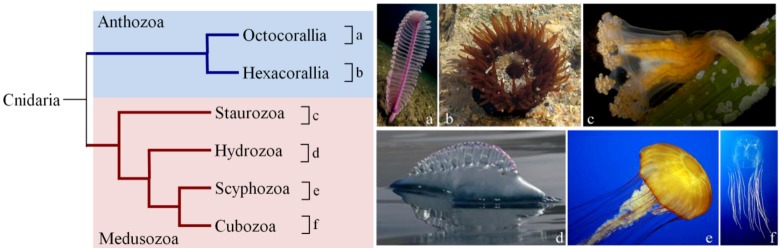
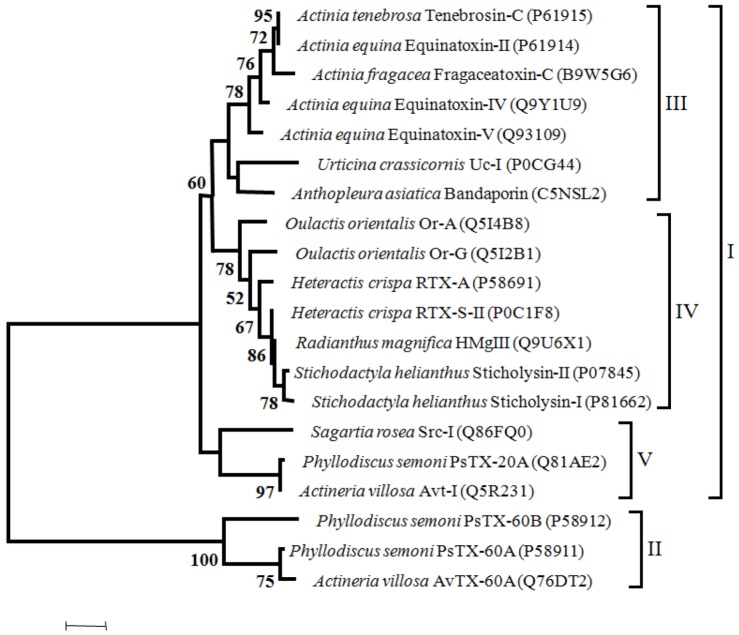
The Cnidaria phylum includes organisms that are among the most venomous animals. The Anthozoa class includes sea anemones, hard corals, soft corals and sea pens. The composition of cnidarian venoms is not known in detail, but they appear to contain a variety of compounds. Currently around 250 of those compounds have been identified (peptides, proteins, enzymes and proteinase inhibitors) and non-proteinaceous substances (purines, quaternary ammonium compounds, biogenic amines and betaines), but very few genes encoding toxins were described and only a few related protein three-dimensional structures are available. Toxins are used for prey acquisition, but also to deter potential predators (with neurotoxicity and cardiotoxicity effects) and even to fight territorial disputes. Cnidaria toxins have been identified on the nematocysts located on the tentacles, acrorhagi and acontia, and in the mucous coat that covers the animal body. Sea anemone toxins comprise mainly proteins and peptides that are cytolytic or neurotoxic with its potency varying with the structure and site of action and are efficient in targeting different animals, such as insects, crustaceans and vertebrates. Sea anemones toxins include voltage-gated Na⁺ and K⁺ channels toxins, acid-sensing ion channel toxins, Cytolysins, toxins with Kunitz-type protease inhibitors activity and toxins with Phospholipase A2 activity. In this review we assessed the phylogentic relationships of sea anemone toxins, characterized such toxins, the genes encoding them and the toxins three-dimensional structures, further providing a state-of-the-art description of the procedures involved in the isolation and purification of bioactive toxins.
Keywords: Cnidaria; phylogeny; sea anemone; toxin; toxin gene.
Figures

Similar articles
-
A RNA-seq approach to identify putative toxins from acrorhagi in aggressive and non-aggressive Anthopleura elegantissima polyps.BMC Genomics. 2015 Mar 21;16(1):221. doi: 10.1186/s12864-015-1417-4. BMC Genomics. 2015. PMID: 25886045 Free PMC article.
-
Sea anemone toxins affecting voltage-gated sodium channels--molecular and evolutionary features.Toxicon. 2009 Dec 15;54(8):1089-101. doi: 10.1016/j.toxicon.2009.02.028. Epub 2009 Mar 5. Toxicon. 2009. PMID: 19268682 Free PMC article. Review.
-
A new toxin from the sea anemone Condylactis gigantea with effect on sodium channel inactivation.Toxicon. 2006 Aug;48(2):211-20. doi: 10.1016/j.toxicon.2006.05.001. Epub 2006 May 19. Toxicon. 2006. PMID: 16814340
-
Novel peptide toxins recently isolated from sea anemones.Toxicon. 2009 Dec 15;54(8):1112-8. doi: 10.1016/j.toxicon.2009.02.031. Epub 2009 Mar 6. Toxicon. 2009. PMID: 19269303 Review.
-
Novel peptide toxins from the sea anemone Stichodactyla haddoni.Peptides. 2008 Apr;29(4):536-44. doi: 10.1016/j.peptides.2007.12.010. Epub 2008 Feb 19. Peptides. 2008. PMID: 18243416
Cited by
-
Response of Cellular Innate Immunity to Cnidarian Pore-Forming Toxins.Molecules. 2018 Oct 4;23(10):2537. doi: 10.3390/molecules23102537. Molecules. 2018. PMID: 30287801 Free PMC article. Review.
-
Identification of a pore-forming protein from sea anemone Anthopleura dowii Verrill (1869) venom by mass spectrometry.J Venom Anim Toxins Incl Trop Dis. 2019 Feb 11;25:e147418. doi: 10.1590/1678-9199-JVATITD-1474-18. eCollection 2019. J Venom Anim Toxins Incl Trop Dis. 2019. PMID: 31131002 Free PMC article.
-
Venom: the sharp end of pain therapeutics.Br J Pain. 2013 Nov;7(4):179-88. doi: 10.1177/2049463713502005. Br J Pain. 2013. PMID: 26516522 Free PMC article.
-
New Sea Anemone Toxin RTX-VI Selectively Modulates Voltage-Gated Sodium Channels.Dokl Biochem Biophys. 2020 Nov;495(1):292-295. doi: 10.1134/S1607672920060071. Epub 2020 Dec 25. Dokl Biochem Biophys. 2020. PMID: 33368037
-
The Transcriptome of the Zoanthid Protopalythoa variabilis (Cnidaria, Anthozoa) Predicts a Basal Repertoire of Toxin-like and Venom-Auxiliary Polypeptides.Genome Biol Evol. 2016 Oct 5;8(9):3045-3064. doi: 10.1093/gbe/evw204. Genome Biol Evol. 2016. PMID: 27566758 Free PMC article.
References
-
- Collins A.G. Recent insights into cnidarian phylogeny. Smithsonian Contrib. Mar. Sci. 2009;38:139–149.
-
- Wikimedia commons. [(acessed on 27 February 2012)]. Available online: http://commons.wikimedia.org/wiki/
Publication types
MeSH terms
Substances
LinkOut - more resources
Full Text Sources
Other Literature Sources
