. 2012 Dec;86(24):13313-23.
doi: 10.1128/JVI.01637-12. Epub 2012 Sep 26.

Conserved region 3 of human papillomavirus 16 E7 contributes to deregulation of the retinoblastoma tumor suppressor

Affiliations

Conserved region 3 of human papillomavirus 16 E7 contributes to deregulation of the retinoblastoma tumor suppressor

Biljana Todorovic et al. J Virol. 2012 Dec.

Abstract

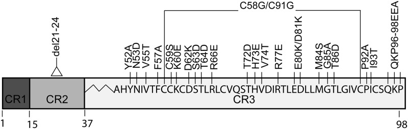
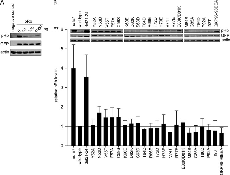
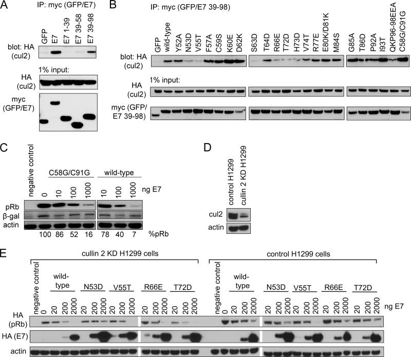
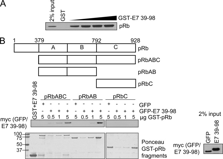
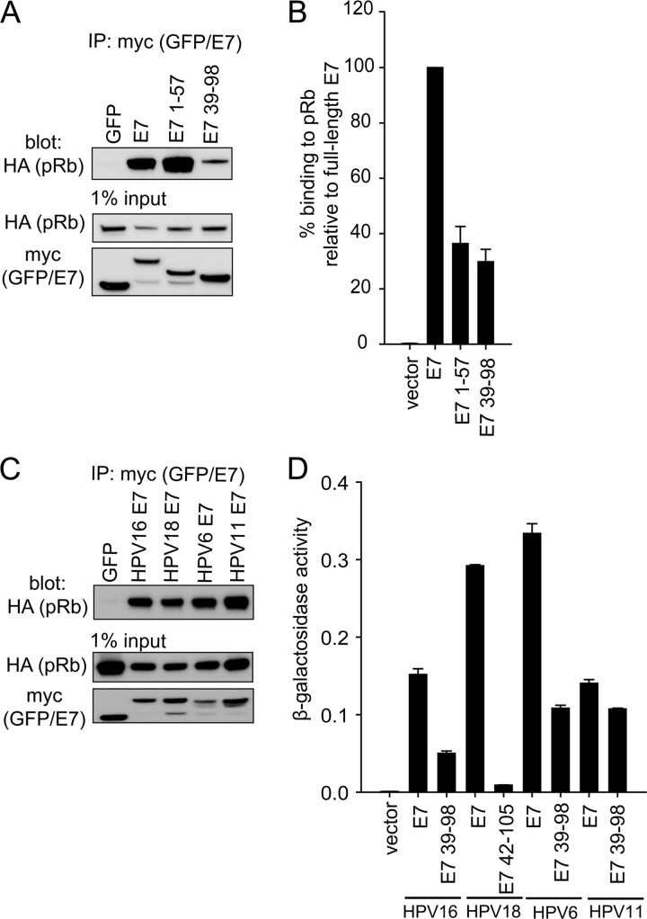
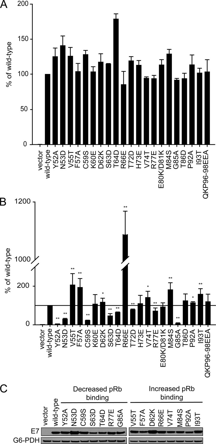
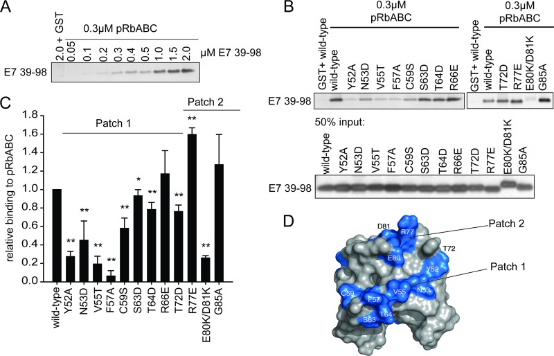
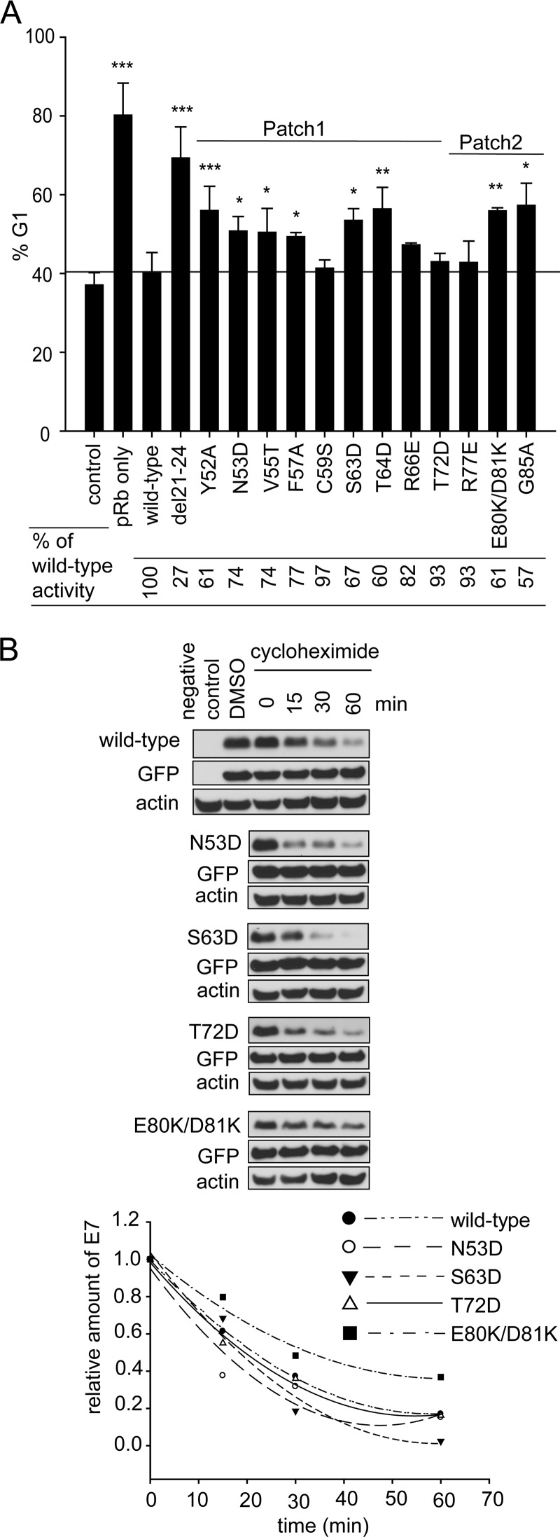
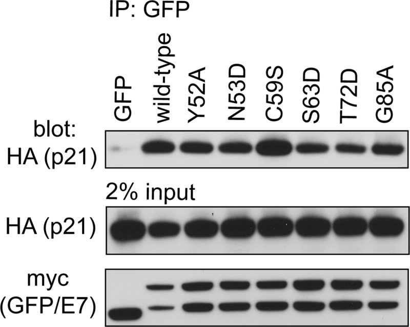
The human papillomavirus (HPV) E7 oncoprotein binds cellular factors, preventing or retargeting their function and thereby making the infected cell conducive for viral replication. A key target of E7 is the product of the retinoblastoma susceptibility locus (pRb). This interaction results in the release of E2F transcription factors and drives the host cell into the S phase of the cell cycle. E7 binds pRb via a high-affinity binding site in conserved region 2 (CR2) and also targets a portion of cellular pRb for degradation via the proteasome. Evidence suggests that a secondary binding site exists in CR3, and that this interaction influences pRb deregulation. Additionally, evidence suggests that CR3 also participates in the degradation of pRb. We have systematically analyzed the molecular mechanisms by which CR3 contributes to deregulation of the pRb pathway by utilizing a comprehensive series of mutations in residues predicted to be exposed on the surface of HPV16 E7 CR3. Despite differences in the ability to interact with cullin 2, all CR3 mutants degrade pRb comparably to wild-type E7. We identified two specific patches of residues on the surface of CR3 that contribute to pRb binding independently of the high-affinity CR2 binding site. Mutants within CR3 that affect pRb binding are less effective than the wild-type E7 in overcoming pRb-induced cell cycle arrest. This demonstrates that the interaction between HPV16 E7 CR3 and pRb is functionally important for alteration of the cell cycle.


Figures

Fig 1
Fig 1
Schematic representation of HPV16 E7 and the position of mutations targeting residues within CR3. The conserved regions (CR1, CR2, and CR3) are depicted as boxes. The amino acid sequences of CR3 and the mutations used in this study are indicated. Also indicated are del21-24, which removes the LxCxE motif in CR2 of E7, and the C58G/C91G mutant, which targets zinc-coordinating cysteine residues within CR3.
Fig 2
Fig 2
All E7 CR3 mutants retain the ability to degrade pRb. (A) Dose-dependent degradation of pRb by wild-type E7 in Saos2 osteosarcoma cells. Saos2 cells were transfected with 1 μg of pRb expression plasmid, 0.1 μg GFP expression plasmid, and the indicated amount of wild-type E7 expression plasmid; 48 h posttransfection cells were collected and samples analyzed by Western blotting for pRb, GFP, and actin levels. (B) pRb degradation by E7 mutants. Full-length wild-type E7 or the indicated mutants were transfected with pRb and GFP expression plasmids at a 1:10 ratio of E7 to pRb, and at 48 h posttransfection levels of pRb were analyzed. The blot is a representative image; the bar graphs represent quantified pRb levels normalized to GFP and actin levels. The relative pRb levels are representative of five independent experiments.
Fig 3
Fig 3
Contribution of cullin 2 complex to E7-induced pRb degradation. (A) CR3 of E7 is necessary and sufficient to bind cullin 2. HT1080 cells were transfected with an equal ratio of hemagglutinin (HA)-tagged cullin 2 expression plasmid to either myc-GFP or myc-GFP-fused E7 fragment (as indicated). Twenty-four h posttransfection, cell lysates were subjected to immunoprecipitation (IP) with anti-myc antibody, followed by Western blotting for cullin 2 with anti-HA antibody. (B) Cullin 2 binding capacity of CR3 mutants. HT1080 cells were cotransfected with HA-tagged cullin 2 and either myc-GFP or myc-GFP-fused E7 39-98 (wild type or indicated mutant). Coimmunoprecipitation was carried out as described above. (C) The C58G/C91G mutant retains the ability to degrade pRb. Saos2 cells were transfected with 1 μg pRb, 0.1 μg β-galactosidase, and the indicated amounts of full-length wild-type or C58G/C91G mutant E7. Samples were collected 48 h posttransfection, and the steady-state levels of pRb were analyzed by Western blotting. %pRb indicates the relative amount of remaining pRb in each lane. (D) Cullin 2 levels in H1299 control and H1299 cullin 2 knockdown (KD) cells. The levels of endogenous cullin 2 present in H1299 control and H1299 cullin 2 KD cells were analyzed using anti-cul2 antibody. (B) Dependence of E7 on cullin 2 for pRb degradation. pRb degradation assays were conducted in cullin 2 KD and control cells for indicated E7 mutants, as described in Materials and Methods.
Fig 4
Fig 4
E7 CR3 interacts with pRb independently of the LxCxE motif in vitro. (A) GST-CR3 associates with pRb from cell lysates. Increasing amounts of GST-CR3 (residues 39 to 98) were incubated with ∼400 μg of Saos2 cell lysate (previously transfected with pRb expression plasmid) and analyzed for the amount of associated pRb via Western blotting. (B) GFP-CR3 (residues 39 to 98) associates with the large and small pockets of pRb. HT1080 cells were transfected with myc-GFP or myc-GFP-fused E7 39-98. Twenty-four h posttransfection, cell lysates were prepared and ∼1 mg was used in each GST pulldown reaction. Five μg of GST was incubated with 1 mg of myc-GFP-E7 39-98 lysate as a control. Five μg of GST-pRbABC (large pocket), GST-pRbAB (small pocket), and GST-pRbC (C terminus of pRb) was incubated with 1 mg of myc-GFP lysate as a control. Increasing amounts of GST-pRb fragments (0.5, 1, or 5 μg) were incubated with 1 mg of myc-GFP-E7 39-98 lysate. Samples were washed and then analyzed by Western blotting for the amount of associated myc-GFP-E7 39-98. The bottom panel is the Ponceau stain of the membrane, illustrating the input levels of GST-pRb fragments. The left side of the Ponceau stain indicates the position and size of the ladder in kDa.
Fig 5
Fig 5
E7 CR3 interacts with pRb independently of the LxCxE motif in vivo. (A) E7 CR3 interacts with pRb in vivo. HT1080 cells were cotransfected with equal amounts of HA-tagged pRb and myc-GFP or myc-GFP-E7 fragments. Twenty-four h posttransfection, cell lysates were prepared and subjected to immunoprecipitation with anti-myc antibody, followed by Western blotting for HA-pRb. (B) HPV16 E7 CR3 interacts with pRb in a yeast two-hybrid assay. Yeast cells were transformed with either empty vector, full-length E7, or the two indicated E7 fragments as prey proteins, along with pRb as bait and a LexA-responsive β-galactosidase reporter plasmid. Following transformation, yeast cells were analyzed for β-galactosidase activity. Data are presented as percent binding relative to that of full-length E7. (C) E7 proteins from other HPV types also associate with pRb. HT1080 cells were cotransfected with equal amounts of HA-tagged pRb and myc-GFP or myc-GFP-E7 from HPV16, HPV18, HPV6, or HPV11. Twenty-four h posttransfection, immunoprecipitation and Western blotting were carried out as described above. (D) CR3 regions from other HPV types also associate with pRb independently from the LxCxE motif. The C-terminal portions of HPV16, HPV18, HPV11, and HPV6 were tested for the ability to associate with pRb similarly to what was described for panel B. Data are represented as experimentally determined β-galactosidase activity.
Fig 6
Fig 6
Identification of CR3 residues involved in binding pRb using the yeast two-hybrid system. (A) Yeast two-hybrid analysis of the binding capabilities of full-length E7 mutants and pRb. Yeast cells were transformed with full-length pRb expression plasmid as bait and full-length wild-type or mutant E7 as prey, in addition to the LexA-responsive β-galactosidase reporter plasmid. Data are shown relative to wild-type E7 and are representative of four independent experiments. (B) Yeast two-hybrid analysis of the binding capabilities of E7 CR3 mutants. Yeast cells were transformed with full-length pRb expression plasmid as bait and wild-type or mutant fragment of E7 spanning residues 39 to 98, in addition to the LexA-responsive β-galactosidase reporter plasmid. Data are represented relative to wild-type E7 39-98. **, P ≤ 0.01; *, P ≤ 0.05. (C) Expression level of E7 CR3 mutants in yeast cells. The same yeast cultures used for the analysis of the pRb-CR3 interaction were also analyzed by Western blotting for E7 CR3 protein expression levels.
Fig 7
Fig 7
Mapping the pRb binding surface on CR3 in vitro. (A) CR3 associates with the large pocket of pRb (pRbABC) in a dose-dependent manner. GST-pRbABC (0.3 μM) was incubated with increasing amounts of purified CR3 (residues 39 to 98), and the amount of associated CR3 was determined by silver staining. (B) Representative gel image of CR3-pRb binding as quantified for panel C. (C) Patch 1 residues most significantly contribute to pRb binding. Purified wild-type CR3 or the indicated mutant was assessed for the ability to bind pRb in a GST pulldown assay using 0.3 μM GST-pRbABC and 0.6 μM CR3. Data are presented relative to wild-type protein and are representative of a minimum of three independent assays. **, P ≤ 0.001; *, P = 0.032. (D) Based on the in vitro analysis, patch 1 and patch 2 residues significantly contributed to pRb binding. Colored in blue are patch 1 residues Y52, N53, V55, F57, C59, S63, T64, and T72, as well as R77, E80, and D81 within patch 2.
Fig 8
Fig 8
CR3-pRb interaction is functionally important for overcoming cell cycle arrest. (A) Mutants with reduced capacity to associate with pRb via CR3 have defects in overcoming cell cycle arrest. Mutants analyzed for pRb binding in vitro were also assessed for their ability to overcome cell cycle arrest induced by reexpression of wild-type pRb in Saos2 cells. The percentage of cells in G1 phase of the cell cycle was determined by flow cytometry. Control cells represent the normally cycling Saos2 population. Saos2 cells expressing pRb only but not E7 are labeled as pRb only. For all other samples, cells express pRb and either the full-length wild-type or indicated mutant E7 protein. Data are representative of a minimum of three independent experiments. ***, P ≤ 0.001; **, P ≤ 0.01; and *, P ≤ 0.05 relative to the wild-type E7 sample. The relative activity of E7 mutants in overcoming cell cycle arrest was calculated and is indicated below the bar graph. (B) Analysis of E7 mutant stability. HT1080 cells were transfected with the expression plasmid for the indicated E7 mutants together with GFP, and 24 h posttransfection they were treated with cycloheximide for 15, 30, or 60 min. The levels of E7 were analyzed by Western blotting. DMSO, dimethylsulfoxide.
Fig 9
Fig 9
Association of E7 mutants with p21Cip1/WAF1. HT1080 cells were cotransfected with expression plasmids for HA epitope-tagged p21 and wild-type full-length E7 or the indicated mutant fused to a myc-GFP tag. Twenty-four h posttransfection, the cells were lysed and immunoprecipitation was performed with anti-GFP antibody, followed by Western blotting.

Similar articles

Cited by

References

    1. Adams A, Gottschling DE, Kaiser CA, Stearns T. 1997. Methods in yeast genetics. Cold Spring Harbor Press, Cold Spring Harbor, NY
    1. Androphy EJ, Hubbert NL, Schiller JT, Lowy DR. 1987. Identification of the HPV-16 E6 protein from transformed mouse cells and human cervical carcinoma cell lines. EMBO J. 6:989–992 - PMC - PubMed
    1. Avvakumov N, Torchia J, Mymryk JS. 2003. Interaction of the HPV E7 proteins with the pCAF acetyltransferase. Oncogene 22:3833–3841 - PubMed
    1. Banks L, et al. 1987. Identification of human papillomavirus type 18 E6 polypeptide in cells derived from human cervical carcinomas. J. Gen. Virol. 68(Pt 5):1351–1359 - PubMed
    1. Barbosa MS, et al. 1990. The region of the HPV E7 oncoprotein homologous to adenovirus E1a and Sv40 large T antigen contains separate domains for Rb binding and casein kinase II phosphorylation. EMBO J. 9:153–160 - PMC - PubMed

Publication types

Substances

LinkOut - more resources