. 2012 Dec;86(24):13174-86.
doi: 10.1128/JVI.02172-12. Epub 2012 Sep 26.

Comprehensive analysis of host cellular interactions with human papillomavirus E6 proteins identifies new E6 binding partners and reflects viral diversity

Affiliations

Comprehensive analysis of host cellular interactions with human papillomavirus E6 proteins identifies new E6 binding partners and reflects viral diversity

Elizabeth A White et al. J Virol. 2012 Dec.

Abstract

We have begun to define the human papillomavirus (HPV)-associated proteome for a subset of the more than 120 HPV types that have been identified to date. Our approach uses a mass spectrometry-based platform for the systematic identification of interactions between human papillomavirus and host cellular proteins, and here we report a proteomic analysis of the E6 proteins from 16 different HPV types. The viruses included represent high-risk, low-risk, and non-cancer-associated types from genus alpha as well as viruses from four different species in genus beta. The E6 interaction data set consists of 153 cellular proteins, including several previously reported HPV E6 interactors such as p53, E6AP, MAML1, and p300/CBP and proteins containing PDZ domains. We report the genus-specific binding of E6s to either E6AP or MAML1, define the specific HPV E6s that bind to p300, and demonstrate several new features of interactions involving beta HPV E6s. In particular, we report that several beta HPV E6s bind to proteins containing PDZ domains and that at least two beta HPV E6s bind to p53. Finally, we report the newly discovered interaction of proteins of E6 of beta genus, species 2, with the Ccr4-Not complex, the first report of a viral protein binding to this complex. This data set represents a comprehensive survey of E6 binding partners that provides a resource for the HPV field and will allow continued studies on the diverse biology of the human papillomaviruses.


Figures

Fig 1
Fig 1
N/Tert-1 cell lines express HA-HPV E6 proteins from 16 virus types. (A) Amino acid sequence alignment of 16 E6 ORFs used in this study. Sequences were aligned by ClustalW and amino acids colored according to the ClustalX color scheme. (B) Western blot of stable E6 expression in N/Tert-HA-E6 stable cell lines, detected with anti-HA antibody. (C) Sequence changes made in HPV18 and 45 E6 ORFs to disrupt the predominant splice donor (SD) site and produce full-length E6. (D) Expression of HPV18 and -45 E6 wild-type (wt) or nonsplice (ns; splice donor site mutated) proteins. N/Tert-1 stable cells were treated with 30 μM MG132 or with DMSO as a control for 4 h and then harvested and analyzed by Western blotting using anti-HA and -actin antibodies.
Fig 2
Fig 2
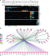
Selected HCIPs of HPV E6s. (A) Heat map representing protein-protein interactions identified by immunoprecipitation-MS/MS and CompPASS analysis when one of 16 unique HPV E6 proteins was used as a bait. Colors in the heat map represent NWD-scores, where an NWD-score ≥ 1 defines a high-confidence interaction. Each column in the heat map represents a replicate experiment. Cells were treated for 4 h with 30 μM MG132 (+) or DMSO control (−) prior to harvest. E6 baits were ordered across the top of the map according to the established HPV phylogeny (6); HCIPs were arranged using a Manhattan distance hierarchical clustering analysis. Selected interactions discussed in the text are shown. (B) Interactome representing HCIPs in complex with various HPV E6 proteins. An interaction is displayed in the heat map when it was detected with two or more HPV E6 bait proteins and had an NWD-score ≥ 1. Dashed lines represent interactions reported in the STRING database.
Fig 3
Fig 3
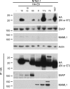
E6AP and MAML1 bind to HPV E6 proteins in a genus-specific manner. N/Tert-1 cells expressing HA-E6 or HPV16 E7-Flag HA (as a control) were subjected to immunoprecipitation with HA antibody. Immunoprecipitates were separated by SDS-PAGE and analyzed by Western blotting using antibodies to HA, actin, E6AP, and MAML1. Top panels, whole-cell lysates. Bottom panels, anti-HA immunoprecipitate (IP: HA).
Fig 4
Fig 4
HPV type-specific interactions with PTPN13 and p300. N/Tert-1 cells expressing HA-E6 or HPV16 E7-Flag HA (as a control) were subjected to immunoprecipitation with HA antibody. Immunoprecipitates were separated by SDS-PAGE and analyzed by Western blotting using antibodies to HA, actin, PTPN13 (A), and p300 (B). Top panels, whole-cell lysates. Bottom panels, anti-HA immunoprecipitate. Cells in panel A were treated with 30 μM MG132 for 4 h prior to harvest. Top panels, whole-cell lysates. Bottom panels, anti-HA immunoprecipitate.
Fig 5
Fig 5
Novel species-specific interactions with genus beta HPV E6 proteins. N/Tert-1 cells expressing HA-E6 or HPV16 E7-Flag HA (as a control) were subjected to immunoprecipitation with HA antibody. Immunoprecipitates were separated by SDS-PAGE and analyzed by Western blotting using antibodies to HA, actin, CNOT1, -2, and -3 (A) or Paxillin and HIF1α (B). Top panels, whole-cell lysates. Bottom panels, anti-HA immunoprecipitate.
Fig 6
Fig 6
HPV16 E6 mutant proteins exhibit distinct binding profiles. (A) Heat map representing protein-protein interactions identified by immunoprecipitation-MS/MS and CompPASS analysis when the wild type (wt) or one of several mutant forms of FlagHA-HPV16 E6 was used as a bait. Colors in the heat map represent NWD-scores, where an NWD-score ≥ 1 defines a high-confidence interaction. Cells were treated for 4 h with 30 μM MG132 (+) or DMSO control (−) prior to harvest. HCIPs were arranged using a Manhattan distance hierarchical clustering analysis. (B) N/Tert-1 cells expressing the wild type or mutant HA-HPV16 E6, FlagHA-HPV16 E6, or HPV16 E7-FlagHA were treated for 4 h with 30 μM MG132 (+) or DMSO control (−), harvested, and subjected to immunoprecipitation with HA antibody. Immunoprecipitates were separated by SDS-PAGE and Western blotted using antibodies to HA, E6AP, p53, Scribble, or actin. Top panels, whole-cell lysates. Bottom panels, anti-HA immunoprecipitate.
Fig 7
Fig 7
E6AP mediates the interaction of E6 with the proteasome. (A) Heat map representing protein-protein interactions identified by immunoprecipitation-MS/MS and CompPASS analysis when one of 16 unique HPV E6 proteins was used as bait. Colors in the heat map represent NWD-scores, where an NWD-score ≥ 1 defines a high-confidence interaction. NWD-scores are the normalized version of the CompPASS WD-score, which in this analysis was considered significant when it was ≥95% of scores representing the interactions in the CompPASS stats table. Each column in the heat map represents a replicate experiment. Cells were treated for 4 h with 30 μM MG132 (+) or DMSO control (−) prior to harvest. E6 baits were ordered across the top of the map according to the established HPV phylogeny (6); HCIPs were arranged using a Manhattan distance hierarchical clustering analysis. Only E6AP and subunits of the proteasome are shown as interactors. (B) Heat map representing protein-protein interactions identified by immunoprecipitation-MS/MS and CompPASS analysis when the wild type or one of several mutant forms of FlagHA-HPV16 E6 was used as bait. Colors in the heat map represent NWD-scores, where an NWD-score ≥ 1 defines a high-confidence interaction. NWD-scores are the normalized version of the CompPASS WD-score, which in this analysis was considered significant when it was ≥95% of scores representing the interactions in the CompPASS stats table. Cells were treated for 4 h with 30 μM MG132 (+) or DMSO control (−) prior to harvest. HCIPs were arranged using a Manhattan distance hierarchical clustering analysis. Only E6AP and subunits of the proteasome are shown as interactors.
Fig 8
Fig 8
p53 is stabilized in cells expressing a subset of the beta HPV E6 proteins. (A) Western blot of p53 expression in N/Tert-HA-E6 stable cell lines. (B) Control cells or N/Tert-1 cells stably expressing HA-HPV16 E6 were treated with 40 μg/ml cycloheximide (CHX) and harvested at the indicated time points. Lysates were analyzed by Western blotting using antibodies to p53, HA, and actin.
Fig 9
Fig 9
p53 is bound by HPV38 and HPV92 E6. N/Tert-1 cells expressing HA-E6 were treated for 4 h with 30 μM MG132 (+) or DMSO control (−) and subjected to immunoprecipitation with HA antibody. Immunoprecipitates were separated by SDS-PAGE and Western blotted using antibodies to HA, E6AP, p53, and actin. Top panels, whole-cell lysates. Bottom panels, anti-HA immunoprecipitate.

Similar articles

Cited by

References

    1. Accardi R, et al. 2006. Skin human papillomavirus type 38 alters p53 functions by accumulation of deltaNp73. EMBO Rep. 7:334–340 - PMC - PubMed
    1. Akgül B, Cooke JC, Storey A. 2006. HPV-associated skin disease. J. Pathol. 208:165–175 - PubMed
    1. Albert TK, et al. 2002. Identification of a ubiquitin-protein ligase subunit within the CCR4-NOT transcription repressor complex. EMBO J. 21:355–364 - PMC - PubMed
    1. Arron ST, Ruby JG, Dybbro E, Ganem D, Derisi JL. 2011. Transcriptome sequencing demonstrates that human papillomavirus is not active in cutaneous squamous cell carcinoma. J. Invest. Dermatol. 131:1745–1753 - PMC - PubMed
    1. Bennett EJ, Rush J, Gygi SP, Harper JW. 2010. Dynamics of cullin-RING ubiquitin ligase network revealed by systematic quantitative proteomics. Cell 143:951–965 - PMC - PubMed

Publication types

Substances