. 2012 Sep 26;32(39):13587-96.
doi: 10.1523/JNEUROSCI.2617-12.2012.

Bcl-x pre-mRNA splicing regulates brain injury after neonatal hypoxia-ischemia

Affiliations

Bcl-x pre-mRNA splicing regulates brain injury after neonatal hypoxia-ischemia

Qingli Xiao et al. J Neurosci. .

Abstract

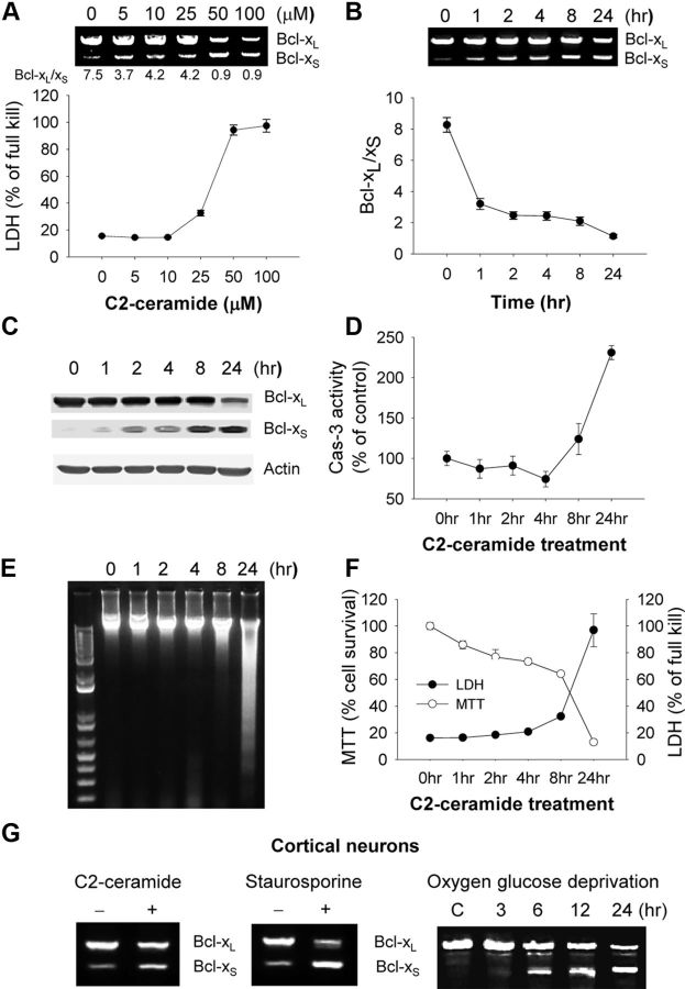
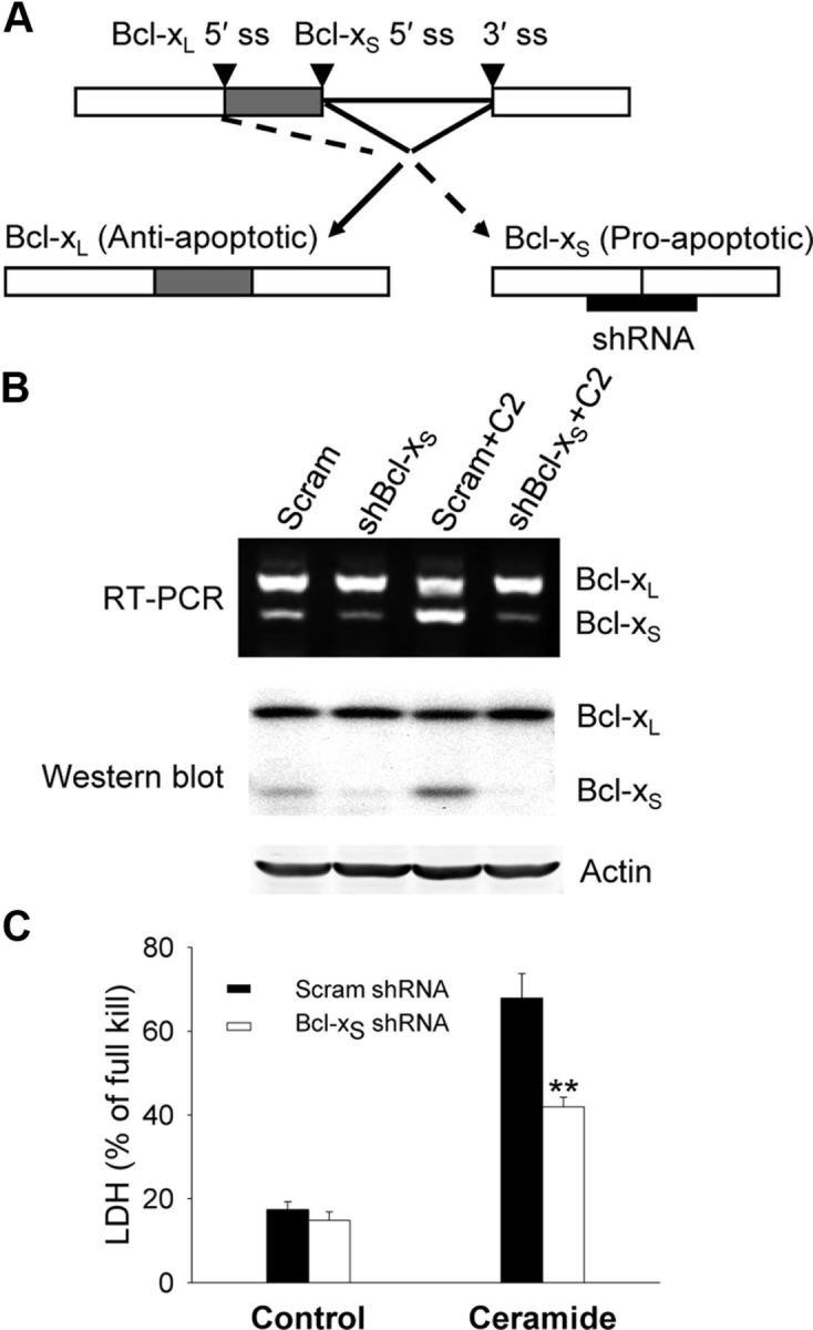
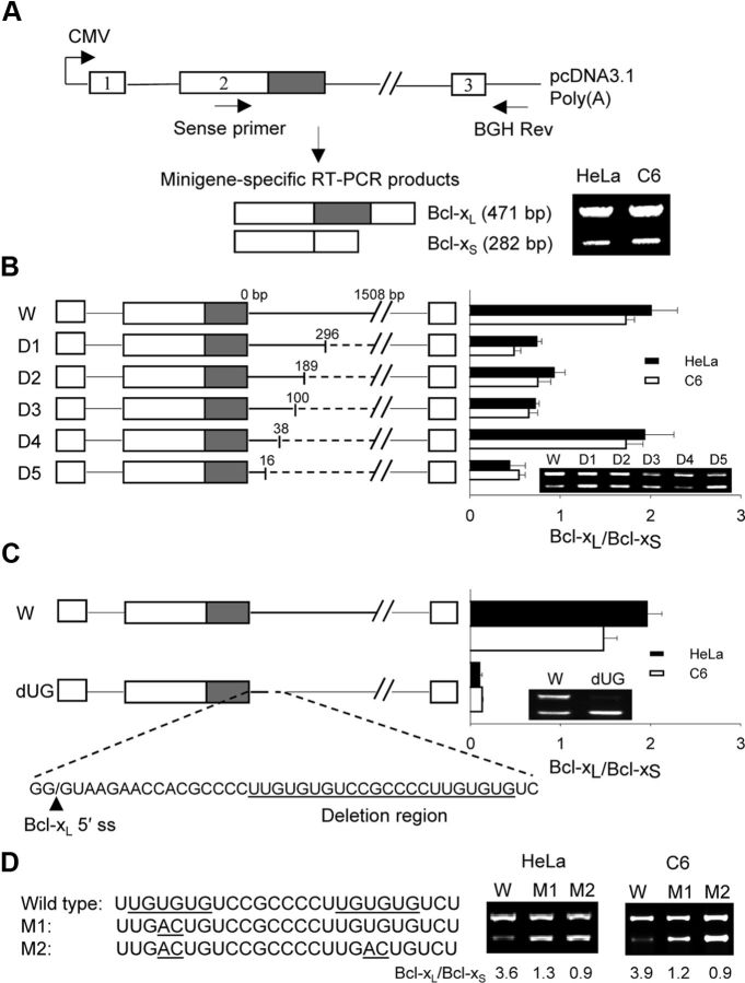
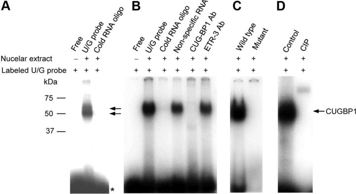
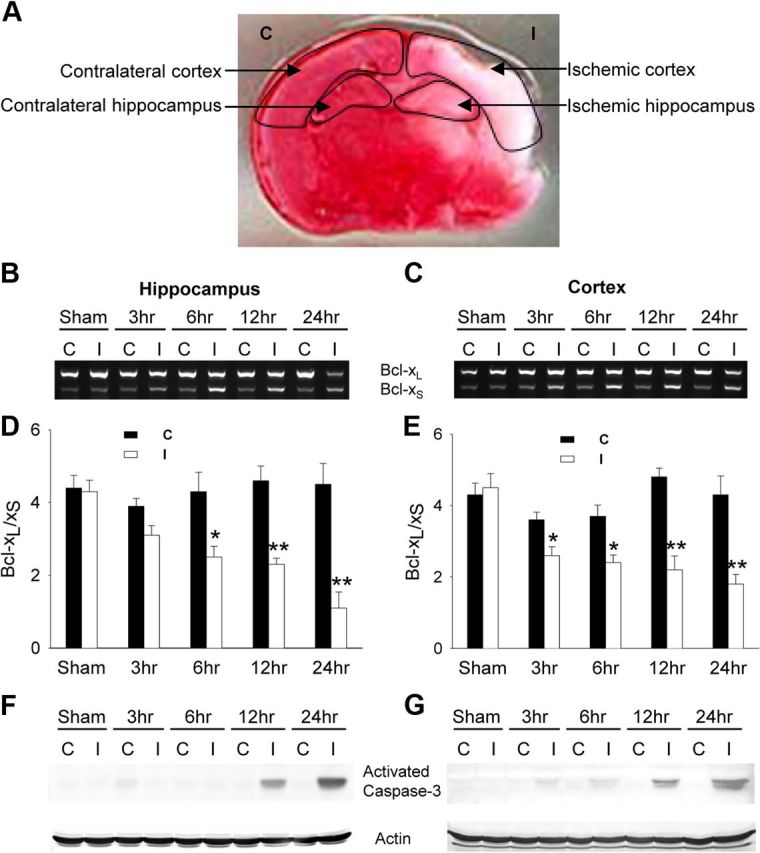
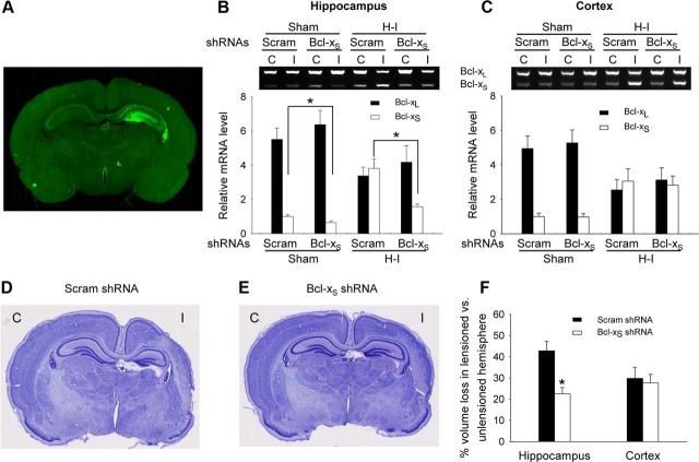
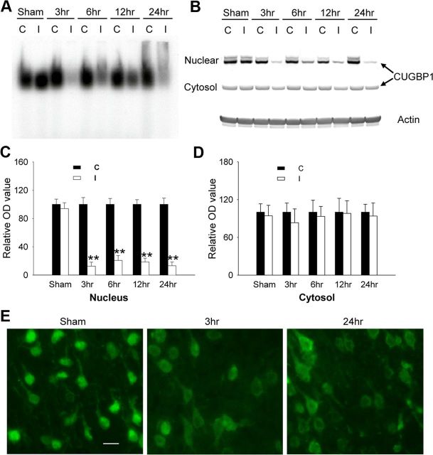
The bcl-x gene appears to play a critical role in regulating apoptosis in the developing and mature CNS and following CNS injury. Two isoforms of Bcl-x are produced as a result of alternative pre-mRNA splicing: Bcl-x(L) (the long form) is anti-apoptotic, while Bcl-x(S) (short form) is pro-apoptotic. Despite the antagonistic activities of these two isoforms, little is known about how regulation of alternative splicing of bcl-x may mediate neural cell apoptosis. Here, we report that apoptotic stimuli (staurosporine or C2-ceramide) reciprocally altered Bcl-x splicing in neural cells, decreasing Bcl-x(L) while increasing Bcl-x(S). Specific knockdown of Bcl-x(S) attenuated apoptosis. To further define regulatory elements that influenced Bcl-x splicing, a Bcl-x minigene was constructed. Deletional analysis revealed several consensus sequences within intron 2 that altered splicing. We found that the splicing factor, CUG-binding-protein-1 (CUGBP1), bound to a consensus sequence close to the Bcl-x(L) 5' splice site, altering the Bcl-x(L)/Bcl-x(S) ratio and influencing cell death. In vivo, neonatal hypoxia-ischemia reciprocally altered Bcl-x pre-mRNA splicing, similar to the in vitro studies. Manipulation of the splice isoforms using viral gene transfer of Bcl-x(S) shRNA into the hippocampus of rats before neonatal hypoxia-ischemia decreased vulnerability to injury. Moreover, alterations in nuclear CUGBP1 preceded Bcl-x splicing changes. These results suggest that alternative pre-mRNA splicing may be an important regulatory mechanism for cell death after acute neurological injury and may potentially provide novel targets for intervention.


Figures

Figure 1.
Figure 1.
Changes in Bcl-x alternative splicing during apoptosis. A, Oligodendrocyte precursor (OP) cells were treated with varying concentrations of C2-ceramide as indicated for 24 h. Total RNA was extracted and analyzed by RT-PCR for alternative splicing of Bcl-x (noted below the gels are the Bcl-xL/Bcl-xS ratios). The bottom graphs show cell death by LDH release. B, OP cells were treated with 50 μm C2-ceramide for varying times as indicated. Total RNA was extracted and analyzed by RT-PCR for alternative splicing of Bcl-x, and Bcl-xL/Bcl-xS ratios were quantified. CF, OP cells were treated with 50 μm C2-ceramide for varying times and immunoblotted for Bcl-xL and Bcl-xS (C), assayed for caspase-3 activity (D), DNA fragmentation (E), and cell survival (MTT) and death (LDH) (F). Data were expressed as mean ± SE of three independent experiments. G, Cortical neurons were treated with 40 μm C2-ceramide or 100 nm staurosporine for 24 h or oxygen glucose deprivation for 90 min, followed by reoxygenation for varying times (3–24 h as indicated). Shown are RT-PCR products showing reciprocal changes in Bcl-x splice isoforms.
Figure 2.
Figure 2.
Bcl-xS knockdown attenuates apoptosis. A, Diagram shows Bcl-x pre-mRNA with mRNA isoforms and shRNA specifically targeted at Bcl-xS. OP cells were infected with a retroviral vector expressing Bcl-xS shRNA or scramble shRNA for 48 h and then treated with C2-ceramide (50 μm) for 16 h as described in Materials and Methods. ss, splice site. B, RT-PCR was performed to analyze alternatively spliced Bcl-x isoforms (top), and total proteins were extracted and analyzed by Western blot for Bcl-xL and Bcl-xS (bottom). C, Cell death was assessed by LDH assay and expressed as mean ± SE of three independent experiments. **p < 0.01 compared to scramble shRNA-treated OP cells.
Figure 3.
Figure 3.
UG rich elements modulate alternative splicing of Bcl-x. A, Shown is the structure of the Bcl-x minigene construct with positions of the primers used to amplify minigene-specific mRNA products. The figure also depicts the PCR fragments obtained from Bcl-x minigene-transfected HeLa and C6 glioma cells. B, Wild-type (W) and truncated minigenes (D1–D5) shown on the left result in dramatically different Bcl-xL/Bcl-xS ratios (right). The numbers above each deletion construct indicate the distance downstream of the Bcl-xL 5′ splice site (ss). C, The wild-type (W) and UG motif-deleted minigenes (22 bp deletion, dUG) alters the ratio of Bcl-xL/Bcl-xS in HeLa and C6 cells, favoring Bcl-xS expression. D, Single (M1) or double (M2) mutations in the UG motifs resulted in a decrease of Bcl-xL/Bcl-xS ratios. Data were expressed as mean ± SE from three independent experiments.
Figure 4.
Figure 4.
CUGBP1 binds to the UG-rich elements of Bcl-x pre-mRNA. Five micrograms of nuclear extracts were prepared from OP cells and subjected to EMSA by reacting with a 32P-labeled UG rich RNA probe. Fiftyfold excess, unlabeled, nonspecific RNA or specific cold RNA samples were included to show specificity of the RNA–protein complex. A, B, UV cross-linking (as described in Materials and Methods) was performed to analyze the molecular weight of trans-acting factor bound to UG probe (A, star (★) indicates free probe). CUGBP1, but not ETR-3 antibodies eliminated the binding complex (B). C, A mutant probe (UG>AC, similar to that used in Fig. 3D) does not form the complex. D, Abrogation of binding is shown when nuclear extracts were treated with calf intestinal alkaline phosphatase (CIP).
Figure 5.
Figure 5.
Alterations in subcellular CUGBP1 distribution affect Bcl-x splicing and cell death. OP cells were treated with C2-ceramide (50 μm) for varying times as indicated. A, Immunocytochemistry reveals that nuclear CUGBP1 rapidly decreases after treatment with C2-ceramide (top, CUGBP1 immunostaining, green; bottom, overlay of CUGBP1 immunostaining, green, and DAPI staining, blue). B, Double-labeling with CUGBP1 immunostaining (red) and TUNEL (green) after C2-ceramide treatment demonstrates that virtually all TUNEL(+) cells had cytoplasmic CUGBP1. C, D, Subcellular localization of CUGBP1 (C) and TUNEL(+) cells (D) were quantified and expressed as mean ± SE. from three independent experiments. **p < 0.01 compared to 0 h control (ANOVA with post hoc Tukey's test). Nuc, Nucleus; Cyto, cytosol. E, Nuclear extracts from OP cells after ceramide treatment demonstrate decreased UG-rich RNA binding using EMSA. F, Western blots from subcellular fractions of OP cells after ceramide treatment (top) confirm the decrease in nuclear CUGBP1. Blots were quantified by densitometry, normalized to actin, and expressed as mean ± SE from three independent experiments (bottom), **p < 0.01 compared to nuclear or cytosol control (ANOVA with post hoc Tukey's test). G, OP cells were transfected with CUGBP1 or vector constructs. H, CUGBP1 was knocked down using RNAi. At 24 h post-transfection, ceramide was added to the cell culture for an additional 16 h. Overexpression and knockdown of CUGBP1 were verified by Western blot (top), alternatively spliced Bcl-x forms were determined by RT-PCR (middle panel), and cell death was assessed by LDH assay (bottom). *p < 0.05 si-CUGBP1 versus si-scramble, **p < 0.01 CUGBP1 construct versus vehicle construct, or si-CUGBP1+ceramide versus si-scramble+ceramide (ANOVA with post hoc Tukey's test). Scale bars, 20 μm.
Figure 6.
Figure 6.
Neonatal hypoxia-ischemia alters Bcl-x alternative splicing. P7 rats underwent unilateral carotid artery ligation and exposure to hypoxia for 2.5 h and were killed at varying times (3, 6, 12, and 24 h) after hypoxia. A, The infarcted area of a representative brain section is shown by TTC staining 24 h after H–I. Arrows indicate contralateral (C) or ischemic (I) hippocampus and cortex used for RNA extraction. B, C, Total RNA was extracted and analyzed by RT-PCR for alternative splicing of Bcl-x. A representative agarose gel of the RT-PCR products from hippocampus (B) or cortex (C) is shown (C, Contralateral hemisphere; I, ischemic hemisphere). D, E, Mean value ± SE of the Bcl-xL/Bcl-xS ratio is from hippocampus (D) and cortex (E) from at least 5 animals per group. *p < 0.05, **p < 0.01 ischemic versus contralateral (ANOVA with post hoc Tukey's test). F, G, Western blots were performed to measure activated caspase-3 from hippocampal (F) and cortical (G) extracts (blots are representative of two independent replicates).
Figure 7.
Figure 7.
Knockdown of Bcl-xS attenuates brain injury following H–I. Lentiviral vectors carrying Bcl-xS shRNA or scrambled (Scram) shRNA were injected into the left hippocampus of P0 rats. A, A representative example of gene transfer is shown by detecting EGFP expression 1 week after injection. B, C, Animals were subjected to H–I at P7 and killed at P14. Bcl-xL and Bcl-xS mRNA levels from the hippocampus (B) and cortex (C) were determined by RT-PCR (mean ± SE from 5 animals). D, E, Representative cresyl-violet-stained brain sections from rats from each group (D, scrambled shRNA: n = 17; E, Bcl-xS shRNA: n = 18) are shown. C, Contralateral hemisphere; I, ischemic hemisphere. F, Bcl-xS shRNA significantly reduced tissue loss in hippocampus where shRNA was targeted but not cortex (mean ± SE; *p < 0.01 Bcl-xS shRNA versus scrambled shRNA).
Figure 8.
Figure 8.
Alterations in subcellular CUGBP1 localization after H–I. A, Cortical nuclear extracts (5 μg) show decreased binding to UG-rich RNA probes after H–I measured via EMSA. BD, Cortical nuclear extracts (5 μg) or cytosolic extracts (20 μg) were subjected to Western blot to measure CUGBP1 levels after H–I, quantified by densitometry, and normalized to actin (B) (mean ± SE of three independent experiments, C, D). C, Contralateral hemisphere; I, ischemic hemisphere.*p < 0.01 ischemic vs contralateral nuclear extracts (ANOVA with post hoc Tukey's test). E, The intracellular localization of CUGBP1 in rat cortex was determined by immunostaining in sham control, 3 and 24 h after H–I. Scale bar, 20 μm.

Similar articles

Cited by

References

    1. Al-Zoubi AM, Efimova EV, Kaithamana S, Martinez O, El-Idrissi Mel-A, Dogan RE, Prabhakar BS. Contrasting effects of IG20 and its splice isoforms, MADD and DENN-SV, on tumor necrosis factor alpha-induced apoptosis and activation of caspase-8 and -3. J Biol Chem. 2001;276:47202–47211. - PubMed
    1. Back SA, Han BH, Luo NL, Chricton CA, Xanthoudakis S, Tam J, Arvin KL, Holtzman DM. Selective vulnerability of late oligodendrocyte progenitors to hypoxia-ischemia. J Neurosci. 2002;22:455–463. - PMC - PubMed
    1. Bae J, Leo CP, Hsu SY, Hsueh AJ. MCL-1S, a splicing variant of the antiapoptotic BCL-2 family member MCL-1, encodes a proapoptotic protein possessing only the BH3 domain. J Biol Chem. 2000;275:25255–25261. - PubMed
    1. Barreau C, Paillard L, Méreau A, Osborne HB. Mammalian CELF/Bruno-like RNA-binding proteins: molecular characteristics and biological functions. Biochimie. 2006;88:515–525. - PubMed
    1. Bingle CD, Craig RW, Swales BM, Singleton V, Zhou P, Whyte MK. Exon skipping in Mcl-1 results in a bcl-2 homology domain 3 only gene product that promotes cell death. J Biol Chem. 2000;275:22136–22146. - PubMed

Publication types

MeSH terms