Brain and testicular tumors in mice with progenitor cells lacking BAX and BAK
- PMID: 22986529
- PMCID: PMC3529761
- DOI: 10.1038/onc.2012.421
Brain and testicular tumors in mice with progenitor cells lacking BAX and BAK
Abstract
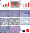
The proapoptotic BCL-2 family proteins BAX and BAK serve as essential gatekeepers of the intrinsic apoptotic pathway and, when activated, transform into pore-forming homo-oligomers that permeabilize the mitochondrial outer membrane. Deletion of Bax and Bak causes marked resistance to death stimuli in a variety of cell types. Bax(-/-)Bak(-/-) mice are predominantly non-viable and survivors exhibit multiple developmental abnormalities characterized by cellular excess, including accumulation of neural progenitor cells in the periventricular, hippocampal, cerebellar and olfactory bulb regions of the brain. To explore the long-term pathophysiological consequences of BAX/BAK deficiency in a stem cell niche, we generated Bak(-/-) mice with conditional deletion of Bax in Nestin-positive cells. Aged Nestin(Cre)Bax(fl/fl)Bak(-/-) mice manifest progressive brain enlargement with a profound accumulation of NeuN- and Sox2-positive neural progenitor cells within the subventricular zone (SVZ). One-third of the mice develop frank masses comprised of neural progenitors, and in 20% of these cases, more aggressive, hypercellular tumors emerged. Unexpectedly, 60% of Nestin(Cre)Bax(fl/fl)Bak(-/-) mice harbored high-grade tumors within the testis, a peripheral site of Nestin expression. This in vivo model of severe apoptotic blockade highlights the constitutive role of BAX/BAK in long-term regulation of Nestin-positive progenitor cell pools, with loss of function predisposing to adult-onset tumorigenesis.
Conflict of interest statement
The authors have no competing financial interests related to the work described in this manuscript.
Figures


Similar articles
-
A role for proapoptotic Bax and Bak in T-cell differentiation and transformation.Blood. 2010 Dec 9;116(24):5237-46. doi: 10.1182/blood-2010-04-279687. Epub 2010 Sep 2. Blood. 2010. PMID: 20813900 Free PMC article.
-
Bak compensated for Bax in p53-null cells to release cytochrome c for the initiation of mitochondrial signaling during Withanolide D-induced apoptosis.PLoS One. 2012;7(3):e34277. doi: 10.1371/journal.pone.0034277. Epub 2012 Mar 29. PLoS One. 2012. Retraction in: PLoS One. 2020 Feb 3;15(2):e0228839. doi: 10.1371/journal.pone.0228839 PMID: 22479585 Free PMC article. Retracted.
-
The proapoptotic activities of Bax and Bak limit the size of the neural stem cell pool.J Neurosci. 2003 Dec 3;23(35):11112-9. doi: 10.1523/JNEUROSCI.23-35-11112.2003. J Neurosci. 2003. PMID: 14657169 Free PMC article.
-
Regulation of mitochondrial morphological dynamics during apoptosis by Bcl-2 family proteins: a key in Bak?Cell Cycle. 2007 Dec 15;6(24):3043-7. doi: 10.4161/cc.6.24.5115. Epub 2007 Oct 2. Cell Cycle. 2007. PMID: 18073534 Review.
-
Physiological and Pharmacological Control of BAK, BAX, and Beyond.Trends Cell Biol. 2016 Dec;26(12):906-917. doi: 10.1016/j.tcb.2016.07.002. Epub 2016 Aug 4. Trends Cell Biol. 2016. PMID: 27498846 Free PMC article. Review.
Cited by
-
Identification of 22 susceptibility loci associated with testicular germ cell tumors.Nat Commun. 2021 Jul 23;12(1):4487. doi: 10.1038/s41467-021-24334-y. Nat Commun. 2021. PMID: 34301922 Free PMC article.
-
Targeting BCL-2 regulated apoptosis in cancer.Open Biol. 2018 May;8(5):180002. doi: 10.1098/rsob.180002. Open Biol. 2018. PMID: 29769323 Free PMC article. Review.
-
Association Between BAK1 Gene rs210138 Polymorphisms and Testicular Germ Cell Tumors: A Systematic Review and Meta-Analysis.Front Endocrinol (Lausanne). 2020 Jan 23;11:2. doi: 10.3389/fendo.2020.00002. eCollection 2020. Front Endocrinol (Lausanne). 2020. PMID: 32038496 Free PMC article.
-
Modeling the function of BAX and BAK in early human brain development using iPSC-derived systems.Cell Death Dis. 2020 Sep 25;11(9):808. doi: 10.1038/s41419-020-03002-x. Cell Death Dis. 2020. PMID: 32978370 Free PMC article.
References
-
- Youle RJ, Strasser A. The BCL-2 protein family: opposing activities that mediate cell death. Nat Rev Mol Cell Biol. 2008;9(1):47–59. Epub 2007/12/22. - PubMed
-
- Tait SW, Green DR. Mitochondria and cell death: outer membrane permeabilization and beyond. Nat Rev Mol Cell Biol. 2010;11(9):621–632. Epub 2010/08/05. - PubMed
-
- Yip KW, Reed JC. Bcl-2 family proteins and cancer. Oncogene. 2008;27(50):6398–6406. Epub 2008/10/29. - PubMed
Publication types
MeSH terms
Substances
Grants and funding
LinkOut - more resources
Full Text Sources
Medical
Molecular Biology Databases
Research Materials

