A genomic regulatory element that directs assembly and function of immune-specific AP-1-IRF complexes
- PMID: 22983707
- PMCID: PMC5789805
- DOI: 10.1126/science.1228309
A genomic regulatory element that directs assembly and function of immune-specific AP-1-IRF complexes
Abstract
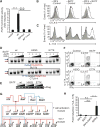
Interferon regulatory factor 4 (IRF4) and IRF8 regulate B, T, macrophage, and dendritic cell differentiation. They are recruited to cis-regulatory Ets-IRF composite elements by PU.1 or Spi-B. How these IRFs target genes in most T cells is enigmatic given the absence of specific Ets partners. Chromatin immunoprecipitation sequencing in T helper 17 (T(H)17) cells reveals that IRF4 targets sequences enriched for activating protein 1 (AP-1)-IRF composite elements (AICEs) that are co-bound by BATF, an AP-1 factor required for T(H)17, B, and dendritic cell differentiation. IRF4 and BATF bind cooperatively to structurally divergent AICEs to promote gene activation and T(H)17 differentiation. The AICE motif directs assembly of IRF4 or IRF8 with BATF heterodimers and is also used in T(H)2, B, and dendritic cells. This genomic regulatory element and cognate factors appear to have evolved to integrate diverse immunomodulatory signals.
Figures


Comment in
-
Immunology. Cooperative transcription factor complexes in control.Science. 2012 Nov 16;338(6109):891-2. doi: 10.1126/science.1231310. Science. 2012. PMID: 23161983 Free PMC article.
Similar articles
-
BATF-JUN is critical for IRF4-mediated transcription in T cells.Nature. 2012 Oct 25;490(7421):543-6. doi: 10.1038/nature11530. Epub 2012 Sep 19. Nature. 2012. PMID: 22992523 Free PMC article.
-
High Amount of Transcription Factor IRF8 Engages AP1-IRF Composite Elements in Enhancers to Direct Type 1 Conventional Dendritic Cell Identity.Immunity. 2020 Oct 13;53(4):759-774.e9. doi: 10.1016/j.immuni.2020.07.018. Epub 2020 Aug 13. Immunity. 2020. PMID: 32795402 Free PMC article.
-
Batf Pioneers the Reorganization of Chromatin in Developing Effector T Cells via Ets1-Dependent Recruitment of Ctcf.Cell Rep. 2019 Oct 29;29(5):1203-1220.e7. doi: 10.1016/j.celrep.2019.09.064. Cell Rep. 2019. PMID: 31665634 Free PMC article.
-
Specificity through cooperation: BATF-IRF interactions control immune-regulatory networks.Nat Rev Immunol. 2013 Jul;13(7):499-509. doi: 10.1038/nri3470. Epub 2013 Jun 21. Nat Rev Immunol. 2013. PMID: 23787991 Review.
-
Direct Inhibition of IRF-Dependent Transcriptional Regulatory Mechanisms Associated With Disease.Front Immunol. 2019 May 24;10:1176. doi: 10.3389/fimmu.2019.01176. eCollection 2019. Front Immunol. 2019. PMID: 31178872 Free PMC article. Review.
Cited by
-
Role of IRF4-Mediated Inflammation: Implication in Neurodegenerative Diseases.Neurol Neurother Open Access J. 2017;2(1):107. doi: 10.23880/nnoaj-16000107. Epub 2017 Jan 12. Neurol Neurother Open Access J. 2017. PMID: 39473489 Free PMC article.
-
Transcriptional rewiring in CD8+ T cells: implications for CAR-T cell therapy against solid tumours.Front Immunol. 2024 Sep 27;15:1412731. doi: 10.3389/fimmu.2024.1412731. eCollection 2024. Front Immunol. 2024. PMID: 39399500 Free PMC article. Review.
-
Optimization of the Irf8 +32-kb enhancer disrupts dendritic cell lineage segregation.Nat Immunol. 2024 Nov;25(11):2043-2056. doi: 10.1038/s41590-024-01976-w. Epub 2024 Oct 7. Nat Immunol. 2024. PMID: 39375550
-
Transcriptional control of interferon-stimulated genes.J Biol Chem. 2024 Sep 12;300(10):107771. doi: 10.1016/j.jbc.2024.107771. Online ahead of print. J Biol Chem. 2024. PMID: 39276937 Free PMC article. Review.
-
Predicting gene expression state and prioritizing putative enhancers using 5hmC signal.Genome Biol. 2024 Jun 3;25(1):142. doi: 10.1186/s13059-024-03273-z. Genome Biol. 2024. PMID: 38825692 Free PMC article.
References
-
- Tamura T, Yanai H, Savitsky D, Taniguchi T. Annu Rev Immunol. 2008;26:535. - PubMed
-
- Lohoff M, Mak TW. Nat Rev Immunol. 2005;5:125. - PubMed
-
- De Silva NS, Simonetti G, Heise N, Klein U. Immunol Rev. 2012;247:73. - PubMed
-
- Klein U, et al. Nat Immunol. 2006;7:773. - PubMed
-
- Sciammas R, et al. Immunity. 2006;25:225. - PubMed
Publication types
MeSH terms
Substances
Associated data
- Actions
Grants and funding
LinkOut - more resources
Full Text Sources
Other Literature Sources
Molecular Biology Databases
Research Materials

