Review
. 2013 Jun;70(12):2083-98.
doi: 10.1007/s00018-012-1153-8. Epub 2012 Sep 15.

Effects of MACPF/CDC proteins on lipid membranes

Affiliations
Review

Effects of MACPF/CDC proteins on lipid membranes

Robert J C Gilbert et al. Cell Mol Life Sci. 2013 Jun.

Abstract

Recent work on the MACPF/CDC superfamily of pore-forming proteins has focused on the structural analysis of monomers and pore-forming oligomeric complexes. We set the family of proteins in context and highlight aspects of their function which the direct and exclusive equation of oligomers with pores fails to explain. Starting with a description of the distribution of MACPF/CDC proteins across the domains of life, we proceed to show how their evolutionary relationships can be understood on the basis of their structural homology and re-evaluate models for pore formation by perforin, in particular. We furthermore highlight data showing the role of incomplete oligomeric rings (arcs) in pore formation and how this can explain small pores generated by oligomers of proteins belonging to the family. We set this in the context of cell biological and biophysical data on the proteins' function and discuss how this helps in the development of an understanding of how they act in processes such as apicomplexan parasites gliding through cells and exiting from cells.


Figures

Fig. 1
Fig. 1
Structure of CDC and MACPF proteins. a Crystal structure of PFO [125] in which the four originally identified domains are labeled D1–D4, but with the MACPF/CDC domain colored pink against the blue of the linking (D2) and C-terminal (D4) domains. The helical regions refolding to form a β sheet are colored orange. b Crystal structure of C8α [13, 34, 35] colored in a way matching PFO: canonical MACPF domain (pink), helices contributing to pore formation (orange)
Fig. 2
Fig. 2
Distribution of MACPF domains. a The distribution of MACPF sequences (PF01823) according to the PFAM database. The associated functions of proteins that contain MACPF domains are also listed when known. b The domain architecture of several examples is presented in the alignment in c, as defined in Pfam. c Alignment of the most conserved part of the MACPF domain. The numbering above the alignment is according to PFN. The numbers in brackets denote the length of the segment that is not conserved between the sequences
Fig. 3
Fig. 3
Structural phylogenetic tree of determined CDC and MACPF structures. a The structures were superimposed and the pairwise all-pairs root mean square deviations were computed using SHP [40, 126] from which evolutionary distances were derived. The tree representation was generated using the programs FITCH and DRAWTREE as part of the PHYLIP package [127]. Structures were displayed using CHIMERA [38] with the PFN structure in blue and each fitted structure in orange. See Table S1 (Supplementary Information, available online) for RMSDs and numbers of structurally equivalent residues. b As a, for the upper segment [13] of each domain only. See Table S2 for RMSDs and numbers of structurally equivalent residues
Fig. 4
Fig. 4
Schematic of gradual evolution from oligomerization with one curvature to oligomerization with another. The sector shapes represent subunits within a curved oligomer such as that formed by CDCs, complement and PFN. CDCs and complement show the lefthand curvature (a); it is proposed [37] that PFN adopts the righthand curvature (c). This would require an essentially no-curvature intermediate (b) which would be inactive and unselectable. It could also have the property of mixed oligomerization (d), which might seem unlikely for a protein, but perhaps no more so than such a switch in handedness of oligomerization such as that proposed between CDCs and complement, and PFN
Fig. 5
Fig. 5
Oligomerization of MACPF/CDC proteins. a Left structure of a perforin pore based on the complement membrane attack complex (MAC) inferred from crystallographic studies of C6 and C8 [13, 34, 35] using the model constructed by Aleshin and colleagues. This model preserves the salt bridge identified in PFN oligomers [30]; the location of the Arg213 side chain is colored blue, and that of the Glu343 side chain red. Right The same model with the MAC superimposed. b Left The PFN cryo-EM map determined by Law et al. [37], colored by height; center top and side views of a surface representation of a map computed for the PFN pore model based on the MAC (a); the similar morphology of a single PFN subunit found in this model compared to the cryo-EM map is outlined with a broken black line in each case; right a surface representation of the MAC model. c Top a comparison of the cryo-EM map [37] (solid surface) and the PFN model based on the MAC structure (mesh) with the PFN structure shown within as a ribbon, arrows representing flexible movements that would improve the fit; bottom rotation of the PFN model provides an improved fit and additional intramolecular flexibility as indicated would improve it still further
Fig. 6
Fig. 6
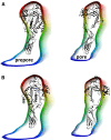
Comparison of prepore and pore subunit profiles. a Prepore (left) and pore (right) subunit profiles as reported previously [31] fitted with the PFN crystal structure with (left, prepore) and without (right, pore) TMH regions of the MACPF/CDC domain. b As a for the orientation proposed by Law et al. [37]
Fig. 7
Fig. 7
Diversity of MACPF/CDC pores. a Images of pores formed by proteins by negative stain electron microscopy for PLY [53], SLO [65], PFO [54], metridiolysin from the sea anemone Metridium senile [67] and PFN [70] and by atomic force microscopy (AFM) for PFO [63] and for the apoptotic protein Bax [95]. Arc pores or other forms of proteolipid complexes are shown by arrowheads. Reprinted from references [53] with permission from The American Society for Biochemistry and Molecular Biology, [54, 67, 70, 95] with permission from Elsevier, [65] with permission from American Society for Microbiology and with permission from Macmillan Publishers Ltd [63]. b Examples of diverse PFN pores formed in 1,2-diphytanoyl-sn-glycerophosphocholine. Large pores with conductance more than 5 nS (above) and small with conductance below 1 nS (below) are shown. The current was measured at a constant voltage of +40 mV. The buffer used was 100 mM KCl, 20 mM HEPES, 100 μM CaCl2, pH 7.4. Conductance histogram of 61 step-like increases recorded from 53 independent single-channel experiments shows two peaks at approximately 500 and 10,000 pS (below)
Fig. 8
Fig. 8
How might arcs form pores? Top left shows a schematic for a ring of MACPF/CDC subunits forming a pore, with TMHs inserted below the ring sitting atop the yellow-colored membrane surface. Top right shows a similar representation for an incomplete ring (an arc of subunits) in which the pore (blue fluid) is formed at the interface between the protein oligomer and the yellow-colored membrane. Below shows a detailed view of the pore formed by an arc with the lipids in a toroidal arrangement, as originally proposed for CDCs by Sucharit Bhakdi and colleagues [65], and as found in Bax- [51] and electrically formed pores [46, 92]
Fig. 9
Fig. 9
Transfer of GrB into the cytosol of target cells. GrB (green circles) is stored together with PFN (gray ovals) in the secretory granules of the effector cells where it is bound to serglycin (star-like shape). Upon target cell recognition, secretory granules are fused with the plasma membrane and the contents are released in the immunological synapse. GrB transfer into the cytosol of the target cells may occur at the level of the plasma membrane through barrel-stave (1) or smaller pores that may contain a lipid component (3). In the “endosomolysis” model (2), GrB is released from giant endosomes, gigantosomes, by the aid of PFN. Endocytosis is facilitated by the cell response due to calcium (black dots) influx through small PFN pores (3) and by active engagement of PFN. Small PFN pores also induce rapid flip-flop of phosphatidylserine (orange squares), as shown for the upper leaflet on the right side of the pore. The fluorescent image shows internal vesicles that resemble gigantosomes in cells, formed in the GUV in the presence of PFN [106]

Similar articles

Cited by

References

    1. Tschopp J, Masson D, Stanley KK. Structural/functional similarity between proteins involved in complement- and cytotoxic T-lymphocyte-mediated cytolysis. Nature. 1986;322:831–834. doi: 10.1038/322831a0. - DOI - PubMed
    1. Rosado CJ, Buckle AM, Law RH, Butcher RE, Kan WT, Bird CH, Ung K, Browne KA, Baran K, Bashtannyk-Puhalovich TA, Faux NG, Wong W, Porter CJ, Pike RN, Ellisdon AM, Pearce MC, Bottomley SP, Emsley J, Smith AI, Rossjohn J, Hartland EL, Voskoboinik I, Trapani JA, Bird PI, Dunstone MA, Whisstock JC. A common fold mediates vertebrate defense and bacterial attack. Science. 2007;317:1548–1551. doi: 10.1126/science.1144706. - DOI - PubMed
    1. Hadders MA, Beringer DX, Gros P. Structure of C8alpha-MACPF reveals mechanism of membrane attack in complement immune defense. Science. 2007;317:1552–1554. doi: 10.1126/science.1147103. - DOI - PubMed
    1. Slade DJ, Lovelace LL, Chruszcz M, Minor W, Lebioda L, Sodetz JM. Crystal structure of the MACPF domain of human complement protein C8 alpha in complex with the C8 gamma subunit. J Mol Biol. 2008;379:331–342. doi: 10.1016/j.jmb.2008.03.061. - DOI - PMC - PubMed
    1. Rosado CJ, Kondos S, Bull TE, Kuiper MJ, Law RHP, Buckle AM, Voskoboinik I, Bird PI, Trapani JA, Whisstock JC, Dunstone MA. The MACPF/CDC family of pore-forming toxins. Cell Microbiol. 2008;10:1765–1774. doi: 10.1111/j.1462-5822.2008.01191.x. - DOI - PMC - PubMed

Publication types

LinkOut - more resources