. 2012;8(8):e1002898.
doi: 10.1371/journal.ppat.1002898. Epub 2012 Aug 30.

Roles of ATM and ATR-mediated DNA damage responses during lytic BK polyomavirus infection

Affiliations

Roles of ATM and ATR-mediated DNA damage responses during lytic BK polyomavirus infection

Mengxi Jiang et al. PLoS Pathog. 2012.

Abstract

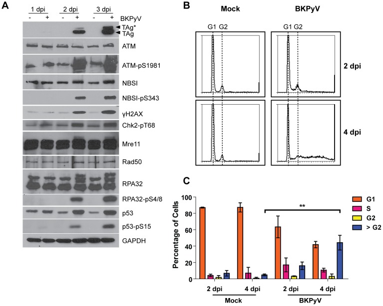
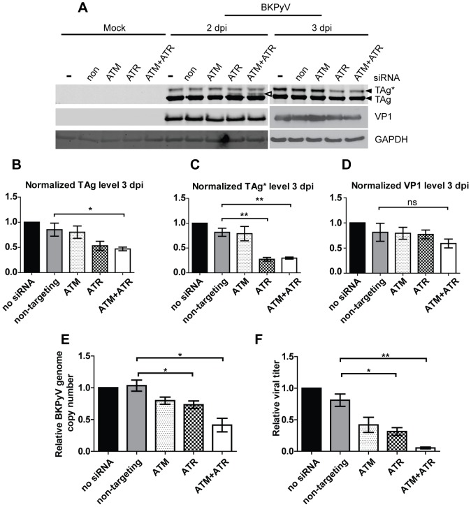
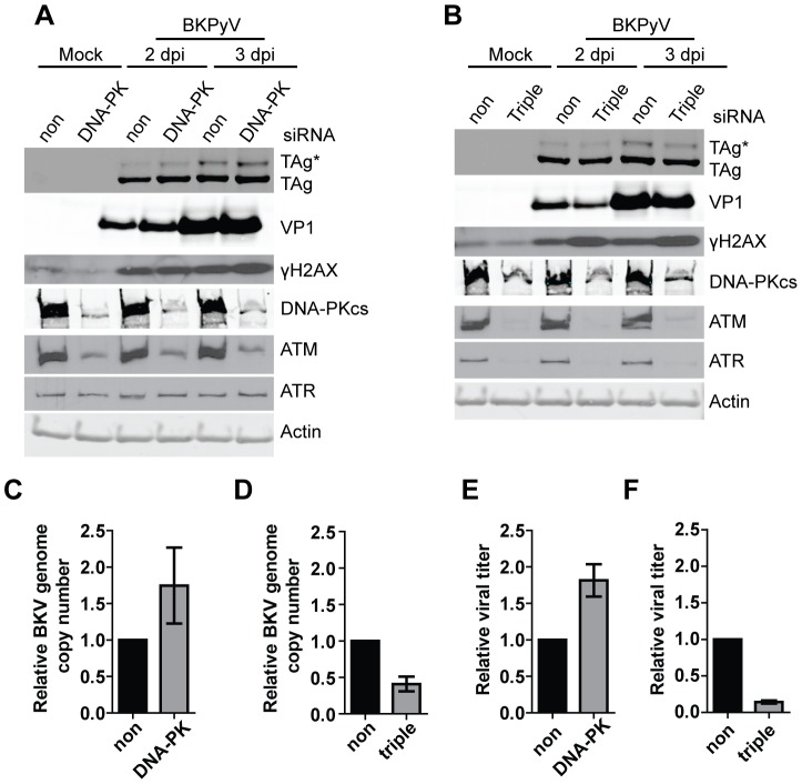
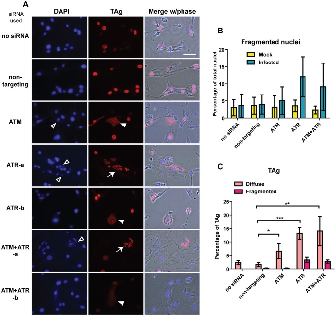
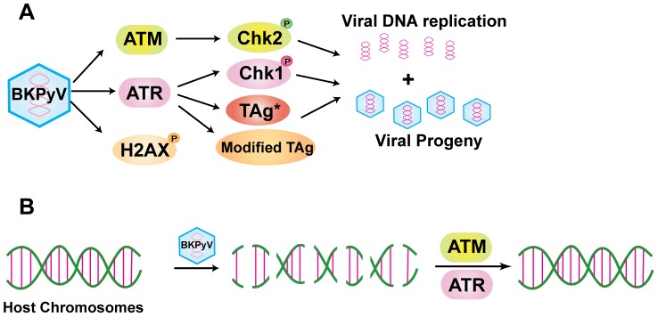
BK polyomavirus (BKPyV) is an emerging pathogen whose reactivation causes severe disease in transplant patients. Unfortunately, there is no specific anti-BKPyV treatment available, and host cell components that affect the infection outcome are not well characterized. In this report, we examined the relationship between BKPyV productive infection and the activation of the cellular DNA damage response (DDR) in natural host cells. Our results showed that both the ataxia-telangiectasia mutated (ATM)- and ATM and Rad-3-related (ATR)-mediated DDR were activated during BKPyV infection, accompanied by the accumulation of polyploid cells. We assessed the involvement of ATM and ATR during infection using small interfering RNA (siRNA) knockdowns. ATM knockdown did not significantly affect viral gene expression, but reduced BKPyV DNA replication and infectious progeny production. ATR knockdown had a slightly more dramatic effect on viral T antigen (TAg) and its modified forms, DNA replication, and progeny production. ATM and ATR double knockdown had an additive effect on DNA replication and resulted in a severe reduction in viral titer. While ATM mainly led to the activation of pChk2 and ATR was primarily responsible for the activation of pChk1, knockdown of all three major phosphatidylinositol 3-kinase-like kinases (ATM, ATR, and DNA-PKcs) did not abolish the activation of γH2AX during BKPyV infection. Finally, in the absence of ATM or ATR, BKPyV infection caused severe DNA damage and aberrant TAg staining patterns. These results indicate that induction of the DDR by BKPyV is critical for productive infection, and that one of the functions of the DDR is to minimize the DNA damage which is generated during BKPyV infection.


Conflict of interest statement

The authors have declared that no competing interests exist.

Figures

Figure 1
Figure 1. BKPyV infection activates the ATM-mediated DDR and induces polyploidy.
(A) RPTE cells were mock infected or infected with BKPyV at an MOI of 5 IU/cell. Total proteins were harvested at the indicated times post infection and probed for the indicated proteins by Western blotting. The two filled arrowheads point to TAg and TAg* as described in Results. (B) RPTE cells were mock infected or infected with BKPyV at an MOI of 5 IU/cell. Cells were fixed at the indicated times post infection and stained with propidium iodide for flow cytometry analysis. Shown are representative histograms of PI intensity. The positions of G1 and G2 peaks are marked. (C) The percentage of cells in each cell cycle phase was calculated using ModFit LT software. Each bar represents the average from three independent experiments and the error bars are the standard deviation (SD) values. **, p<0.01 (all statistical analyses were performed using a two-tailed and unpaired Student's t-test).
Figure 2
Figure 2. ATM is not required for TAg expression but is required for optimal BKPyV replication.
RPTE cells were transfected with no siRNA or the indicated siRNAs followed by infection with BKPyV at an MOI of 0.5 IU/cell at 3 dpt. Non, non-targeting siRNA. (A) Total proteins were harvested at the indicated times post infection and probed for TAg, ATM, ATM-pS1981, and actin by Western blotting. (B) Low molecular weight DNA was extracted from the cells and BKPyV viral DNA was quantified using real-time PCR. All data were normalized to mitochondrial DNA present in the same sample (mitochondrial DNA level was not affected by BKPyV infection; data not shown), and “no siRNA 3 dpi” was arbitrarily set to 1. One representative experiment from two independent experiments is shown; triplicate samples were analyzed in the same assay. Error bars are the SD values. (C) Viral lysates were harvested from the same cells at 2 dpi, and quantified using an IU assay. “no siRNA” was arbitrarily set to 1. Each bar represents the average from three independent experiments and the error bars are the SD values. *, p<0.05. (D) Total proteins were harvested as in (A) and probed for the indicated DDR proteins by Western blotting.
Figure 3
Figure 3. ATR-mediated DDR is activated during BKPyV infection.
(A) Total proteins were harvested as described in Figure 1 and probed for the indicated proteins by Western blotting. (B) Cells were transfected with indicated siRNAs and infected as described in Figure 2. Total proteins were harvested at 3 dpi and probed for the indicated proteins.
Figure 4
Figure 4. The effect of ATR and ATM knockdown on BKPyV infection.
(A) Cells were transfected with indicated siRNAs and infected with BKPyV as described in Figure 2. Total proteins were harvested at the indicated times post infection and probed for the indicated proteins by Western blotting. The open arrowhead points to a third TAg band that appeared in ATR single knockdown and ATM+ATR double knockdown cells at 2 dpi. (B–D) Quantification of the protein bands using the Odyssey Infrared Imaging System. All data were normalized to actin present in the same sample. “no siRNA” was arbitrarily set to 1 in each set. ns, not statistically significant. (E) BKPyV viral DNA and (F) viral titer were quantified as described in Figure 2B and 2C. “no siRNA” was arbitrarily set to 1 in each set. Each bar represents the average from three independent experiments and the error bars are the SD values. *, p<0.05; **, p<0.01.
Figure 5
Figure 5. The Chk1 inhibitor UCN-01 blocks BKPyV infection.
RPTE cells were infected with BKPyV at an MOI of 0.5 IU/cell. At 1 dpi, UCN-01 was added to the cells and total proteins (A), viral DNA (B), and viral lysate (C) were harvested at 2 dpi and analyzed as in Figure 2. Each bar represents the average from three independent experiments and the error bars are the SD values.
Figure 6
Figure 6. γH2X is activated in ATM, ATR, DNA-PKcs triple knockdown cells.
Cells were transfected with indicated siRNAs. (A) DNA-PKcs single knockdown and (B) ATM, ATR, DNA-PK triple knockdown. Total proteins (A, B), viral DNA (C, D), and viral lysate (E, F) were harvested at the indicated times post infection and analyzed as in Figure 2. Each bar represents the average from three independent experiments and the error bars are the SD values.
Figure 7
Figure 7. ATM and ATR knockdown leads to aberrant DAPI and TAg staining patterns during BKPyV infection.
(A) Cells were transfected with indicated siRNAs and infected with BKPyV as described in Figure 2. Cells were fixed at 3 dpi and stained for DAPI (blue) and TAg (red). Shown are representative epifluorescence pictures and merges with phase contrast pictures. Open arrowheads, fragmented DAPI staining. Filled arrowheads, diffuse TAg staining. Arrows, fragmented TAg. “ATR-a” and “ATR-b” are two different fields of view to show fragmented and diffuse TAg staining, respectively. Similarly, “ATM+ATR-a” and “ATM+ATR-b” are two fields. Scale bar, 50 µm. Bar graphs show the quantitation of fragmented nuclei (B) and aberrant TAg staining (C). Each bar represents the average from three independent experiments (at least 500 nuclei and 100 TAg-positive cells were scored in each sample per independent experiment), and the error bars are the SD values. *, p<0.05; **, p<0.01; ***, p<0.001.
Figure 8
Figure 8. ATM and ATR knockdown results in severe chromosome damage in BKPyV-infected cells.
Cells were transfected with indicated siRNAs and infected with BKPyV as described in Figure 2. At 3 dpi, cells were fixed and metaphase chromosomes were prepared as described in the Materials and Methods. (A) Representative pictures of normal chromosomes, chromosomes that contain breaks (arrows point to chromosome breaks), and shattered chromosomes. Scale bar, 20 µm. At least 50 metaphase chromosomes were scored from each sample per independent experiment. Bar graphs show the quantitation of the average number of breaks per cell (among all the non-shattered metaphases) (B) and the percentage of shattered metaphases of all the infected samples (C). Each bar represents the average from three independent experiments and the error bars are the SD values. **, p<0.01.
Figure 9
Figure 9. Model of the roles of the DDR during BKPyV infection.
(A) Upon BKPyV infection, both the ATM- and ATR-mediated DDR are activated. ATM mainly contributes to the activation of pChk2, whereas ATR mainly activates pChk1. Both ATM and ATR contribute to BKPyV DNA replication and infectious progeny production. ATR may also affect the modification status of TAg. Although γH2AX is induced by BKPyV, the molecular trigger for this induction is unknown. (B) BKPyV infection induces extensive chromosome damage, which is efficiently repaired with the aid of both ATM and ATR.

Similar articles

Cited by

References

    1. Gardner SD, Field AM, Coleman DV, Hulme B (1971) New human papovavirus (B.K.) isolated from urine after renal transplantation. Lancet 1: 1253–1257. - PubMed
    1. Jiang M, Abend JR, Johnson SF, Imperiale MJ (2009) The role of polyomaviruses in human disease. Virology 384: 266–273. - PMC - PubMed
    1. Bennett SM, Broekema NM, Imperiale MJ (2012) BK Polyomavirus: Emerging Pathogen. Microbes Infect 14: 672–83. - PMC - PubMed
    1. Imperiale MJ, Major EO (2007) Polyomaviruses. In: Knipe DM, Howley PM, editors. Fields Virology. Fifth ed. Philadelphia, PA: Lippincott Williams & Wilkins. pp. 2263–2298.
    1. Fanning E, Zhao K (2009) SV40 DNA replication: from the A gene to a nanomachine. Virology 384: 352–359. - PMC - PubMed

Publication types

MeSH terms