Review
. 2012 Sep;44(10):759-65.
doi: 10.1055/s-0032-1321866. Epub 2012 Sep 5.

Prkar1a in the regulation of insulin secretion

Affiliations
Review

Prkar1a in the regulation of insulin secretion

M A Hussain et al. Horm Metab Res. 2012 Sep.

Abstract

The incidence of type 2 diabetes mellitus (T2DM) is rapidly increasing worldwide with significant consequences on individual quality of life as well as economic burden on states' healthcare costs. While origins of the pathogenesis of T2DM are poorly understood, an early defect in glucose-stimulated insulin secretion (GSIS) from pancreatic β-cells is considered a hallmark of T2DM. Upon a glucose stimulus, insulin is secreted in a biphasic manner with an early first-phase burst of insulin, which is followed by a second, more sustained phase of insulin output. First phase insulin secretion is diminished early in T2DM as well is in subjects who are at risk of developing T2DM. An effective treatment of T2DM with incretin hormone glucagon-like peptide-1 (GLP-1) or its long acting peptide analogue exendin-4 (E4), restores first-phase and augments second-phase glucose stimulated insulin secretion. This effect of incretin action occurs within minutes of GLP-1/E4 infusion in T2DM humans. An additional important consideration is that incretin hormones augment GSIS only above a certain glucose threshold, which is slightly above the normal glucose range. This ensures that incretin hormones stimulate GSIS only when glucose levels are high, while they are ineffective when insulin levels are below a certain threshold. Activation of the GLP-1 receptor, which is highly expressed on pancreatic β-cells, stimulates 2 -distinct intracellular signaling pathways: a) the cAMP-protein kinase A branch and b) the cAMP-EPAC2 (EPAC=exchange protein activated by cAMP) branch. While the EPAC2 branch is considered to mediate GLP-1 effects on first-phase GSIS, the PKA branch is necessary for the former branch to be active. However, how these 2 branches interplay and converge and how their effects on insulin secretion and insulin vesicle exocytosis are coordinated is poorly understood.Thus, at the outset of our studies we have a poorly understood intracellular interplay of cAMP-dependent signaling pathways, which - when stimulated - restore glucose-dependent first phase and augment second phase insulin secretion in the ailing β-cells of T2DM.


Conflict of interest statement

Conflict of Interest

The authors have no conflict of interest to disclose.

Figures

Fig. 1
Fig. 1
Prkar1a ablation in pancreatic islets. Immunoblot (left) with densitometric analysis (right) of total islet protein from wt-prkar1a, hetprka1a, Δ-prkar1a mice. Specific Prkar1a ablation is detectable, while other prkar subtypes and Pkac remain unchanged. Total prkar1a reduction reflects predominant prkar1a expression in β-cells. Prkar1a expression is approximately 50 % reduced in het-prkar1a islets and 90 % reduced in Δ-rkar1a islets. CREB phosphorylation increases with reduced Prkar1a abundance. Reproduced with kind permission of Elsevier.
Fig. 2
Fig. 2
Glucose stimulated insulin secretion in Δ-prkar1a, het-prkar1a, and wt-prkar1a mice in vivo and from respective mouse islets in vitro. a Plasma glucose levels during an ipGTT in littermates of indicated genotypes. Δ-Prkar1a mice have markedly diminished glucose excursion during ipGTT but do not exhibit baseline or post glucose hypoglycemia (*p < 0.05). b Plasma glucose levels during ipITT in littermates of indicated genotypes. No difference is seen in insulin sensitivity among the different genotypes (*p < 0.05). c Serum insulin levels during ipGTT in littermates of indicated genotypes. All animals have similar baseline insulin levels. Δ-Prkar1a mice have markedly increased glucose stimulated insulin levels, predominantly during the initial phases of ipGTT (*p < 0.05). Reproduced with kind permission of Elsevier.
Fig. 3
Fig. 3
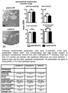
Δ-Prkar1a mice do not show any change in islet mass or β-cell proliferation. Top panel: Representative photomicrographs of Δ-prkar1a and control pancreata and morphometric analyses of pancreatic β-cells. Bottom panel: Pancreas morphometric analyses of control, heterezygote and Δ-prkar1a mice confirm no changes in islet cell mass in Δ-prkar1a mice. Reproduced with kind permission of Elsevier.
Fig. 4
Fig. 4
Oral GTT after in humans with inactivating PRKAR1A mutations and controls. a Subjects with a PRKAR1A mutation exhibit normal fasting glucose levels but reduced glucose excursion after an oral glucose load. b In subjects with a PRKAR1A mutation serum insulin levels were not different at baseline, and reached a higher peak with increased overall insulin secretion. c Table summarizing fasting glucose and insulin levels as well as area under the glucose and insulin curves shown in a and b (*p < 0.05). Reproduced with kind permission of Elsevier.
Fig. 5
Fig. 5
Representative electron microscopic images of islets (A, B: 50 000 × magnification of transmission EM; C, D Immuno EM microscopy for insulin detection, 50 000 × magnification) of wt-prkar1a (top) and Δ-prkar1a (bottom) littermates. Δ-Prkar1a islets exhibit increased vesicle size in proximity of intraislet capillaries, while dense cores containing insulin are unchanged. Panels A and B show capillaries (denoted by “c”) with insulin vesicles along the capillary border (arrowheads). E Dense core size distribution in percent of total vesicles viewed. No difference between the various genotypes is observed (mean ± SEM). F Size distribution of insulin vesicles within 1 000 nm of capillaries in percent of total vesicles observed. Δ-Prkar1a exhibit significantly larger vesicles (mean ± SEM, * signifies p < 0.05). G Number of insulin vesicles aligned along intra-islet capillary/ 10 µm of plasma membrane length. Δ-Prkar1a β-cells show significantly more vesicles adjacent to capillaries (mean ± SEM, * signifies p < 0.05). Reproduced with kind permission of Elsevier.
Fig. 6
Fig. 6
a Immunohistochemical staining of mouse pancreas sections. Co-immunostaining with insulin (green) and with nonspecific antibody (top) or snapin-specific antibody (bottom) (red). Nuclear counterstain with DAPI (blue). Separate pseudocolored images are shown with digitally merged image on bottom right panel, respectively. Snapin immunoreactivity co-localizes with insulin immunoreactivity in pancreatic islets. b Co-immunoprecipitation for snapin serine phosphorylation in mouse islets treated with E4 without or with PKA specific inhibition with myr-PKI. E4 stimulates snapin phosphorylation, which is inhibited by adding myr-PKI. Immunoblot for 10 % of protein input at bottom. c Co-immunoprecipitation for snapin serine phosphorylation in human islets treated with E4 (10 nM) without or with PKA specific inhibition with myr-PKI. E4 stimulates snapin phosphorylation, which is inhibited by adding myr-PKI. Immunoblot for 10 % of protein input at bottom. Reproduced with kind permission of Elsevier.
Fig. 7
Fig. 7
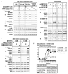
PKA mediated phosphorylation of snapin maps to serine 50. Snapin S50 phosphorylation increases interaction with secretory vesicle-associated proteins SNAP-25, EPAC2 and collectrin. Snapin interaction with VAMP2 is glucose dependent. E4 stimulated SNAP-25 interaction with EPAC2 is not PKA mediated. Overexpression in islets of snapin S50D potentiates GSIS. a Transiently transfected INS1 832/13 cells expressing C-terminal FLAG tagged WT, S50A or S50D snapin were treated with PBS (vehicle) E4 (10 nM), myr-PKI (10 nM), treated E4 + myr-PKI, or transfected with Pkac followed by IP for FLAG or SNAP-25 and IB for phosphoserine or interacting proteins. Snapin serine phosphorylation occurs in WT snapin and not in S50A and S50D mutants by E4 and Pkac action. E4 effect is inhibited by myr-PKI. Snapin interaction with SNAP-25, collectrin or EPAC2 occurs only with phosphorylated WT snapin (by E4 or Pkac) or with snapin S50D as does SNAP-25 interaction with collectrin. SNAP-25 interaction with EPAC2 occurs with E4 in a PKA-independent manner and not inhibited by myr-PKI. b Transiently transfected INS1 832/13 cells expressing C-terminal FLAG tagged WT, S50A or S50D snapin cultured in low (3 mM) or high (10 mM) glucose and co-IP/IB as in a. Snapin S50D mutant binds SNAP-25, collectrin and EPAC2 in both low and high glucose. Snapin interaction with VAMP2 occurs at elevated glucose levels only. SNAP-25-VAMP2 interaction occurs only with snapin S50D and at elevated glucose levels. c Isolated C57Bl/6 mouse islets cultured in low (3 mM) or high (10 mM) glucose and treated with PBS, E4 (10 nM), myr-PKI (10 nM) and E4 + myr-PKI followed by co-IP/IB as in a. Snapin serine phosphorylation is stimulated by E4 in a PKA dependent manner and increases snapin interaction with SNAP-25, collectrin and EPAC2 independently of glucose levels. Snapin-VAMP2 interaction is stimulated by E4 in PKA dependent manner only with high glucose. E4 stimulates SNAP25-EPAC2 interaction in a glucose- and PKA-independent manner. SNAP-25-VAMP2 interaction is stimulated by E4 in a glucose and PKA-dependent manner. PKA activity is verified by phosphorylation of CREB at serine 133 (p-CREB). d Perifusion studies of C57Bl/6 mouse islets in low (3 mM) followed by high (10 mM) glucose concentrations. Islets were treated with either PBS (inverted triangle), E4 (10 nM) (upright triangle) during perifusion, or had been transduced with control (circle) or snapin S50D (square) expressing adenovirus. Table below the curve summarizes area under the curves for first and second phase insulin secretion (*p < 0.05 vs. vehicle). Reproduced with kind permission of Elsevier.
Fig. 8
Fig. 8
Impaired GSIS in DIO diabetic mouse islets is corrected with snapin S50D. a Immunoblot of islet proteins from C57Bl/6 mice on normal chow and high fat diet. b Mapping of snapin serine-O-GlcNAcylation. DIO diabetic islets were transduced with adenovirus expressing C-terminal FLAG-tagged wild-type, S50A and S50D snapin isoforms. Immunoprecipitation with FLAG antibody followed by immunoblot was performed. Representative immunoblot is shown. Bar graph indicates densitometric analysis of 3 separate studies. c Time course of snapin phosphorylation after E4 treatment of cultured islets of diet induced diabetic mice. d Perifusion studies of DIO mouse islets in low (3 mM) followed by high (10 mM) glucose concentrations. Islets were treated with either PBS (inverted triangle), E4 (10 nM) (upright triangle) during perifusion, or had been transduced with control (circle) or snapin S50D (square) expressing adenovirus. Table below the curve summarizes area under the curves for first and second phase insulin secretion (*p < 0.05 vs. vehicle). Reproduced with kind permission of Elsevier.

Similar articles

Cited by

References

    1. Nolan CJ, Damm P, Prentki M. Type 2 diabetes across generations: from pathophysiology to prevention and management. Lancet. 2011;378:169–181. - PubMed
    1. Del Prato S, Tiengo A. The importance of first-phase insulin secretion: implications for the therapy of type 2 diabetes mellitus. Diabetes Metab Res Rev. 2001;17:164–174. - PubMed
    1. Gerich JE. Is reduced first-phase insulin release the earliest detectable abnormality in individuals destined to develop type 2 diabetes? Diabetes. 2002;51(Suppl 1):S117–S121. - PubMed
    1. Lillioja S, Mott DM, Howard BV, Bennett PH, Yki-Jarvinen H, Frimond D, Nyomba BL, Zurlo F, Swinburn B, Bogardus C. Impaired glucose tolerance as a disorder of insulin action. Longitudinal and cross-sectional studies in Pima Indians. N Engl J Med. 1988;318:1217–1225. - PubMed
    1. Vaag A, Henriksen JE, Madsbad S, Holm N, Beck-Nielsen H. Insulin secretion, insulin action, and hepatic glucose production in identical twins discordant for non-insulin-dependent diabetes mellitus. J Clin Invest. 1995;95:690–698. - PMC - PubMed

Publication types

MeSH terms

Substances