

TGF-β - an excellent servant but a bad master
- PMID: 22943793
- PMCID: PMC3494542
- DOI: 10.1186/1479-5876-10-183
TGF-β - an excellent servant but a bad master
Abstract
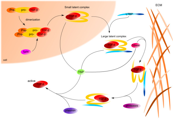
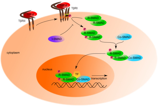
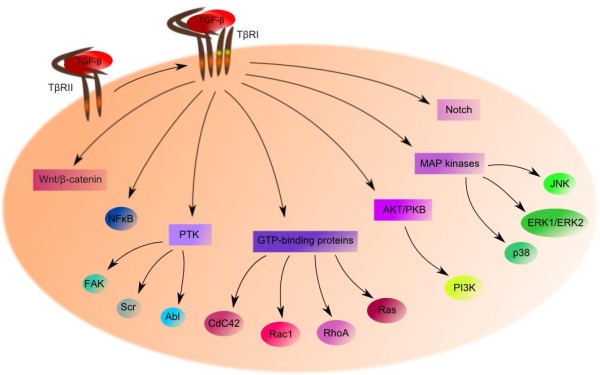
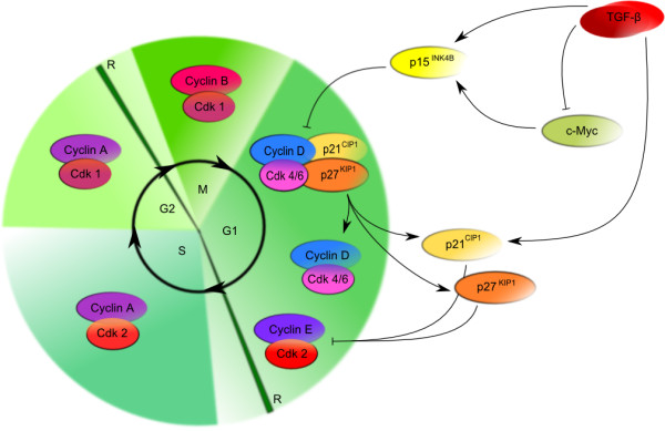
The transforming growth factor (TGF-β) family of growth factors controls an immense number of cellular responses and figures prominently in development and homeostasis of most human tissues. Work over the past decades has revealed significant insight into the TGF-β signal transduction network, such as activation of serine/threonine receptors through ligand binding, activation of SMAD proteins through phosphorylation, regulation of target genes expression in association with DNA-binding partners and regulation of SMAD activity and degradation. Disruption of the TGF-β pathway has been implicated in many human diseases, including solid and hematopoietic tumors. As a potent inhibitor of cell proliferation, TGF-β acts as a tumor suppressor; however in tumor cells, TGF-β looses anti-proliferative response and become an oncogenic factor. This article reviews current understanding of TGF-β signaling and different mechanisms that lead to its impairment in various solid tumors and hematological malignancies.
Figures
Similar articles
-
Mechanisms of TGF-beta signaling from cell membrane to the nucleus.Cell. 2003 Jun 13;113(6):685-700. doi: 10.1016/s0092-8674(03)00432-x. Cell. 2003. PMID: 12809600 Review.
-
Regulation of transforming growth factor-beta signaling.Mol Cell Biol Res Commun. 2001 Nov;4(6):321-30. doi: 10.1006/mcbr.2001.0301. Mol Cell Biol Res Commun. 2001. PMID: 11703090 Review.
-
Signal transduction by transforming growth factor-beta: a cooperative paradigm with extensive negative regulation.J Cell Biochem Suppl. 1998;30-31:111-22. J Cell Biochem Suppl. 1998. PMID: 9893262 Review.
-
Transforming growth factor-β signalling: role and consequences of Smad linker region phosphorylation.Cell Signal. 2013 Oct;25(10):2017-24. doi: 10.1016/j.cellsig.2013.06.001. Epub 2013 Jun 11. Cell Signal. 2013. PMID: 23770288 Review.
-
Transforming Growth Factor-β Promotes Liver Tumorigenesis in Mice via Up-regulation of Snail.Gastroenterology. 2017 Nov;153(5):1378-1391.e6. doi: 10.1053/j.gastro.2017.07.014. Epub 2017 Jul 20. Gastroenterology. 2017. PMID: 28734833
Cited by
-
Simvastatin downregulates expression of TGF-βRII and inhibits proliferation of A549 cells via ERK.Tumour Biol. 2015 Jun;36(6):4819-24. doi: 10.1007/s13277-015-3134-7. Epub 2015 Jan 29. Tumour Biol. 2015. PMID: 25631750
-
Knockdown of SIGLEC1 inhibits osteogenic differentiation to alleviate ankylosing spondylitis progression by suppressing the TGF-β1/SMAD signaling pathway.Naunyn Schmiedebergs Arch Pharmacol. 2025 Mar;398(3):2933-2944. doi: 10.1007/s00210-024-03456-2. Epub 2024 Sep 21. Naunyn Schmiedebergs Arch Pharmacol. 2025. PMID: 39305328
-
Circular RNA CREBBP modulates cartilage degradation by activating the Smad1/5 pathway through the TGFβ2/ALK1 axis.Exp Mol Med. 2022 Oct;54(10):1727-1740. doi: 10.1038/s12276-022-00865-2. Epub 2022 Oct 12. Exp Mol Med. 2022. PMID: 36224344 Free PMC article.
-
Emerging trends and research foci of epithelial-mesenchymal transition in gliomas: A scientometric analysis and review.Front Oncol. 2022 Oct 20;12:1015236. doi: 10.3389/fonc.2022.1015236. eCollection 2022. Front Oncol. 2022. PMID: 36338770 Free PMC article. Review.
-
Reduced Expression of YAP in Dermal Fibroblasts is Associated with Impaired Wound Healing in Type 2 Diabetic Mice.Tissue Eng Regen Med. 2017 Jan 17;14(1):49-55. doi: 10.1007/s13770-016-0019-9. eCollection 2017 Feb. Tissue Eng Regen Med. 2017. PMID: 30603461 Free PMC article.
References
-
- Derynck R. The TGF-β Family.: Cold Spring Harbor Laboratory. 2008. Press.
Publication types
MeSH terms
Substances
LinkOut - more resources
Full Text Sources
Other Literature Sources
Research Materials
Miscellaneous
