

The endoplasmic reticulum stress transducer BBF2H7 suppresses apoptosis by activating the ATF5-MCL1 pathway in growth plate cartilage
- PMID: 22936798
- PMCID: PMC3476286
- DOI: 10.1074/jbc.M112.373746
The endoplasmic reticulum stress transducer BBF2H7 suppresses apoptosis by activating the ATF5-MCL1 pathway in growth plate cartilage
Abstract
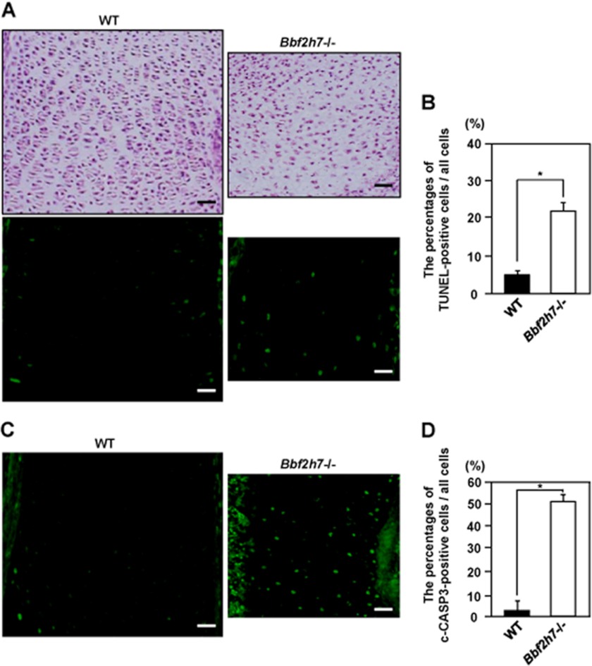
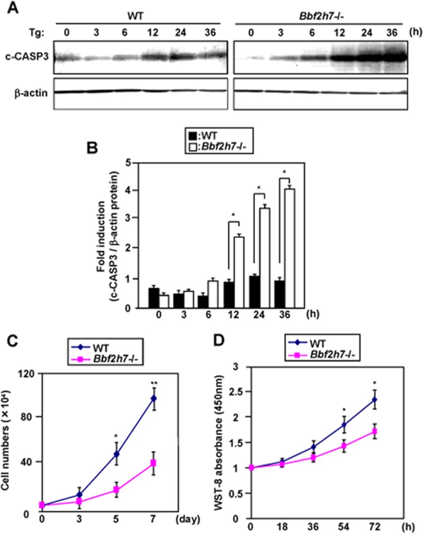
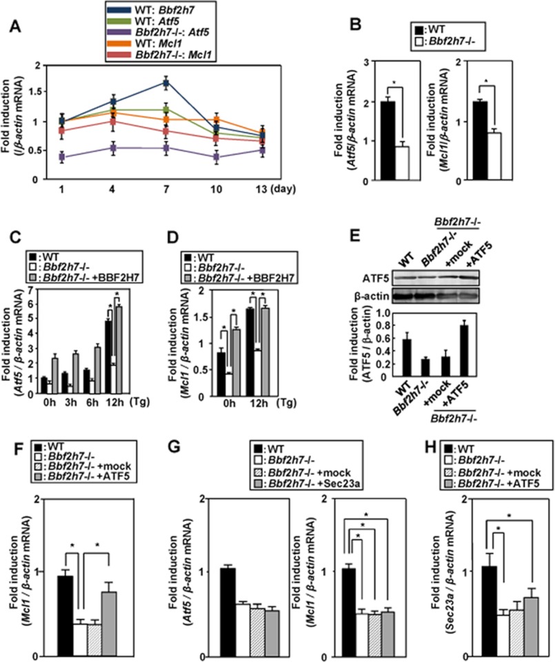
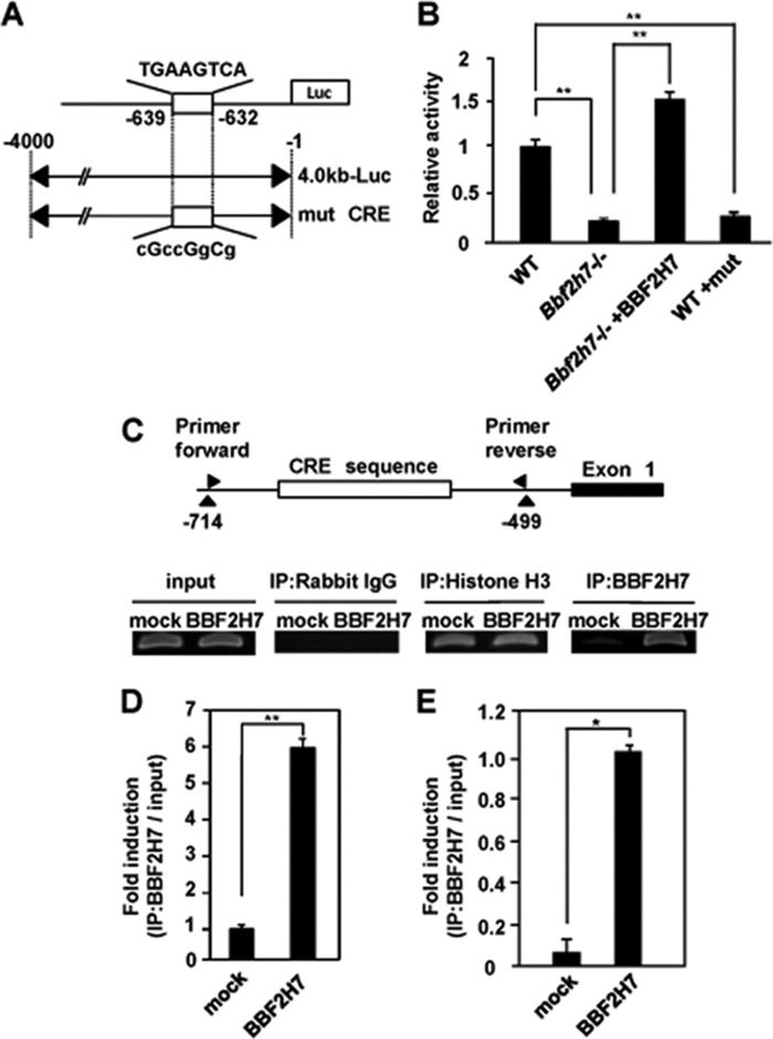
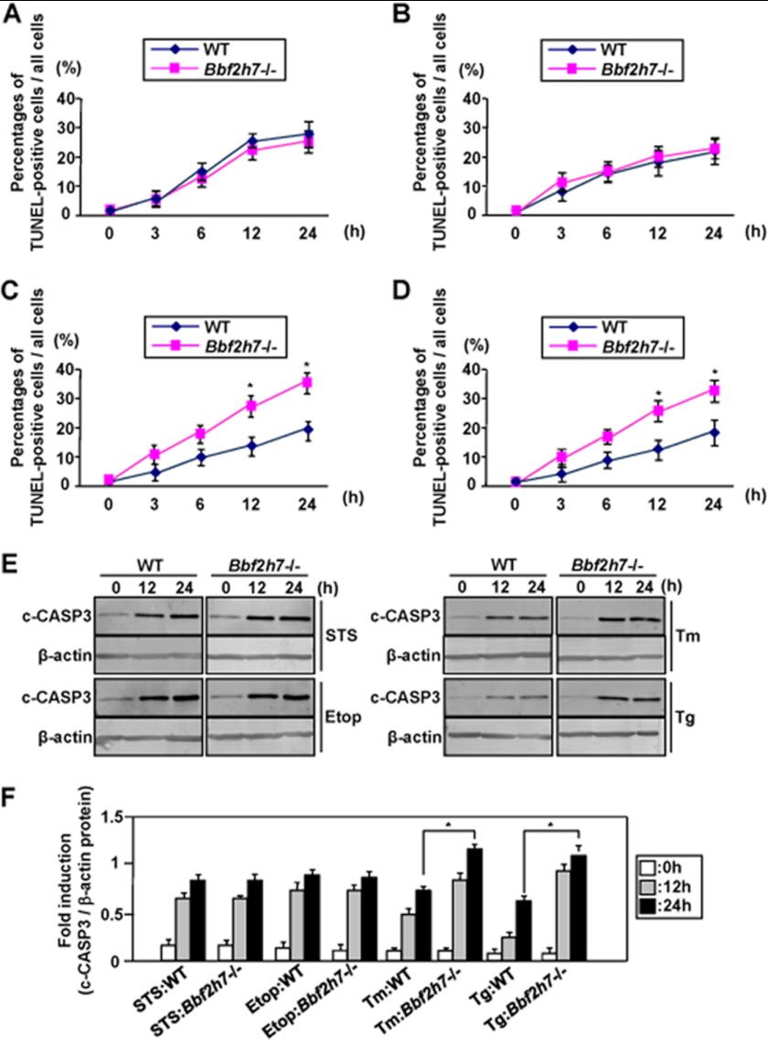
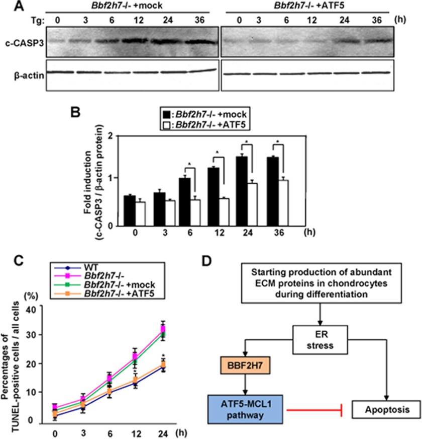
BBF2H7 (box B-binding factor 2 human homolog on chromosome 7) is a basic leucine zipper transmembrane transcription factor that belongs to the cyclic AMP-responsive element-binding protein (CREB)/activating transcription factor (ATF) family. This novel endoplasmic reticulum (ER) stress transducer is localized in the ER and is cleaved in its transmembrane region in response to ER stress. BBF2H7 has been shown to be expressed in proliferating chondrocytes in cartilage during the development of long bones. The target of BBF2H7 is Sec23a, one of the coat protein complex II components. Bbf2h7-deficient (Bbf2h7(-/-)) mice exhibit severe chondrodysplasia, with expansion of the rough ER in proliferating chondrocytes caused by impaired secretion of extracellular matrix (ECM) proteins. We observed a decrease in the number of proliferating chondrocytes in the cartilage of Bbf2h7(-/-) mice. TUNEL staining of the cartilage showed that apoptosis was promoted in Bbf2h7(-/-) chondrocytes. Atf5 (activating transcription factor 5), another member of the CREB/ATF family and an antiapoptotic factor, was also found to be a target of BBF2H7 in chondrocytes. ATF5 activated the transcription of Mcl1 (myeloid cell leukemia sequence 1), which belongs to the antiapoptotic B-cell leukemia/lymphoma 2 family, to suppress apoptosis. Finally, we found that the BBF2H7-ATF5-MCL1 pathway specifically suppressed ER stress-induced apoptosis in chondrocytes. Taken together, our findings indicate that BBF2H7 is activated in response to ER stress caused by synthesis of abundant ECM proteins and plays crucial roles as a bifunctional regulator to accelerate ECM protein secretion and suppress ER stress-induced apoptosis by activating the ATF5-MCL1 pathway during chondrogenesis.
Figures
Similar articles
-
Regulation of endoplasmic reticulum stress response by a BBF2H7-mediated Sec23a pathway is essential for chondrogenesis.Nat Cell Biol. 2009 Oct;11(10):1197-204. doi: 10.1038/ncb1962. Epub 2009 Sep 20. Nat Cell Biol. 2009. PMID: 19767744
-
Master regulator for chondrogenesis, Sox9, regulates transcriptional activation of the endoplasmic reticulum stress transducer BBF2H7/CREB3L2 in chondrocytes.J Biol Chem. 2014 May 16;289(20):13810-20. doi: 10.1074/jbc.M113.543322. Epub 2014 Apr 6. J Biol Chem. 2014. PMID: 24711445 Free PMC article.
-
Chondrocyte proliferation regulated by secreted luminal domain of ER stress transducer BBF2H7/CREB3L2.Mol Cell. 2014 Jan 9;53(1):127-39. doi: 10.1016/j.molcel.2013.11.008. Epub 2013 Dec 12. Mol Cell. 2014. PMID: 24332809
-
[Bone and Calcium Research Update 2015. The regulation of proliferation and differentiation by the ER stress transducer in chondrocytes].Clin Calcium. 2015 Jan;25(1):29-36. Clin Calcium. 2015. PMID: 25530520 Review. Japanese.
-
Physiological unfolded protein response regulated by OASIS family members, transmembrane bZIP transcription factors.IUBMB Life. 2011 Apr;63(4):233-9. doi: 10.1002/iub.433. Epub 2011 Mar 24. IUBMB Life. 2011. PMID: 21438114 Review.
Cited by
-
Endoplasmic Reticulum Stress and Unfolded Protein Response in Cartilage Pathophysiology; Contributing Factors to Apoptosis and Osteoarthritis.Int J Mol Sci. 2017 Mar 20;18(3):665. doi: 10.3390/ijms18030665. Int J Mol Sci. 2017. PMID: 28335520 Free PMC article. Review.
-
DNA methyltransferase 3 beta mediates the methylation of the microRNA-34a promoter and enhances chondrocyte viability in osteoarthritis.Bioengineered. 2021 Dec;12(2):11138-11155. doi: 10.1080/21655979.2021.2005308. Bioengineered. 2021. PMID: 34783292 Free PMC article.
-
Expression of activating transcription factor 5 (ATF5) is mediated by microRNA-520b-3p under diverse cellular stress in cancer cells.PLoS One. 2020 Jun 30;15(6):e0225044. doi: 10.1371/journal.pone.0225044. eCollection 2020. PLoS One. 2020. PMID: 32603335 Free PMC article.
-
Advancements in Activating Transcription Factor 5 Function in Regulating Cell Stress and Survival.Int J Mol Sci. 2022 Jun 27;23(13):7129. doi: 10.3390/ijms23137129. Int J Mol Sci. 2022. PMID: 35806136 Free PMC article. Review.
-
SUMO2/3 modification of activating transcription factor 5 (ATF5) controls its dynamic translocation at the centrosome.J Biol Chem. 2018 Feb 23;293(8):2939-2948. doi: 10.1074/jbc.RA117.001151. Epub 2018 Jan 11. J Biol Chem. 2018. PMID: 29326161 Free PMC article.
References
-
- Schröder M., Kaufman R. J. (2005) ER stress and the unfolded protein response. Mutat Res. 569, 29–63 - PubMed
-
- Calfon M., Zeng H., Urano F., Till J. H., Hubbard S. R., Harding H. P., Clark S. G., Ron D. (2002) IRE1 couples endoplasmic reticulum load to secretory capacity by processing the XBP-1 mRNA. Nature 415, 92–96 - PubMed
Publication types
MeSH terms
Substances
LinkOut - more resources
Full Text Sources
Molecular Biology Databases
