. 2013 Feb;62(2):321-35.
doi: 10.1007/s00262-012-1342-1. Epub 2012 Aug 26.

Inhibitor of apoptosis protein (IAP) antagonists demonstrate divergent immunomodulatory properties in human immune subsets with implications for combination therapy

Affiliations

Inhibitor of apoptosis protein (IAP) antagonists demonstrate divergent immunomodulatory properties in human immune subsets with implications for combination therapy

Ashley J Knights et al. Cancer Immunol Immunother. 2013 Feb.

Abstract

Inhibitor of apoptosis proteins (IAPs) are critical in regulating apoptosis resistance in cancer. Antagonists of IAPs, such as LCL161, are in clinical development and show promise as anti-cancer agents for solid and hematological cancers, with preliminary data suggesting they may act as immunomodulators. IAP antagonists hypersensitize tumor cells to TNF-α-mediated apoptosis, an effect that may work in synergy with that of cancer vaccines. This study aimed to further investigate the immunomodulatory properties of LCL161 on human immune subsets. T lymphocytes treated with LCL161 demonstrated significantly enhanced cytokine secretion upon activation, with little effect on CD4 and CD8 T-cell survival or proliferation. LCL161 treatment of peripheral blood mononuclear cells significantly enhanced priming of naïve T cells with synthetic peptides in vitro. Myeloid dendritic cells underwent phenotypic maturation upon IAP antagonism and demonstrated a reduced capacity to cross-present a tumor antigen-based vaccine. These effects are potentially mediated through an observed activation of the canonical and non-canonical NF-κB pathways, following IAP antagonism with a resulting upregulation of anti-apoptotic molecules. In conclusion, this study demonstrated the immunomodulatory properties of antagonists at physiologically relevant concentrations and indicates their combination with immunotherapy requires further investigation.


Conflict of interest statement

The authors declare that they have no conflict of interest.

Figures

Fig. 1
Fig. 1
Significant increase in the frequency of cytokine-secreting T cells and the duration of secretion following treatment with the IAP antagonist LCL161. Production of the cytokines IL-2 and TNF-α by purified CD4+ and CD8+ T cells was assessed by ICS flow cytometry at 24, 48 and 72 h following activation with anti-CD3/CD28 micro-particles and titrating concentrations of LCL161. a A representative example for IL-2 production by CD4 +T cells from 3 donors is shown. Percentage of cytokine-producing cells (y-axis) is shown as a fraction of total CD3+CD4+ T cells in the FACS lymphocyte gate. Each point represents the mean from triplicate wells for each donor, with the grand mean and SEM shown at each concentration of LCL161 (x-axis). b To normalize for inter-donor variation, the fold increase in percentage of cytokine-producing cells was calculated for each concentration in relation to the untreated control and plotted for each time point. Mean values with SEM from the results obtained with 6 donors are shown for IL-2 (upper histograms) and TNF-α (lower histograms) production in CD4+ (left histograms) and CD8+ (right histograms) T cells. One-way ANOVA with a Dunnett’s post-test against the untreated control was performed, *P < 0.05, **P < 0.01, ***P < 0.001
Fig. 2
Fig. 2
A significant increase in the frequency of antigen-specific T cells stimulated in in vitro peptide sensitization assays is mediated by IAP antagonist treatment. The effects of LCL161 treatment on in vitro stimulation of peptide-specific CD8+ T cells were assessed 10 days after stimulation of 1 × 106 PBMC with an HLA-A2-restricted synthetic peptide for either Flu-M1 (58–66), EBV BMLF1 (280–288) or melan-A (26–35). Specific cells were detected using fluorescently labeled recombinant HLA-A2 MHC peptide multimers containing the relevant peptide. a Representative contour plots are shown for each peptide with titrating concentrations of LCL161. The percentage of MHC/peptide multimer-positive (y-axis) CD8+ (x-axis) CD3+ T cells in the FACS lymphocyte gate is shown. b Percentage of tetramer + CD8+ cells (y-axis) are shown for each peptide and LCL161 concentration (x-axis). Each point represents a mean value from multiple replicate stimulations performed with 5 donors (Flu, EBV BMLF1) or 6 donors (melan-A) and the grand mean (horizontal bar) with SEM is indicated. One-way ANOVA with a Dunnett’s post-test against the untreated control was performed, *P < 0.05, ***P < 0.001
Fig. 3
Fig. 3
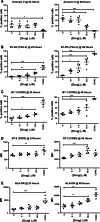
IAP antagonism induces phenotypic maturation of monocyte-derived dendritic cells. The effects of LCL161 treatment on maturation of immature monocyte-derived dendritic cells were assessed in vitro 24 h (left scatter plots) and 48 h (right scatter plots) following treatment with LCL161. MoDCs were treated on day 4 following differentiation from monocytes, seeded in triplicate wells per concentration and treated with LCL161 at the concentrations indicated (x-axis). Soluble CD40L was used as a maximal control for induction of maturation. Each point represents the mean value obtained from triplicate wells per donor. Horizontal bars indicate the grand mean derived from the results obtained with 4 donors, and error bars show the SEM. a For annexin V, staining was performed in combination with a viability dye. Ann-V-positive, viability dye negative viable cells were counted as apoptotic cells (y-axis). (B/C) For B7-DC and B7-1, the percentage of viable (Live/Dead fluorescent dye negative) positive cells (y-axis) within the monocyte gate of the FACS plot is indicated. (D/E) For B7-2 and HLA-DR, the mean fluorescence intensity (MFI) of the positive cells (>95 %) is indicated (y-axis) for the viable cells within the monocyte gate of the FACS plot. One-way ANOVA with a Dunnett’s post-test against the untreated control was performed, *P < 0.05, **P < 0.01, ***P < 0.001
Fig. 4
Fig. 4
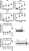
Exposure of moDC to the IAP antagonist LCL161 activates the canonical and non-canonical NF-κB pathways. MoDCs were left untreated (UT) or treated with 10 μM LCL161 and assessed for the activation of the NF-κB pathway by Western blot analysis. Whole cell lysates were prepared 30 min and 2, 4, 6 and 24 h following drug treatment. CD40L treatment for 24 h represents the positive control. Equal quantities of protein were evaluated on a 4–12 % SDS-PAGE gel, transferred to membrane and probed with antibodies to IκBα (a, b) phospho-p65/Rel-A (c, d) and p52/p100 (e, f); antibodies to β-tublin (a, b) and β-actin (c–f) were used for reference. Chemiluminescent imaging was used for (b), and near-infrared fluorescence detection was used for (d) and (f). a and c show quantified percentage expression levels as a percentage of the respective untreated control, whereas e shows a ratio for expression levels of p52 to p100 at each time point; all values were first normalized to the respective house keeping protein (β-tublin and β-actin). a, c, e Each point represents the result per donor with the mean (horizontal bar) and SEM shown from 4 donors. One-way ANOVA with a Dunnett’s post-test against the untreated control was performed, *P < 0.05, **P < 0.01, ***P < 0.001
Fig. 5
Fig. 5
Treatment of moDC with the IAP antagonist LCL161 induces expression of anti-apoptotic factors. MoDCs were left untreated (UT) or treated with 10 μM LCL161 and assessed for expression of anti-apoptotic molecules by qPRC and Western blot analysis. Whole cell lysates and mRNA were prepared from cells treated for 2, 4, 6 and 24 h. CD40L treatment for 24 h represents the positive control. Untreated cells were cultured in parallel for 24 h. a Semi-quantitative RT-PCR was performed on cDNA from the treated imoDC with specific primer pairs for cIAP1, cIAP2, Bcl-2 and Bcl-xL using SYBRgreen chemistry. Relative transcript copy numbers are expressed as copy number of NY-ESO-1 transcripts per β-actin transcript using 2(β—actin CT—gene of interest CT). For Western blot analysis, equal quantities of protein were loaded and ran on 4–12 % SDS-PAGE gels, transferred to membrane and probed for the indicated proteins. Antibodies to cIAP2 (b) and Bcl-xL (c) were used at a 1:500 dilution. Near-infrared fluorescence detection was used with the appropriate secondary antibodies, and membranes were imaged accordingly. For each condition, values were normalized to the expression of β-actin and quantified percentage expression levels are shown as a percentage of the respective untreated control. Each point represents the result per donor with the mean (horizontal bar) and SEM shown from 4 donors. One-way ANOVA with a Dunnett’s post-test against the untreated control was performed, *P < 0.05, **P < 0.01, ***P < 0.001
Fig. 6
Fig. 6
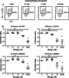
IAP antagonists significantly decrease cross-presentation of tumor antigen by monocyte-derived dendritic cells. MoDCs plated in triplicate were treated for 24 or 48 h with titrating concentrations of LCL161 or LBW242, and rNY-ESO-1/ISCOMATRIX was added for an additional 18 h following treatment. An antigen-specific CD8+ T-cell clone was used to assess cross-presentation of MHC-class I-associated epitopes in a standard recognition assay measuring specific cytokine secretion by the T cells upon activation. a An NY-ESO-1-specific (157–165/HLA-A2) CD8+ T-cell clone was incubated for 4 h with the previously treated and antigen-pulsed DC, and the percentage of activated, IFN-γ-positive CD8+ CD3+ cells (y-axis) within the lymphocyte FACS gate (x-axis) was assessed at each drug concentration used to treat the moDC (indicated above contour plots). b All values were normalized against the untreated control to compensate for inter-donor variation in the % IFN-γ+ cells responding within the untreated group, and therefore allowing assessment of drug effect. This value is referred to as an ‘activation index’ (y-axis) and indicates relative activation (IFN-γ positivity) of the specific T-cell clone following a 4-h incubation with the 24- or 48-h drug-treated and antigen-pulsed DC at the concentration indicated (x-axis) for LCL161 (upper scatter plots) and LBW242 (lower scatter plots). Each point represents the mean from triplicate results per donor, per concentration with the grand mean (horizontal bar) and SEM shown from 4 donors. One-way ANOVA with a Dunnett’s post-test against the untreated control was performed, *P < 0.05, ***P < 0.001

Similar articles

Cited by

References

    1. Hodi FS, O’Day SJ, McDermott DF, et al. Improved survival with ipilimumab in patients with metastatic melanoma. N Engl J Med. 2010;363:711–723. doi: 10.1056/NEJMoa1003466. - DOI - PMC - PubMed
    1. Kantoff PW, Higano CS, Shore ND, et al. Sipuleucel-T immunotherapy for castration-resistant prostate cancer. N Engl J Med. 2010;363:411–422. doi: 10.1056/NEJMoa1001294. - DOI - PubMed
    1. Heine A, Held SA, Bringmann A, Holderried TA, Brossart P. Immunomodulatory effects of anti-angiogenic drugs. Leukemia. 2011;25:899–905. doi: 10.1038/leu.2011.24. - DOI - PubMed
    1. Ott PA, Adams S. Small-molecule protein kinase inhibitors and their effects on the immune system: implications for cancer treatment. Immunotherapy. 2011;3:213–227. doi: 10.2217/imt.10.99. - DOI - PMC - PubMed
    1. Du C, Fang M, Li Y, Li L, Wang X. Smac, a mitochondrial protein that promotes cytochrome c-dependent caspase activation by eliminating IAP inhibition. Cell. 2000;102:33–42. doi: 10.1016/S0092-8674(00)00008-8. - DOI - PubMed

Publication types

MeSH terms