. 2012 Oct;23(20):4065-78.
doi: 10.1091/mbc.E12-04-0263. Epub 2012 Aug 23.

Myo1c binding to submembrane actin mediates insulin-induced tethering of GLUT4 vesicles

Affiliations

Myo1c binding to submembrane actin mediates insulin-induced tethering of GLUT4 vesicles

Shlomit Boguslavsky et al. Mol Biol Cell. 2012 Oct.

Abstract

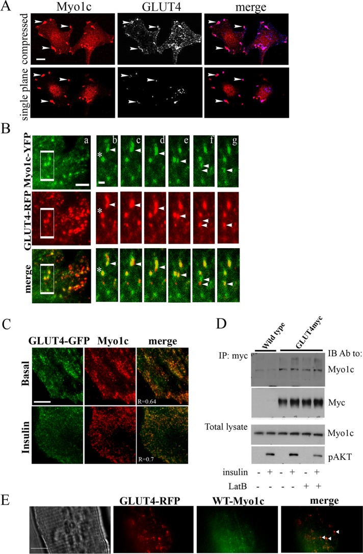
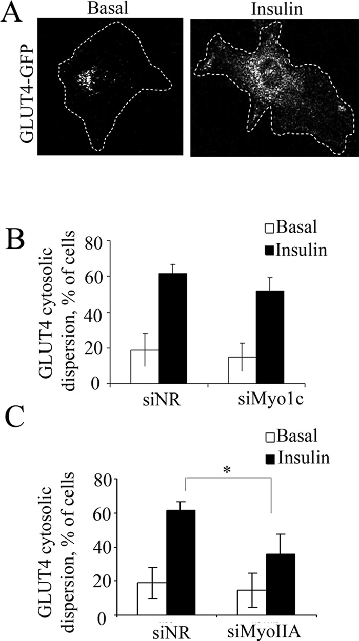
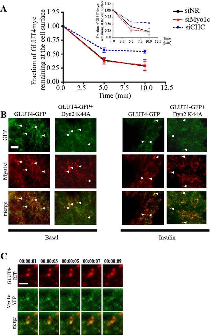
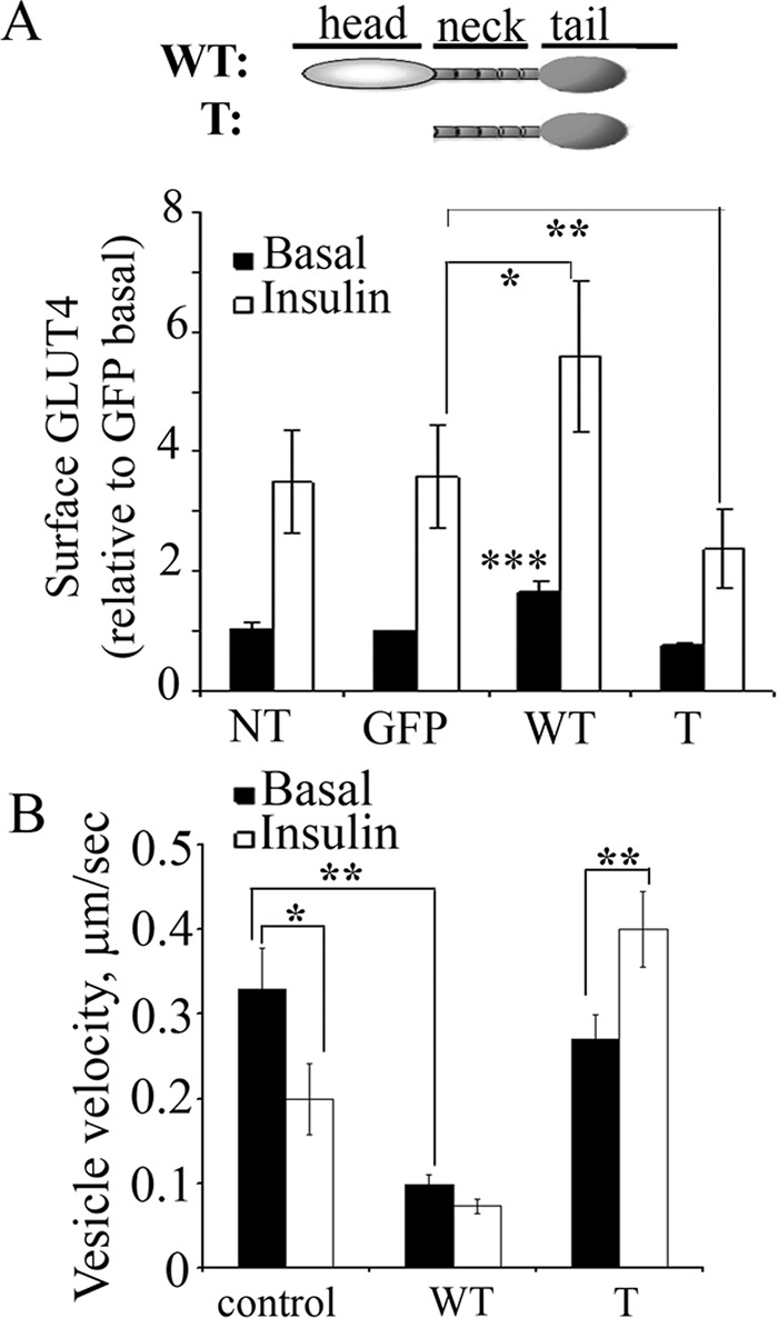
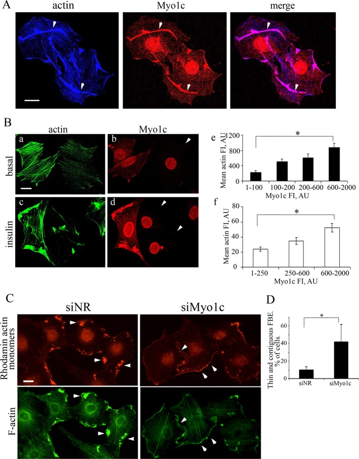
GLUT4-containing vesicles cycle between the plasma membrane and intracellular compartments. Insulin promotes GLUT4 exocytosis by regulating GLUT4 vesicle arrival at the cell periphery and its subsequent tethering, docking, and fusion with the plasma membrane. The molecular machinery involved in GLUT4 vesicle tethering is unknown. We show here that Myo1c, an actin-based motor protein that associates with membranes and actin filaments, is required for insulin-induced vesicle tethering in muscle cells. Myo1c was found to associate with both mobile and tethered GLUT4 vesicles and to be required for vesicle capture in the total internal reflection fluorescence (TIRF) zone beneath the plasma membrane. Myo1c knockdown or overexpression of an actin binding-deficient Myo1c mutant abolished insulin-induced vesicle immobilization, increased GLUT4 vesicle velocity in the TIRF zone, and prevented their externalization. Conversely, Myo1c overexpression immobilized GLUT4 vesicles in the TIRF zone and promoted insulin-induced GLUT4 exposure to the extracellular milieu. Myo1c also contributed to insulin-dependent actin filament remodeling. Thus we propose that interaction of vesicular Myo1c with cortical actin filaments is required for insulin-mediated tethering of GLUT4 vesicles and for efficient GLUT4 surface delivery in muscle cells.


Figures

FIGURE 1:
FIGURE 1:
Myo1c and GLUT4 colocalize at the plasma membrane and on mobile intracellular vesicles. (A) Myo1c colocalizes with surface GLUT4 in insulin-induced ruffles. L6-GLUT4myc myoblasts were treated with insulin (100 nM, 10 min). Surface-localized GLUT4myc was labeled with anti-myc prior to permeabilization and Myo1c was then labeled after permeabilization. Shown are representative confocal images (of three experiments) acquired from a single focal plane 4–6 μm above the coverslip (to capture insulin-induced ruffles that may protrude 2–3 μm above the dorsal focal plane of the cell) and a combined image (compressed) of all focal planes. Arrowheads indicate Myo1c and GLUT4 sites of colocalization. In the merged image, blue artificial color represents GLUT4 staining. Scale bar: 10 μm. (B) Myo1c localizes to dynamic vesicles and colocalizes with GLUT4-positive vesicles at the TIRF zone. WT L6 myoblasts were transiently cotransfected with GLUT4-RFP (shown in red) and Myo1c-YFP (shown in green), incubated in serum-free media for 3 h, and imaged live using multicolor TIRF microscopy (Movie S1, a–c). Panel (a) shows a single frame of a representative cell in the basal state. The outlined region in panel (a) is magnified in panels (b–g), which are individual frames of this region captured 2-s apart. One stationary vesicle in which the two proteins colocalize is marked by an asterisk. Instances of colocalization on mobile vesicles are indicated by arrowheads. Scale bars: 5 and 0.8 μm (insets). (C) Endogenous Myo1c (red) colocalizes with GLUT4-GFP (green) vesicles. WT L6 myoblasts were transfected with GLUT4-GFP and treated without/with insulin as in (A); this was followed by immunofluorescence labeling of endogenous Myo1c and then multicolor TIRF microscopy imaging of fixed cells. The extent of colocalization was quantified using the Pearson's r (R) as described in Materials and Methods. Scale bar: 5 μm. (D) Endogenous Myo1c coimmunoprecipitates with GLUT4. Lysates of serum-starved L6-GLUT4myc or WT L6 myoblasts were pretreated with/without 1 μM latrunculin B (LatB) for 1 h; this was followed by insulin stimulation as in (A). GLUT4 immunoprecipitates (IP) were immunoblotted (IB) with Myo1c antibody or anti-Myc antibody (GLUT4myc). Shown is one representative blot out of three independent experiments. (E) Unstimulated FDB fibers expressing GLUT4-RFP and WT-Myo1c-YFP were fixed but not permeabilized. A representative TIRF image from three independent experiments is illustrated in differential image contrast (DIC), red and green channels, and as a merged image. Arrowheads mark the colocalization of the two proteins on punctate vesicular structures in the submembrane region of the muscle fiber. Scale bar: 100 μm.
FIGURE 2:
FIGURE 2:
Insulin-stimulated GLUT4 translocation to the plasma membrane is reduced by Myo1c knockdown. (A) Efficient siRNA-mediated knockdown of Myo1c (200 nM, oligo 1) as indicated by immunoblotting. siNR was used as a negative control, and expression of ERK was not affected. Shown is one representative blot out of three independent experiments. L6-GLUT4myc myoblasts were transfected with siNR or Myo1c siRNA (siMyo1c) and treated with/without insulin as in Figure 1. (B and C) WT L6 myoblasts transfected with siNR or siMyo1c oligo 1 (B, n = 4) or oligo 2 (C, n = 2), were transiently transfected with GLUT4myc-GFP. Forty-eight hours after transfection, the cells were treated with/without insulin; this was followed by staining of surface GLUT4myc and endogenous Myo1c as in Figure 1B. Knockdown cells with low expression of Myo1c were further analyzed for surface GLUT4 fluorescence intensity (at least 20 cells per condition). Quantification of fold increases in surface GLUT4myc relative to the siNR basal is presented (mean ± SE, #, p < 0.05, *, p = 0.08). (D) L6-GLUT4myc myoblasts stably expressing AS160 were transfected with siNR or siMyo1c and treated with/without insulin as above. Shown is one representative immunoblot for native and phospho-Akt and phospho-AS160 out of three independent experiments. (E) The pAS160/AS160 ratio from (D) is calculated from three independent experiments.
FIGURE 3:
FIGURE 3:
Myo1c is not required for cytosolic dispersion of GLUT4 to the cell periphery. (A) Representative images of the typical perinuclear localization of GLUT4myc-GFP in basal (left) and cytosolic localization in the insulin-stimulated state (right). (B and C) WT L6 myoblasts transfected with siNR, siMyo1c (B), or siMyoIIA (C), were transiently transfected with GLUT4myc-GFP. After 24 h, cells were transferred to glass coverslips and allowed to recover for 24–48 h. Cells were then treated with/without insulin for 20 min; this was followed by staining of endogenous Myo1c (B, n = 4) or MyoIIA (C, n = 3). Knockdown cells (at least 20 cells per condition) with low expression of Myo1c (B) or MyoIIA (C) were selected for further analysis of the percent of cells with cytosolic dispersion of GLUT4myc-GFP (mean ± SD, *, p < 0.01) as described in Materials and Methods.
FIGURE 4:
FIGURE 4:
Myo1c is not required for GLUT4 internalization and associates with GLUT4 exocytic vesicles. (A) The fraction of GLUT4myc remaining at the surface at two times of internalization in L6 myoblasts transfected with siNR, siMyo1c, or siRNA to clathrin heavy chain (CHC). Main panel: insulin-stimulated cells; inset: unstimulated cells. Data are the means ± SE representative of three independent experiments. (B) Endogenous Myo1c (red) colocalizes with GLUT4myc-GFP (green) vesicles following inhibition of GLUT4myc internalization. L6 myoblasts were transfected with either a 5:1 M ratio of Dyn2 K44A to GLUT4myc-GFP or GLUT4myc-GFP alone and treated without/with insulin (100 nM, 20 min) at 24 h after transfection; this was followed by immunofluorescence labeling of endogenous Myo1c and imaging by multicolor TIRF microscopy. Arrowheads indicate examples of colocalization. Scale bar: 3 μm. (C) A GLUT4-RFP fusion event is illustrated. WT L6 myoblasts were transiently cotransfected with GLUT4-RFP (red) and Myo1c-YFP (green) and imaged live by multicolor TIRF microscopy (see also Movie S2, a–c). Individual frames shown were captured 2-ms apart. A vesicle containing GLUT4-RFP and Myo1-YFP spreads laterally as it collapses onto the plasma membrane. Scale bars: 2.6 μm.
FIGURE 5:
FIGURE 5:
Myo1c binding to actin immobilizes submembrane GLUT4 and underlies the contribution of the motor protein to GLUT4 translocation. (A) Cell surface GLUT4myc was measured in individual L6-GLUT4myc myoblasts transfected with YFP-conjugated Myo1c-WT or Myo1c-T (a mutant unable to bind actin), and the results are presented as fold increases in surface GLUT4myc relative to a GFP-transfected basal control (mean ± SE, n = 3, *, p = 0.1, **, p = 0.19, ***, p < 0.05). NT, not treated. (B) WT L6 cells were cotransfected with GLUT4-RFP and either GFP (control, n = 6), WT-Myo1c-YFP (WT, n = 4), or Myo1c-T-YFP (T, n = 4). Two-color TIRF microscopy was used to visualize and image the traffic of GLUT4-RFP in each condition for 15 s before and 15 s after insulin stimulation (100 nM, 10min). Data represent the mean vesicle velocity (μm/s) calculated for at least 100 different tracks in each condition acquired by imaging 10–12 time points/s. Individual vesicles were tracked for 15 s or until the vesicle disappeared from the TIRF zone (mean ± SE, *, p < 0.05, **, p < 0.01).
FIGURE 6:
FIGURE 6:
Myo1c knockdown prevents proper actin filament reorganization. (A) Myo1c colocalizes with remodeled actin in insulin-induced ruffles. L6-GLUT4myc myoblasts were treated with insulin as in Figure 1; this was followed by labeling with phalloidin and Myo1c-specific antibody. Arrowheads mark sites of colocalization. A representative image of three independent experiments is shown. Scale bar: 10 μm. (B, a–d) Filamentous actin is reduced in cells depleted of Myo1c. L6-GLUT4myc myoblasts were transfected with siNR or siMyo1c and treated with/without insulin; this was followed by costaining of Myo1c and F-actin. Arrowheads mark cells with efficient Myo1c expression. Representative images of three independent experiments are shown. Scale bar: 10 μm. (B, e and f) Quantification of actin fluorescence intensity as a function of Myo1c expression. The fluorescence intensity of actin and Myo1c in basal (B, e, n = 82) or insulin-stimulated (B, f, n = 78) control and Myo1c-depleted cells was analyzed using ImageJ. Myo1c (or actin) fluorescence intensities were calculated from images acquired with the same exposure times for control and Myo1c knockdown cells (*, p < 0.01). (C) Detection of actin FBEs by imaging of rhodamine-labeled actin monomers (red) in permeabilized siNR- or siMyo1c-treated WT L6 cells after insulin (100 nM, 10 min) stimulation. F-actin was visualized with Alexa Fluor 488–phalloidin (green). Images are representative of three experiments. Note that rhodamine-actin–labeled peripheral structures in Myo1c knockdown cells are thin and long (marked by arrowheads) compared with thick and short ruffles in control cells. Scale bar: 20 μm. (D) Quantification of the percent of cells with thin, elongated FBE (n = 3, *, p < 0.05).

Similar articles

Cited by

References

    1. Adams RJ, Pollard TD. Binding of myosin I to membrane lipids. Nature. 1989;340:565–568. - PubMed
    1. Antonescu CN, Diaz M, Femia G, Planas JV, Klip A. Clathrin-dependent and independent endocytosis of glucose transporter 4 (GLUT4) in myoblasts: regulation by mitochondrial uncoupling. Traffic. 2008;9:1173–1190. - PubMed
    1. Barylko B, Jung G, Albanesi JP. Structure, function, and regulation of myosin 1C. Acta Biochim Pol. 2005;52:373–380. - PubMed
    1. Bayer KU, De Koninck P, Leonard AS, Hell JW, Schulman H. Interaction with the NMDA receptor locks CaMKII in an active conformation. Nature. 2001;411:801–805. - PubMed
    1. Bayer KU, LeBel E, McDonald GL, O'Leary H, Schulman H, De Koninck P. Transition from reversible to persistent binding of CaMKII to postsynaptic sites and NR2B. J Neurosci. 2006;26:1164–1174. - PMC - PubMed

Publication types

MeSH terms