. 2012 Oct 12;287(42):35444-35453.
doi: 10.1074/jbc.M112.394544. Epub 2012 Aug 23.

Histone deacetylase-1 (HDAC1) is a molecular switch between neuronal survival and death

Affiliations

Histone deacetylase-1 (HDAC1) is a molecular switch between neuronal survival and death

Farah H Bardai et al. J Biol Chem. .

Abstract

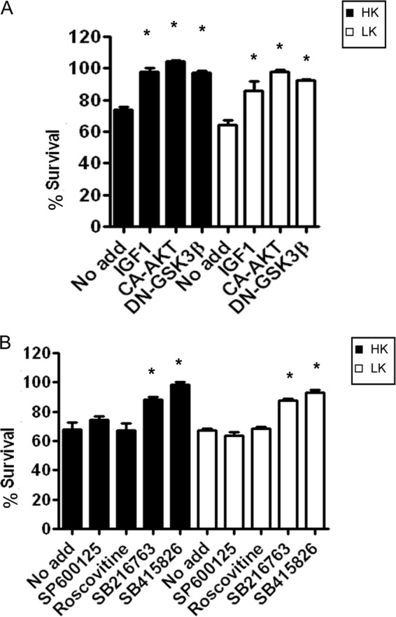
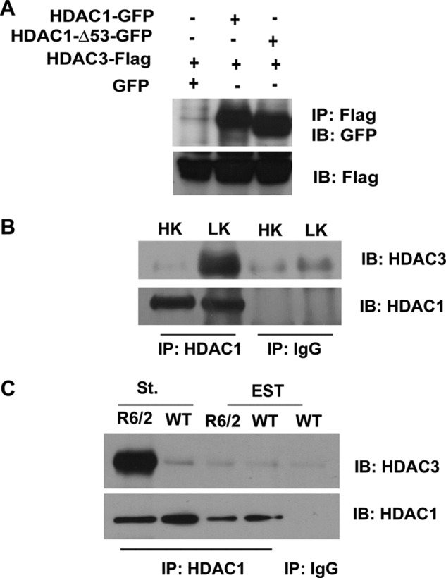
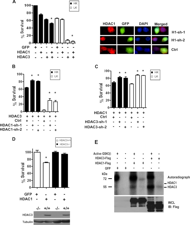
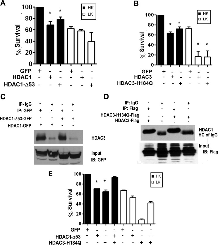
Both neuroprotective and neurotoxic roles have previously been described for histone deacetylase-1 (HDAC1). Here we report that HDAC1 expression is elevated in vulnerable brain regions of two mouse models of neurodegeneration, the R6/2 model of Huntington disease and the Ca(2+)/calmodulin-dependent protein kinase (CaMK)/p25 double-transgenic model of tauopathic degeneration, suggesting a role in promoting neuronal death. Indeed, elevating HDAC1 expression by ectopic expression promotes the death of otherwise healthy cerebellar granule neurons and cortical neurons in culture. The neurotoxic effect of HDAC1 requires interaction and cooperation with HDAC3, which has previously been shown to selectively induce the death of neurons. HDAC1-HDAC3 interaction is greatly elevated under conditions of neurodegeneration both in vitro and in vivo. Furthermore, the knockdown of HDAC3 suppresses HDAC1-induced neurotoxicity, and the knockdown of HDAC1 suppresses HDAC3 neurotoxicity. As described previously for HDAC3, the neurotoxic effect of HDAC1 is inhibited by treatment with IGF-1, the expression of Akt, or the inhibition of glycogen synthase kinase 3β (GSK3β). In addition to HDAC3, HDAC1 has been shown to interact with histone deacetylase-related protein (HDRP), a truncated form of HDAC9, whose expression is down-regulated during neuronal death. In contrast to HDAC3, the interaction between HDRP and HDAC1 protects neurons from death, an effect involving acquisition of the deacetylase activity of HDAC1 by HDRP. We find that elevated HDRP inhibits HDAC1-HDAC3 interaction and prevents the neurotoxic effect of either of these two proteins. Together, our results suggest that HDAC1 is a molecular switch between neuronal survival and death. Its interaction with HDRP promotes neuronal survival, whereas interaction with HDAC3 results in neuronal death.


Figures

FIGURE 1.
FIGURE 1.
Induction of HDAC1 expression in vulnerable brain region in mouse models of neurodegenerative disease. A, Western blot analysis of lysates from the striatum (St.) or extra-striatal tissue (EST) of R6/2 mice at 6, 9, and 13 weeks were analyzed for expression of HDAC1 or tubulin as loading control. B, in the CaMK/p25 transgenic line, p25-GFP expression is driven through a doxycycline-inducible CaMK promoter. Left panel, expression of p25-GFP transgene is observed only when its expression is induced and in the cortex and hippocampus where the CaMK promoter is active. Right panel, HDAC1 expression is elevated in cortex and hippocampus, but not in cerebellum. Lysates for both panels were from mice in which transgene expression was induced for 8 weeks at which time neurodegeneration is obvious in the cortex and hippocampus (our observations and Ref. 23). Tg-ON, transgenic mice taken off the doxycycline-containing food; Tg-OFF, transgenic mice kept on the food. C, lysates from CGN cultures treated with either HK or LK for 6, 9, 12, or 15 h were subjected to Western blotting and probed for HDAC1. The blot was reprobed with tubulin to show equal loading. The lack of alterations in HDAC1 expression was confirmed in multiple experiments. D, cultured cortical neurons were either left untreated or treated with HCA for 3, 6, 9, 12, or 15 h, and the lysates were subjected to Western blotting and probed for HDAC1. The blot was reprobed with tubulin to show equal loading. The absence of major alterations in HDAC1 expression was confirmed in additional experiments.
FIGURE 2.
FIGURE 2.
Elevated HDAC1 is toxic to neurons. A, CGNs were transfected with either GFP or HDAC1 and then switched to HK or LK medium 8 h later. Viability of transfected neurons was assessed after 24 h. Viability is normalized to cultures transfected with GFP and treated with HK. B, cortical neurons were transfected with GFP or HDAC1 for 6 h and were either left untreated (Unt) or treated with HCA. Viability of transfected neurons was assessed after 14 h and is normalized to GFP-transfected untreated cultures. Error bars indicate S.D.
FIGURE 3.
FIGURE 3.
HDAC1 toxicity can be prevented by inhibiting GSK3β or by activating the PI3K-AKT pathway. A, CGNs were transfected with HDAC1 and treated with HK or LK medium 8 h later. The cultures were either left with no additives (No add) or treated with IGF-1, or co-transfected with CA-Akt or DN-GSK3β. Viability was assessed 24 h later and normalized to cultures transfected with GFP and treated with HK. B, CGNs were transfected with HDAC1 and treated with HK or LK medium 8 h later. The cultures were either left with no additives or supplemented with JNK inhibitor (SP600125), CDK inhibitor (roscovitine), or two different GSK3β inhibitors (SB216763 and SB415826). Viability was assessed 24 h later and normalized to cultures transfected with GFP and treated with HK. Error bars indicate S.D.
FIGURE 4.
FIGURE 4.
Full-length HDAC1 and HDAC1-Δ53 interact with HDAC3. A, HEK293 cells were co-transfected with FLAG-tagged HDAC3 and GFP, HDAC1, or HDAC1-Δ53 for 24 h, after which lysates were collected and immunoprecipitation (IP) was performed using FLAG antibody. Lysates were subjected to Western blot (IB) analysis and probed with GFP antibody. The same membrane was then reprobed with FLAG antibody to show the immunoprecipitated HDAC3 protein. B, CGNs were treated with either HK or LK for 6 h. Lysates were collected, and immunoprecipitation was performed using either HDAC1 or IgG antibody as negative control. The immunoprecipitate was subjected to Western blot analysis and probed with HDAC3 antibody. The membrane was reprobed with HDAC1 antibody to show pulldown of HDAC1. C, lysates from the striatum (St.) and extra-striatal tissue (EST) of R6/2 mice or their wild-type (WT) littermates were used to immunoprecipitate HDAC1 and were then run on a Western gel and probed with HDAC3 antibody. The membrane was reprobed with HDAC1 to show pulldown of the endogenous protein.
FIGURE 5.
FIGURE 5.
HDAC1 and HDAC3 cooperate to produce toxicity. A, CGNs transfected with GFP, HDAC1, HDAC3, or co-transfected with HDAC1 and HDAC3 were treated with HK or LK medium for 24 h. The viability of cells was assessed after immunocytochemistry and normalized to GFP-transfected cultures in HK. The panel on the right shows knockdown of HDAC1 by the two shRNAs in CGNs co-transfected with GFP and either control (Ctrl) or one of two HDAC1 shRNAs (H1-sh-1 or H1-sh-2). Cells expressing shRNA are identified based on GFP staining and knockdown of endogenous HDAC1evaluated using an HDAC1 antibody. B, CGNs were co-transfected with HDAC3 and either control (Ctrl) or one of two HDAC1 shRNAs (HDAC1-sh1 or HDAC1-sh2) and then treated with HK or LK medium for 24 h, after which cells were fixed, subjected to immunocytochemistry, and assessed for viability. Viability was normalized to GFP-transfected cultures in HK. C, CGNs were co-transfected with HDAC1 and either control (Ctrl) or one of two HDAC3 shRNAs (HDAC3-sh1 or HDAC3-sh2) for 48 h and then treated with HK or LK medium for 24 h after which cells were fixed, subjected to immunocytochemistry, and assessed for viability. D, upper panel, cortical neurons cultured from Nes-Cre Hdac3−/− mice or from their wild-type littermates were transfected with either GFP or HDAC1, and viability was assessed after 20 h. Lower panel, whole brain lysates from Hdac3−/− conditional KO mice (−/−) and wild-type littermates (+/+) were analyzed by Western blot using an HDAC3 antibody. The membrane was reprobed with tubulin. Error bars in panels A–D indicate S.D. E, lysates from HEK293 cells transfected with GFP or FLAG-tagged HDAC1 or HDAC3 were immunoprecipitated using either GFP or FLAG antibodies. The immunoprecipitated proteins were used in an in vitro kinase assay with or without active GSK3β as indicated. Autoradiograph shows a phosphorylated band in the HDAC3 lane, but no band is discernible for HDAC1. WCL, whole cell lysate; IB, immunoblot.
FIGURE 6.
FIGURE 6.
Deacetylase activity of either HDAC1 or HDAC3 Is sufficient for neurotoxicity. A, CGNs transfected with GFP, HDAC1, or HDAC1-Δ53 were treated with HK or LK medium after 8 h. Viability was assessed 24 h later. B, CGNs transfected with GFP, HDAC3, or HDAC3-H134Q were treated with HK or LK medium 8 h later for 24 h, and viability was assessed. C, HEK293 cells were transfected with either GFP-tagged HDAC1 or HDAC1-Δ53. The proteins were immunoprecipitated (IP) using either GFP or IgG antibody and analyzed by Western blotting (IB) with HDAC3 antibody. Both HDAC1 and HDAC1-Δ53 pull down endogenous HDAC3. The lower panel shows the input probed with GFP antibody showing overexpressed HDAC1 proteins. D, lysates from HEK293 cells transfected with FLAG-tagged HDAC3 or HDAC3-H134Q were immunoprecipitated with either FLAG or IgG antibody and analyzed by Western blotting with HDAC1 antibody. Both HDAC3 and HDAC3-H134Q pull down HDAC1. The lower panel shows the input probed with FLAG antibody showing overexpressed HDAC3 proteins. E, CGNs were transfected with GFP, HDAC1-Δ53, or HDAC3-H134Q or co-transfected with HDAC1-Δ53 and HDAC3-H134Q and treated with HK or LK medium after 8 h for 24 h, after which viability was quantified. Error bars in panels A, B, and E indicate S.D.
FIGURE 7.
FIGURE 7.
HDRP competes with HDAC3 for binding to HDAC1 and can reduce HDAC1 and HDAC3 toxicity. A, HEK293 cells were transfected with either GFP or HDRP-FLAG for 36 h. Endogenous HDAC3 was immunoprecipitated, and the immunoprecipitate (IP) was subjected to Western blotting and probed with HDAC1 antibody. The membrane was reprobed with HDAC3 antibody to show equal pulldown. The lower two panels show input lanes with overexpressed HDRP and equal endogenous HDAC1 expression. WCL, whole cell lysate. B, HEK293 cells were co-transfected with HDRP-FLAG and GFP, HDAC3-FLAG, HDAC1-Δ53-GFP, or HDAC1-GFP for 36 h. Immunoprecipitation was carried out using either GFP or HDAC3 antibody, and the immunoprecipitates were subjected to Western blot analysis and probed with FLAG antibody. The membrane was reprobed with GFP to show pulldown of the HDAC1 proteins. The bottom panel shows pulldown of FLAG-tagged HDAC3. C, CGNs were transfected with HDAC1, HDAC3, HDAC1-Δ53, or HDRP or co-transfected HDRP with HDAC1, HDAC3, or HDAC1-Δ53. The cultures were switched to HK or LK medium 8 h later, and viability was assessed 24 h later. Error bars indicate S.D.

Similar articles

Cited by

References

    1. Yang X. J., Seto E. (2008) The Rpd3/Hda1 family of lysine deacetylases: from bacteria and yeast to mice and men. Nat. Rev. Mol. Cell Biol. 9, 206–218 - PMC - PubMed
    1. Haberland M., Montgomery R. L., Olson E. N. (2009) The many roles of histone deacetylases in development and physiology: implications for disease and therapy. Nat. Rev. Genet. 10, 32–42 - PMC - PubMed
    1. D'Mello S. R. (2009) Histone deacetylases as targets for the treatment of human neurodegenerative diseases. Drug News Perspect. 22, 513–524 - PMC - PubMed
    1. Kazantsev A. G., Thompson L. M. (2008) Therapeutic application of histone deacetylase inhibitors for central nervous system disorders. Nat. Rev. Drug Discov. 7, 854–868 - PubMed
    1. Sleiman S. F., Basso M., Mahishi L., Kozikowski A. P., Donohoe M. E., Langley B., Ratan R. R. (2009) Putting the “HAT” back on survival signaling: the promises and challenges of HDAC inhibition in the treatment of neurological conditions. Expert Opin. Investig. Drugs 18, 573–584 - PMC - PubMed

Publication types

MeSH terms