

CD160 and PD-1 co-expression on HIV-specific CD8 T cells defines a subset with advanced dysfunction
- PMID: 22916009
- PMCID: PMC3420930
- DOI: 10.1371/journal.ppat.1002840
CD160 and PD-1 co-expression on HIV-specific CD8 T cells defines a subset with advanced dysfunction
Abstract
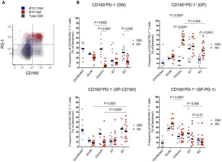
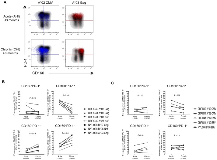
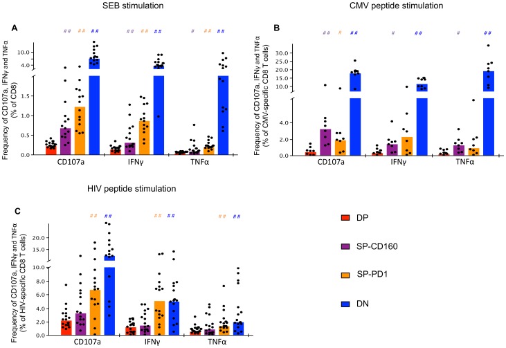
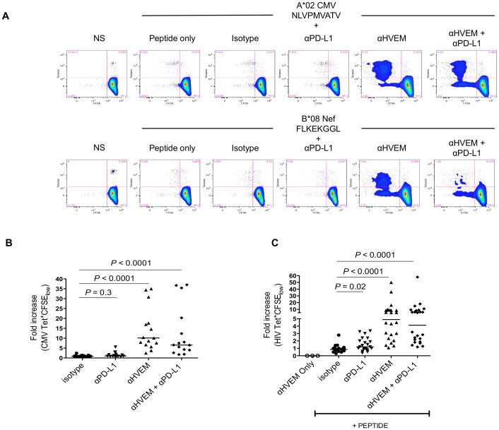
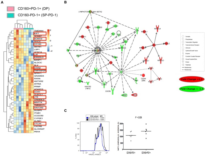
Chronic viral infections lead to persistent CD8 T cell activation and functional exhaustion. Expression of programmed cell death-1 (PD-1) has been associated to CD8 T cell dysfunction in HIV infection. Herein we report that another negative regulator of T cell activation, CD160, was also upregulated on HIV-specific CD8 T lymphocytes mostly during the chronic phase of infection. CD8 T cells that expressed CD160 or PD-1 were still functional whereas co-expression of CD160 and PD-1 on CD8 T cells defined a novel subset with all the characteristics of functionally exhausted T cells. Blocking the interaction of CD160 with HVEM, its natural ligand, increased HIV-specific CD8 T cell proliferation and cytokine production. Transcriptional profiling showed that CD160(-)PD-1(+)CD8 T cells encompassed a subset of CD8(+) T cells with activated transcriptional programs, while CD160(+)PD-1(+) T cells encompassed primarily CD8(+) T cells with an exhausted phenotype. The transcriptional profile of CD160(+)PD-1(+) T cells showed the downregulation of the NFκB transcriptional node and the upregulation of several inhibitors of T cell survival and function. Overall, we show that CD160 and PD-1 expressing subsets allow differentiating between activated and exhausted CD8 T cells further reinforcing the notion that restoration of function will require multipronged approaches that target several negative regulators.
Conflict of interest statement
The authors have declared that no competing interests exist.
Figures
Similar articles
-
CD160 Plays a Protective Role During Chronic Infection by Enhancing Both Functionalities and Proliferative Capacity of CD8+ T Cells.Front Immunol. 2020 Sep 11;11:2188. doi: 10.3389/fimmu.2020.02188. eCollection 2020. Front Immunol. 2020. PMID: 33072082 Free PMC article.
-
CD160 isoforms and regulation of CD4 and CD8 T-cell responses.J Transl Med. 2014 Sep 2;12:217. doi: 10.1186/s12967-014-0217-y. J Transl Med. 2014. PMID: 25179432 Free PMC article.
-
Antibodies targeting BTLA or TIM-3 enhance HIV-1 specific T cell responses in combination with PD-1 blockade.Clin Immunol. 2017 Oct;183:167-173. doi: 10.1016/j.clim.2017.09.002. Epub 2017 Sep 4. Clin Immunol. 2017. PMID: 28882621
-
Coinhibitory receptors and CD8 T cell exhaustion in chronic infections.Curr Opin HIV AIDS. 2014 Sep;9(5):439-45. doi: 10.1097/COH.0000000000000088. Curr Opin HIV AIDS. 2014. PMID: 25010894 Review.
-
The CD160, BTLA, LIGHT/HVEM pathway: a bidirectional switch regulating T-cell activation.Immunol Rev. 2009 May;229(1):244-58. doi: 10.1111/j.1600-065X.2009.00783.x. Immunol Rev. 2009. PMID: 19426226 Review.
Cited by
-
PD-1 coinhibitory signals: the link between pathogenesis and protection.Semin Immunol. 2013 Oct 31;25(3):219-27. doi: 10.1016/j.smim.2013.02.002. Epub 2013 Mar 31. Semin Immunol. 2013. PMID: 23548749 Free PMC article. Review.
-
TIM-3 does not act as a receptor for galectin-9.PLoS Pathog. 2013 Mar;9(3):e1003253. doi: 10.1371/journal.ppat.1003253. Epub 2013 Mar 21. PLoS Pathog. 2013. PMID: 23555261 Free PMC article.
-
HIV-specific CD8+ T cells from elite controllers are primed for survival.J Virol. 2013 May;87(9):5170-81. doi: 10.1128/JVI.02379-12. Epub 2013 Feb 28. J Virol. 2013. PMID: 23449791 Free PMC article.
-
CD160 Plays a Protective Role During Chronic Infection by Enhancing Both Functionalities and Proliferative Capacity of CD8+ T Cells.Front Immunol. 2020 Sep 11;11:2188. doi: 10.3389/fimmu.2020.02188. eCollection 2020. Front Immunol. 2020. PMID: 33072082 Free PMC article.
-
Patients with HIV-associated cancers have evidence of increased T cell dysfunction and exhaustion prior to cancer diagnosis.J Immunother Cancer. 2022 Apr;10(4):e004564. doi: 10.1136/jitc-2022-004564. J Immunother Cancer. 2022. PMID: 35470232 Free PMC article.
References
-
- Saksena NK, Wu JQ, Potter SJ, Wilkinson J, Wang B (2008) Human immunodeficiency virus interactions with CD8+ T lymphocytes. Curr HIV Res 6: 1–9. - PubMed
-
- Gougeon ML, Montagnier L (1999) Programmed cell death as a mechanism of CD4 and CD8 T cell deletion in AIDS. Molecular control and effect of highly active anti-retroviral therapy. Ann N Y Acad Sci 887: 199–212. - PubMed
-
- Trautmann L, Said EA, Halwani R, Janbazian L, Chomont N, et al. (2007) Programmed death 1: a critical regulator of T-cell function and a strong target for immunotherapies for chronic viral infections. Curr Opin HIV AIDS 2: 219–227. - PubMed
Publication types
MeSH terms
Substances
Grants and funding
LinkOut - more resources
Full Text Sources
Other Literature Sources
Medical
Research Materials
