. 2012 Oct;18(10):1862-74.
doi: 10.1261/rna.034520.112. Epub 2012 Aug 21.

Mechanistic characterization of the 5'-triphosphate-dependent activation of PKR: lack of 5'-end nucleobase specificity, evidence for a distinct triphosphate binding site, and a critical role for the dsRBD

Affiliations

Mechanistic characterization of the 5'-triphosphate-dependent activation of PKR: lack of 5'-end nucleobase specificity, evidence for a distinct triphosphate binding site, and a critical role for the dsRBD

Rebecca Toroney et al. RNA. 2012 Oct.

Abstract

The protein kinase PKR is activated by RNA to phosphorylate eIF-2α, inhibiting translation initiation. Long dsRNA activates PKR via interactions with the dsRNA-binding domain (dsRBD). Weakly structured RNA also activates PKR and does so in a 5'-triphosphate (ppp)-dependent fashion, however relatively little is known about this pathway. We used a mutant T7 RNA polymerase to incorporate all four triphosphate-containing nucleotides into the first position of a largely single-stranded RNA and found absence of selectivity, in that all four transcripts activate PKR. Recognition of 5'-triphosphate, but not the nucleobase at the 5'-most position, makes this RNA-mediated innate immune response sensitive to a broad array of viruses. PKR was neither activated in the presence of γ-GTP nor recognized NTPs other than ATP in activation competition and ITC binding assays. This indicates that the binding site for ATP is selective, which contrasts with the site for the 5' end of ppp-ssRNA. Activation experiments reveal that short dsRNAs compete with 5'-triphosphate RNAs and heparin for activation, and likewise gel-shift assays reveal that activating 5'-triphosphate RNAs and heparin compete with short dsRNAs for binding to PKR's dsRBD. The dsRBD thus plays a critical role in the activation of PKR by ppp-ssRNA and even heparin. At the same time, cross-linking experiments indicate that ppp-ssRNA interacts with PKR outside of the dsRBD as well. Overall, 5'-triphosphate-containing, weakly structured RNAs activate PKR via interactions with both the dsRBD and a distinct triphosphate binding site that lacks 5'-nucleobase specificity, allowing the innate immune response to provide broad-spectrum protection from pathogens.


Figures

FIGURE 1.
FIGURE 1.
Protein and RNA constructs used in this study. (A) Schematic of PKR primary sequence. The N-terminal dsRNA binding domain (dsRBD, also referred to as P20), composed of two tandem dsRNA-binding motifs (dsRBM 1 and 2), and the catalytic C-terminal kinase domain are indicated. The positions of point mutations used in this study are indicated. The double-mutant PKR (dmPKR) contains both K60A and K150A mutations, and the K296R mutation in the kinase domain renders PKR catalytically inactive. (B) Experimentally determined secondary structure of ssRNA-47 (Nallagatla et al. 2007). The starting nucleotide is indicated in boldface; G is shown, which is typical of transcripts made from WT T7 polymerase, but RNAs were also transcribed containing pppA, pppC, and pppU using mutant T7 polymerase. (C) Sequence of ssRNA-20. Secondary structure prediction via free energy minimization (mFold) indicates that this RNA is essentially completely unstructured. (D) Experimentally determined secondary structure of ss-dsRNA (9,11) (Zheng and Bevilacqua 2004).
FIGURE 2.
FIGURE 2.
All ssRNA-47 5′-triphosphate-starting nucleotides activate PKR. (A) Verification of starting nucleotide identity. RNA was transcribed from DNA templates coding for starting the transcript with pppG, pppA, pppC, and pppU, in the presence of [γ-32P]ATP or [γ-32P]GTP. The expected starting RNA nucleotide based on template (nt. 1: pppX) is indicated. Percent incorporation was calculated by normalizing the counts at the indicated mobility “ssRNA-47” to the counts from the pppA lane for [γ-32P]ATP incorporation, or the pppG lane for [γ-32P]GTP incorporation. 7 M urea gel is shown. (B) Activation of PKR by ssRNA-47 with pppG, pppA, pppU, and pppC. RNA concentrations were 0.31, 0.63, 1.3, 2.5, 5.0, and 10 μM for pppG- and pppA-ssRNA-47; 0.15, 0.31, 0.63, 1.3, 2.5, 5.0, and 10 μM for pppU-ssRNA-47; and 0.15, 0.31, 0.63, 1.3, 2.5, and 5.0 μM for pppC-ssRNA-47. Phosphorylation activities are provided under the gels and were normalized to the dsRNA-79 lane in the top gel. (C) Graphical representation of phosphorylation activities from panel B as a function of RNA concentration. (D) Activation of PKR by A-ssRNA-47 and G-ssRNA-47 are both dependent on a 5′-triphosphate. RNAs starting with 5′-triphosphate were generated by in vitro T7 transcription, while RNAs starting with 5′-OH-G and 5′-OH-A were chemically synthesized. Phosphorylation activities are provided under the gel and were normalized to the dsRNA-79 lane. For both panels B and D, 10% SDS-PAGE gels are shown, with the position of phosphorylated PKR (p-PKR) indicated.
FIGURE 3.
FIGURE 3.
The ATP binding site has high specificity: activation assays. (A) Only ATP supports PKR phosphorylation. Activation of PKR by dsRNA-79 using [γ-32P]GTP or [γ-32P]ATP as the phosphate source. RNA concentrations are provided. In the “γ-GTP” lanes, activation assays were performed as per standard assay conditions (see Materials and Methods) with the following exceptions: 100 μM GTP was used instead of ATP, and 0.1 or 1.5 μCi/μL [γ-32P]GTP was added instead of 1.5 μCi/μL [γ-32P]ATP. In the “γ-ATP” lanes, standard assay conditions were used. (B) NTP-competition assays reveal that only free ATP competes with ATP for activation. PKR activation by dsRNA-79 or pppG-ssRNA-47 was assayed in the presence of increasing concentrations of unlabeled ATP, GTP, CTP, and UTP. The concentrations of dsRNA-79 and pppG-ssRNA-47 were 0.1 and 2.5 μM, respectively. The concentrations of each NTP were 0.1, 0.5, 1, and 2 mM. (All of these are added concentrations and are in the background of 100 μM ATP.) The “no-RNA” and “no-competitor-NTP” lanes are included as negative and positive controls, respectively. Phosphorylation activities were normalized to the “no-competitor-NTP” lane in the middle gel. For both A and B, a 10% SDS-PAGE gel is shown, with the position of phosphorylated PKR (p-PKR) indicated. (C) Graphical representation of phosphorylation activities from panel B.
FIGURE 4.
FIGURE 4.
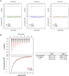
The ATP binding site has high affinity: ITC experiments. ITC titration curves for NTP binding to K296R. (A) GTP (green trace), UTP (blue trace), and CTP (orange trace) binding to K296R. Titration of each NTP into buffer is included (black traces). NTP traces are offset from buffer traces by ∼0.1 μcal/sec in raw data (upper panels) for clarity. Legends are provided in plots. (B) ATP binding to K296R (red trace). Titration of ATP into buffer is included (black trace). ATP titration curve is fitted to a two-site binding model (see Materials and Methods), and thermodynamic parameters are provided to the right of the plot. The major contribution is with site 1 (n = 1.32 ± 0.09) and a Kd of 19 ± 1.8 μM. Note that uncertainty in ΔH is due to the lack of a good lower baseline, which is common with micromolar Kd experiments. However, this does not affect the Kd value because that is mainly based on the slope of the transition region.
FIGURE 5.
FIGURE 5.
PKR activation by ppp-ssRNA and heparin is inhibited by dsRNA, but not by short ppp-ssRNA. RNA-competition assays. (A) Competition for dsRNA-79-mediated PKR activation by short dsRNA and ssRNA. PKR activation by dsRNA-79 was assayed in the presence of increasing concentrations dsRNA-20 and ppp-ssRNA-20. The concentration of dsRNA-79 was held constant at 0.1 μM, and concentrations of dsRNA-20 and ppp-ssRNA-20 were 0.1, 1, 2.5, and 5 μM. (B) Competition for ppp-ssRNA-47-mediated PKR activation by short dsRNA and ssRNA. PKR activation was assayed as in A, with the concentration of ssRNA-47 held constant at 2.5 μM. (C) Competition for heparin-mediated PKR activation by short dsRNA and ssRNA. PKR activation was assayed as in A, with the concentration of heparin (average molecular weight of 6 kDa) held constant at 75 μg/mL, and concentrations of competitor RNAs at 0.1, 1, 2.5, 5, and 10 μM. For all panels, 10% SDS-PAGE gels are shown, with the position of phosphorylated PKR (p-PKR) indicated. The “no-RNA” and “no-competitor” lanes are included as negative and positive controls, respectively. Activation in the absence of added RNA was negligible. Phosphorylation activities are provided under the gels and were normalized to the 0.1 μM competitor lane for each set of competitors; activities were normalized in this manner because in some cases the presence of competitor at low concentrations appeared to stimulate the reaction.
FIGURE 6.
FIGURE 6.
Photochemical cross-linking reveals interaction of ppp-ssRNA-47 with PKR regions outside the dsRBD. 4thioU-substituted ppp-ssRNA-47 was incubated with wild-type PKR, P20, or dmPKR and exposed to 365-nm light for 0, 10, 20, or 30 min and analyzed by 7% denaturing (7 M urea) PAGE. The positions of free RNA and cross-linked products are indicated. Protein dependence of cross-linked products was confirmed by the absence of products upon irradiating 4-thioU-ppp-ssRNA-47 in buffer alone (first three lanes).
FIGURE 7.
FIGURE 7.
Competition for P20 binding by dsRNA, ssRNA, and heparin by EMSAs. A trace amount of p*dsRNA-20 was incubated with P20 in the presence of various unlabeled competitors and analyzed by 10% native PAGE. Top strand (TS) dsRNA-20 was 5′-32P-labeled and annealed to excess unlabeled bottom strand (BS). Formation of annealed p*dsRNA-20 duplex was confirmed by the microshift of p*TS in the presence of BS (lane 2). A no-competitor mobility shift was detected upon addition of 3 μM P20, with slight formation of a second complex (lane 3). In the remaining lanes, 3 μM P20 bound to trace p*dsRNA-20 was challenged with either unlabeled RNA or heparin competitor. Competitor RNA concentrations were 5 and 10 μM, and heparin concentrations were 10, 100, 1000, and 2000 μg/mL. Mobility of p*dsRNA-20, free and bound to P20 complexes, is indicated.

Similar articles

Cited by

References

    1. Anderson BR, Muramatsu H, Jha BK, Silverman RH, Weissman D, Kariko K 2010. Nucleoside modifications in RNA limit activation of 2′-5′-oligoadenylate synthetase and increase resistance to cleavage by RNase L. Nucleic Acids Res 39: 9329–9338 - PMC - PubMed
    1. Anderson E, Pierre-Louis WS, Wong CJ, Lary JW, Cole JL 2011. Heparin activates PKR by inducing dimerization. J Mol Biol 413: 973–984 - PMC - PubMed
    1. Armstrong RN, Kondo H, Kaiser ET 1979. Cyclic AMP-dependent ATPase activity of bovine heart protein kinase. Proc Natl Acad Sci 76: 722–725 - PMC - PubMed
    1. Bevilacqua PC, Cech TR 1996. Minor-groove recognition of double-stranded RNA by the double-stranded RNA-binding domain from the RNA-activated protein kinase PKR. Biochemistry 35: 9983–9994 - PubMed
    1. Bieger CD, Nierlich DP 1989. Distribution of 5′-triphosphate termini on the mRNA of Escherichia coli. J Bacteriol 171: 141–147 - PMC - PubMed

Publication types

MeSH terms

LinkOut - more resources