. 2012 Oct 17;31(20):3976-90.
doi: 10.1038/emboj.2012.235. Epub 2012 Aug 21.

A new class of carriers that transport selective cargo from the trans Golgi network to the cell surface

Affiliations

A new class of carriers that transport selective cargo from the trans Golgi network to the cell surface

Yuichi Wakana et al. EMBO J. .

Abstract

We have isolated a membrane fraction enriched in a class of transport carriers that form at the trans Golgi network (TGN) and are destined for the cell surface in HeLa cells. Protein kinase D (PKD) is required for the biogenesis of these carriers that contain myosin II, Rab6a, Rab8a, and synaptotagmin II, as well as a number of secretory and plasma membrane-specific cargoes. Our findings reveal a requirement for myosin II in the migration of these transport carriers but not in their biogenesis per se. Based on the cargo secreted by these carriers we have named them CARTS for CARriers of the TGN to the cell Surface. Surprisingly, CARTS are distinct from the carriers that transport vesicular stomatitis virus (VSV)-G protein and collagen I from the TGN to the cell surface. Altogether, the identification of CARTS provides a valuable means to understand TGN to cell surface traffic.


Conflict of interest statement

The authors declare that they have no conflict of interest.

Figures
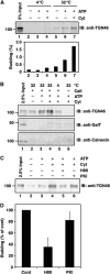
Figure 1
Figure 1
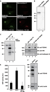
Biogenesis of the TGN46 containing transport carriers. (A, B) Permeabilized HeLa cells were incubated under conditions indicated. The reaction mixture was centrifuged at low speed (10 000 g for 10 min) and the supernatant was further centrifuged at high speed (100 000 g for 1 h). The high speed pellet was western blotted with antibodies against TGN46, GalT, and calnexin. The input in the figure refers to permeabilized HeLa cells used as starting material for these experiments. (C, D) The permeabilized HeLa cells were pretreated with H89 or PKI and then incubated with rat liver cytosol (Cyt) and an ATP regenerating system. The high speed pellet was western blotted with anti-TGN46. (D) Quantification of TGN46 in the high speed pellet. The average values of three independent experiments are shown (mean±s.d.). The control (Cont) refers to the permeabilized HeLa cells incubated with DMSO, ATP regenerating system and rat liver cytosol. Figure source data can be found with the Supplementary data.
Figure 2
Figure 2
Immunoisolation of the TGN46 containing transport carriers. (A) The anti-TGN46 antibody recognizes the cytoplasmic tail of TGN46. HeLa cells were permeabilized with digitonin, fixed with paraformaldehyde, incubated with or without Triton X-100 (TX-100), and then visualized with affinity purified anti-TGN46 and anti-mannosidase II antibodies. Bar, 10 μm. (B) HeLa cells were transfected with control (Cont) siRNA or siRNA oligos specific for TGN46. This procedure was repeated after 24 h. Seventy-two hours after the first transfection the cells were lysed and the lysates were western blotted with the anti-TGN46 antibody. (C) Permeabilized HeLa cells were incubated with an ATP regenerating system and rat liver cytosol at 32°C. The low speed supernatant of the reaction mixture was incubated with or without anti-TGN46 antibody and the immunoprecipitates were western blotted with anti-TGN46 antibody. An aliquot of the low speed supernatant was centrifuged at high speed (100 000 g for 1 h) and the pellet was analysed (Total transport carriers (TC)). The single and double stars denote immunoglobulin heavy chain and a non-specific polypeptide, respectively. (D) Permeabilized HeLa cells were incubated with an ATP regenerating system and rat liver cytosol at 4 or 32°C. Immunoprecipitates were western blotted with indicated antibodies. (E) Low speed supernatant of reaction mixture was prepared from HeLa-ssHRP cells and subjected to the immunoprecipitation in the presence or absence of TX-100. The HRP activity in immunoprecipitated materials was quantified by chemiluminescence. The average values of two experiments are shown (mean±s.d.). (F) Immunoisolated TGN46 containing membranes were released from the magnetic beads by incubation with 100 mM sodium carbonate (pH 11.5) and collected by high speed centrifugation (100 000 g for 1 h). The membranes were western blotted with indicated antibodies: single and double stars denote immunoglobulin heavy chain and a non-specific polypeptide, respectively. Figure source data can be found with the Supplementary data.
Figure 3
Figure 3
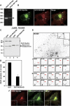
PKD is required for PAUF secretion. (A) PAUF cDNA was amplified from HeLa RNA by RT–PCR. Two sets of primers were designed to synthesize cDNA fragments of 378 and 401 bp, respectively, in length from mRNA. (B) PAUF-MycHis and GST-PKD2-KD were co-expressed in HeLa cells and visualized by fluorescence microscopy with anti-Myc and anti-GST antibodies, respectively. Arrowheads indicate tubules attached to the TGN. Bar, 10 μm. (C, D) PAUF-MycHis was co-expressed with GST (control) or GST-PKD2-KD in HeLa cells. The secretion of PAUF-MycHis was monitored by western blotting the cell lysate (lanes 1 and 2) and the medium (lanes 3 and 4) with anti-His antibody: stars denote a non-specific polypeptide. (D) Quantification of PAUF secretion. The average values of three independent experiments are shown (mean±s.d.). (E) Live-cell imaging of CARTS trafficking. HeLa cells expressing PAUF-mRFP were imaged at 440 ms intervals for ∼3 min. The kinetics of the disappearance of PAUF containing membranes in the areas marked I and II are shown in detail in the respective lower panels. The time (seconds) is indicated in each panel. Bars, 5 μm. G, Golgi. N, Nucleus. See also Supplementary Movie S1. (F) PAUF-MycHis and lysozyme C-3 × FLAG were co-expressed in HeLa cells and visualized by fluorescence microscopy with anti-Myc and anti-FLAG antibodies, respectively. High magnifications of small punctate elements are shown in the inset. Bars, 10 and 5 μm (inset). Figure source data can be found with the Supplementary data.
Figure 4
Figure 4
Ultrastructure of CARTS. (AD) HeLa cells were transfected with PAUF-MycHis and on the following day they were incubated at 20°C for 5 h in the presence of cycloheximide. The cells were shifted to 37°C to release PAUF-MycHis from the Golgi membranes and fixed at either 15 min (A) or 60 min (B, C). In a parallel incubation, cells were incubated with tannic acid post 20°C block to prevent fusion with the plasma membrane (B). Cells were then fixed and prepared for immuno-EM as described in Materials and methods. Arrows in all panels indicate CARTS labelled with the anti-Myc antibody; empty arrows show unlabelled clathrin-coated structures, and the sites of CARTS fusion with the plasma membrane are marked by arrowheads. (D) Quantitation of the number of CARTS and their corresponding size. Bar, 200 nm.
Figure 5
Figure 5
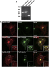
Syt II is contained in CARTS. (A) Syt II cDNA was amplified from HeLa RNA by RT–PCR. Three sets of primers were designed to synthesize cDNA fragments of 486, 499, and 227 bp, respectively, in length from mRNA. (B) Syt II-3 × FLAG was expressed alone (top row) or in combination with GST-PKD2-KD (middle row) or PAUF-MycHis (bottom row) in HeLa cells. The cells were double stained with anti-FLAG and anti-TGN46, anti-GST, or anti-Myc antibodies. The boxed areas are enlarged in the insets. Arrowheads indicate tubules attached to the TGN. Bars, 10 and 5 μm (inset).
Figure 6
Figure 6
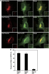
CARTS contain Rab6a and Rab8a. (A) HeLa cells were transfected with plasmids for PAUF-MycHis and Rab6a-GFP, Rab8a-GFP, or Rab11a-GFP and visualized by fluorescence microscopy. High magnifications of small punctate elements are shown in the inset. Bars, 10 and 5 μm (inset). (B) Quantification of co-localization of Rabs with CARTS. Rab6a-GFP: n=452 punctate elements in 6 cells, Rab8a-GFP: n=360 punctate elements in 5 cells, and Rab11a-GFP: n=364 punctate elements in 5 cells (mean±s.d.).
Figure 7
Figure 7
Myosin II is required for PAUF secretion. (A) Myosin IIa co-localization with PKD-KD at the TGN. HeLa cells co-expressing GFP-Myosin IIa and GST-PKD2-KD were visualized with fluorescence microscopy. Enlarged images of the perinuclear region (boxed) are shown in the lower panels. Arrowheads indicate the small punctate elements containing both GFP-Myosin IIa and GST-PKD2-KD. Bars, 10 μm. (B, C) HeLa cells were transfected with control (Cont) siRNA or siRNA oligos specific for myosin IIa. Forty-eight hours later, the cells were transfected with a plasmid for PAUF-MycHis. The secretion of PAUF-MycHis was monitored by western blotting the cell lysate and the medium with anti-His antibody. (D, E) The secretion of PAUF-MycHis from HeLa cells treated with DMSO (control) or 100 μM blebbistatin (Bleb) was monitored as described above. (C, E) Quantification of PAUF secretion. The amount of secreted PAUF was normalized with the expression level. The average values of three independent experiments are shown (mean±s.d.). Figure source data can be found with the Supplementary data.
Figure 8
Figure 8
Myosin II is not required for the biogenesis of CARTS. (A) The starting materials for the biogenesis of CARTS, permeabilized HeLa cells and rat liver cytosol, were western blotted with anti-myosin II antibody. (B, C) The reaction mixture for the biogenesis of CARTS was incubated with DMSO (control), 100 μM blebbistatin (Bleb), or 500 nM latrunculin A (Lat A). The high speed pellet containing CARTS was western blotted with anti-TGN46 antibody. (C) Quantification of CARTS formation. The average values of three independent experiments are shown (mean±s.d.). (D, E) HeLa cells expressing PAUF-MycHis were incubated at 20°C for 2 h in the presence of 20 μg/ml cycloheximide. After pretreatment with DMSO or 100 μM Bleb for 15 min, the cells were shifted to 32°C. After 1 h incubation at 32°C, the cells were fixed and stained with anti-Myc antibody. Bars, 10 μm. (E) Quantification of CARTS. The number of CARTS in DMSO-treated cells (n=15) and blebbistatin-treated cells (n=15) was counted. The average number of CARTS per cell is shown (mean±s.d.). Figure source data can be found with the Supplementary data.
Figure 9
Figure 9
CARTS exclude collagen I and VSV-G. (A) PAUF-MycHis and GFP-collagen I or PAUF-mRFP expressing HeLa cells were incubated at 20°C for 2 h in the presence of 20 μg/ml cycloheximide and then shifted to 32°C for 15 min. The cells were visualized with fluorescence microscopy. (B) PAUF-MycHis and VSV-G-GFP expressing HeLa cells were incubated at 40°C overnight, after which they were incubated at 20°C for 2 h in the presence of 20 μg/ml cycloheximide and then shifted to 32°C for 15 min. The cells were visualized with fluorescence microscopy. High magnifications of small punctate elements are shown in the inset. Bars, 10 and 5 μm (inset).

Comment in

Similar articles

Cited by

References

    1. Banting G, Ponnambalam S (1997) TGN38 and its orthologues: roles in post-TGN vesicle formation and maintenance of TGN morphology. Biochim Biophys Acta 1355: 209–217 - PubMed
    1. Bard F, Casano L, Mallabiabarrena A, Wallace E, Saito K, Kitayama H, Guizzunti G, Hu Y, Wendler F, Dasgupta R, Perrimon N, Malhotra V (2006) Functional genomics reveals genes involved in protein secretion and Golgi organization. Nature 439: 604–607 - PubMed
    1. Bard F, Malhotra V (2006) The formation of TGN-to-plasma-membrane transport carriers. Annu Rev Cell Dev Biol 22: 439–455 - PubMed
    1. Barlowe C, Orci L, Yeung T, Hosobuchi M, Hamamoto S, Salama N, Rexach MF, Ravazzola M, Amherdt M, Schekman R (1994) COPII: a membrane coat formed by Sec proteins that drive vesicle budding from the endoplasmic reticulum. Cell 77: 895–907 - PubMed
    1. Borner GH, Antrobus R, Hirst J, Bhumbra GS, Kozik P, Jackson LP, Sahlender DA, Robinson MS (2012) Multivariate proteomic profiling identifies novel accessory proteins of coated vesicles. J Cell Biol 197: 141–160 - PMC - PubMed

Publication types

MeSH terms

LinkOut - more resources