. 2012 Sep;15(9):1272-80.
doi: 10.1038/nn.3185. Epub 2012 Aug 19.

K-ATP channels in dopamine substantia nigra neurons control bursting and novelty-induced exploration

Affiliations

K-ATP channels in dopamine substantia nigra neurons control bursting and novelty-induced exploration

Julia Schiemann et al. Nat Neurosci. 2012 Sep.

Abstract

Phasic activation of the dopamine (DA) midbrain system in response to unexpected reward or novelty is critical for adaptive behavioral strategies. This activation of DA midbrain neurons occurs via a synaptically triggered switch from low-frequency background spiking to transient high-frequency burst firing. We found that, in medial DA neurons of the substantia nigra (SN), activity of ATP-sensitive potassium (K-ATP) channels enabled NMDA-mediated bursting in vitro as well as spontaneous in vivo burst firing in anesthetized mice. Cell-selective silencing of K-ATP channel activity in medial SN DA neurons revealed that their K-ATP channel-gated burst firing was crucial for novelty-dependent exploratory behavior. We also detected a transcriptional upregulation of K-ATP channel and NMDA receptor subunits, as well as high in vivo burst firing, in surviving SN DA neurons from Parkinson's disease patients, suggesting that burst-gating K-ATP channel function in DA neurons affects phenotypes in both disease and health.


Figures

Figure 1
Figure 1. In vivo firing characteristics and differences of burst properties of identified DA neurons in the SN and VTA.
(a) Left, in vivo single-unit recording of spontaneous activity of l-SN DA neuron and schematic spike train representation. Burst discharges as defined by 80/160-ms criterion are highlighted by green bars. Top right, corresponding interspike interval (ISI) histogram for >10 min of continuous activity. Inset, extracellularly recorded triphasic action potential (averaged waveform). The bursty-oscillatory pattern was reflected in the bimodal ISI histogram and by the prominent initial peak in the ACH (bottom right; gray lines indicate raw data and the black line represents the smoothed ACH fit; the green line is the GLO model fit; Supplementary Note). Bottom row, single-cell labeling, multi-immunofluorescence and confocal microscopy verified the DA phenotype and anatomical position of the recorded neuron in l-SN (dorsal tier, vertical line separates l-SN and m-SN). Shown is a neurobiotin-filled cell (green) expressing tyrosine hydroxylase (TH, blue). (b,c) In vivo single-unit recording of a neurobiotin-filled m-SN DA neuron with a bursty-irregular firing mode (b) and of a lateral VTA DA neuron with single spike-irregular mode (c). Data are presented as in a. (d) Mean firing frequencies for identified VTA, m-SN and l-SN DA neurons. (e) Relative distributions of four firing patterns (Supplementary Fig. 1) defined by ACH- and GLO-based classification. (f) Percentage of spikes fired in bursts. (g) Distinct region-selective burst properties. Bursts lasted longer in l-SN than m-SN and intraburst frequencies were faster in VTA DA neurons. *P < 0.05. For detailed in vivo properties and neurochemical subtypes, see Supplementary Table 1 and Supplementary Figure 1. Data are presented as mean ± s.e.m.; n is the number of identified neurons (d,f,g).
Figure 2
Figure 2. K-ATP channels selectively control in vivo burst firing in m-SN DA neurons.
(a) In vivo single-unit activity of m-SN DA neuron recorded in anesthetized wild-type (WT) mice. The bursty-oscillatory pattern was evident in the sample trace, raster plot, biphasic ISI histogram (inset, single action potential) and ACH (with GLO model fit, colors as in Fig. 1). Bottom, the recorded neuron was neurochemically identified by neurobiotin filling combined with tyrosine hydroxylase expression. (b) In vivo single-unit activity of a m-SN DA neuron in a Kir6.2−/− mouse displaying single spike-oscillatory firing. Data are presented as in a. See Supplementary Figure 3 for original recordings of l-SN and VTA DA neurons in wild-type and Kir6.2−/− mice. (c) Mean firing frequencies for m-SN and l-SN DA neurons in wild-type and Kir6.2−/− mice (in c,d, mean ± s.e.m.; n, number of identified neurons). (d) Spikes that fired in bursts were selectively reduced (3.3-fold) in m-SN DA neurons in Kir6.2−/− compared with wild type. **P < 0.01. (e) The decrease of burst activity in Kir6.2−/− m-SN DA neurons was confirmed by changes in ACH- and GLO-based classifications. Pattern distributions differed significantly between m-SN Kir6.2−/− and wild type (*P < 0.05). See Supplementary Table 2a for data and statistics. (f) Functional burst map. Wild-type (left) and Kir6.2−/− (right) DA neurons were plotted according to their position in the SN (bregma, −3.08 mm). Symbol size scales with %SFB. Note the clustering of non-bursting m-SN DA neurons in Kir6.2−/− compared with wild type.
Figure 3
Figure 3. Co-activation of K-ATP channels and NMDA receptors is sufficient to induce bursting in vitro.
(a) In vitro cell-attached recording (voltage clamp), raster plot and ACH of synaptically isolated wild-type m-SN DA neuron. Co-application of NMDA and NN414 (K-ATP-channel opener) enhanced spike rate and induced pacemaker-burst switch with 80/160-ms bursts (green bars). Whole-cell recording of same cell (current clamp) to monitor subthreshold membrane potentials. Note bursts on depolarizing waves separated by hyperpolarizations. Inset, burst in higher temporal resolution. (b) In vitro cell-attached recording of a Kir6.2−/− m-SN DA neuron (presented as in a). Co-application of NMDA and NN414 increased firing rate but not bursting. Note continuous single-spike oscillatory firing, in both on-cell and whole-cell recording of same cell. Brain atlas shows rostromedial SN (blue), where all in vitro recordings were performed. (c) Mean firing frequencies for m-SN DA neurons in wild type and Kir6.2−/−. NMDA and NN414 significantly enhanced in vitro frequencies (wild type, 2.4 ± 0.2 and 7.7 ± 0.3 Hz, P < 0.0001, t = 17.83, n = 8 recorded cells, N = 8 animals; Kir6.2−/−, 2.1 ± 0.3 and 6.1 ± 0.2 Hz, P < 0.0001, t = 8.72; in c,d, n = 8, N = 8; paired t tests, mean ± s.e.m.). ***P < 0.001. (d) %SFB was significantly increased after NMDA + NN414 in wild type (1.1 ± 0.4 and 54.5 ± 9.9%, P = 0.001, t = 5.29), but not in Kir6.2−/− (1.1 ± 0.6 and 6.4 ± 3.9%, P = 0.24, t = 1.29) m-SN DA cells. See Supplementary Figure 4a for reversible pharmacological induction of K-ATP-gated bursting. (e) K-ATP and NMDA receptor co-activation-mediated bursting in wild-type m-SN DA neurons was confirmed by respective changes in ACH-based classifications. Pattern distributions differed significantly for wild type (P < 0.001, χ2 = 16), but not Kir6.2−/− (P = 0.2, χ2 = 3.69; Pearson’s Chi-squared test).
Figure 4
Figure 4. K-ATP channel activation shifts maximal firing frequencies induced by current injection into the burst range in m-SN DA neurons in vitro.
(a) In vitro whole-cell patch-clamp recording (current clamp) of a m-SN DA wild-type neuron showing spike discharges in response to consecutive depolarizing current steps from −70 mV (increments of 50 pA, 2-s current pulse, 225 ms shown with time relative to stimulus onset). K-ATP channels were activated by the selective SUR1-K-ATP-channel opener NN414 (10 μM). Right, post initial-spike time histogram. Current steps of increasing amplitude triggered high-frequency, burst-like discharges. The gray line at 80 ms after the initial spike indicates the threshold for burst onset as defined in vivo. Note the high spike probability in this period. (b,c) Current-clamp recordings (presented as in a) of a wild-type m-SN DA neuron in the presence of 300 μM tolbutamide (b) and of a Kir6.2−/− m-SN DA neuron (c). Note the prolonged intervals in the absence of K-ATP channels. (d) Activation of K-ATP channels in vitro induced fast frequencies between the initial and the first somatodendritic spike. K-ATP-triggered burst-like discharges (19.3 ± 1.5 Hz, n = 7, N = 5) were clearly faster than the in vivo burst threshold (gray line at 12.5 Hz). In contrast, pharmacological or genetic inactivation of K-ATP channels using tolbutamide or Kir6.2−/− mice resulted in significantly slower discharge rates (11.4 ± 0.6 Hz, n = 8, N = 4; 11.6 ± 1.9 Hz, n = 8, N = 2; respectively; P = 0.001, F = 9.87, one-way ANOVA with Bonferroni post-tests). **P < 0.01. Data are presented as mean ± s.e.m., n, number of recorded neurons; N, number of mice.
Figure 5
Figure 5. Virus-mediated expression of dominant-negative Kir6.2 pore mutant induced selective functional silencing of K-ATP channels in SN DA neurons.
(a) rAAV2-coded HA-tagged dominant-negative (Kir6.2G132S-HA or Kir6.2_DN) and wild-type Kir6.2 subunits (Kir6.2_WT, for control) were used. (b,c) DAB (b) and fluorescent labeling (c) immunocytochemistry revealed selective and efficient expression of HA-tagged Kir6.2_DN subunits (white) across the entire m-SN and l-SN and in somatodendritic domains of single DA neurons (that is, tyrosine hydroxylase positive, blue). Inset in c highlights a selectively transduced tyrosine hydroxylase-positive neuron in the SNr. Right, confocal images showing an overlay of HA and tyrosine hydroxylase signals at a higher magnification. (d) Quantification of transduction efficiency (tyrosine hydroxylase and HA double-positive neurons) across the entire SN (Kir6.2_WT and Kir6.2_DN; n, total number of SN neurons counted, N = 6 mice). Nigral tyrosine hydroxylase-positive neurons expressed mutant and wild-type Kir6.2-HA constructs to a similar degree. In contrast, only a minor fraction of VTA DA neurons was transduced (Kir6.2_DN, 32.9% TH+ HA+, 57.6% TH+ HA, 9.5% TH HA+, n = 575; Kir6.2_WT, 34.9%, 38%, 27.1%; n = 939, N = 6 mice; data not shown and Supplementary Figs. 5 and 6). (e) Suppression of K-ATP-mediated currents by Kir6.2_DN. K-ATP washout currents were absent in vitro during whole-cell patch-clamp recordings of Kir6.2_DN-transduced SN DA cells. Kir6.2_WT expression did not affect K-ATP channel activation (steady-state washout currents: Kir6.2_DN, 0.5 ± 6.3 pA, n = 6 recorded neurons; Kir6.2_WT, 118.7 ± 39.7 pA, n = 4; P = 0.014 (U = 0.0), Mann-Whitney U test; mean ± s.e.m., N = 8 mice). Tyrosine hydroxylase and HA co-expression was confirmed by neurobiotin-filling and confocal analyses (data not shown).
Figure 6
Figure 6. Cell-selective silencing of K-ATP channels in SN DA neurons using virally mediated gene transfer is sufficient to prevent burst firing in m-SN neurons.
(a) In vivo single-unit activity of a m-SN DA neuron transduced with rAAV2 constructs expressing wild-type Kir6.2 subunits (Kir6.2_WT). Similar to m-SN DA wild-type cells (Figs. 1b and 2a), it fired in a bursty pattern as shown in raw trace, raster plot, ISI histogram (inset, single action potential) and ACH with GLO-model fit. Neurobiotin-labeling (green) of the recorded neuron identified transduction with HA-tagged Kir6.2_WT constructs (white) and coexpression of tyrosine hydroxylase (blue). (b) Selective silencing of postsynaptic K-ATP channels by expression of Kir6.2_DN in m-SN DA neurons prevented burst firing. Data are presented as in a. The neurobiotin-filled neuron coexpressed HA-tagged Kir6.2_DN and tyrosine hydroxylase and was located in the m-SN. Although mean firing frequencies were similar, the percentage of spikes fired in bursts was significantly reduced (2.4-fold, P = 0.033) in Kir6.2_DN compared with Kir6.2_WT (see Supplementary Table 3). (c) Decrease of burst activity in Kir6.2_DN m-SN DA neurons compared with Kir6.2_WT. ACH- and GLO-based pattern classification differed significantly between dominant negative and wild type (ACH, P = 0.041; GLO, P = 0.053). Data are presented as mean ± s.e.m. (n, number of identified neurons, see Supplementary Table 3). *P < 0.05. (d) Overlaid plot of mean average spike shapes of m-SN neurons expressing Kir6.2_WT (blue, s.e.m.) and Kir6.2_DN (green, s.e.m.). Note the prolonged action potential duration in Kir6.2_DN compared with Kir6.2_WT. For comparison, spike durations from wild-type and Kir6.2−/− mice are plotted (gray; P = 0.016, F = 2.6, one-way ANOVA with Bonferroni post test, **P < 0.01).
Figure 7
Figure 7. K-ATP channels in m-SN DA neurons are necessary for novelty-dependent exploratory behavior.
(a) Locomotion in novel open field (track length per min). Note the decreased locomotion (minutes 1–2) in Kir6.2−/− (856 ± 141 cm, N = 17 mice) compared with the increased locomotion in wild type (1,230 ± 45 cm, N = 20, P = 0.011, t = 2.69, F = 8.26). In contrast, no significant difference in cumulative locomotion was observed in minutes 3–20 (P = 0.27, t = 1.12, F = 7.4). All data given as mean ± s.e.m. and analyzed with unpaired t tests. *P < 0.05. (b) Rearings in the novel open field (n per minute). The numbers of rearings were significantly smaller in Kir6.2−/− (201 ± 15) than in wild type (290 ± 9, P < 0.0001, t = 5.16, F = 2.42). ***P < 0.001. (c) Immunohistochemical confirmation of SN-selective bilateral transduction of DA neurons in mice for open field testing (13-14 d after injection). HA-tagged Kir6.2_DN viral constructs (white) were expressed in most SN tyrosine hydroxylase-positive cells (blue). Expression was either targeted at the entire SN (m+l-SN, left) or restricted to l-SN (control, right). (d) Locomotion in novel open field using mice with bilateral K-ATP channel silencing across the whole SN or in just the l-SN. Note the absence of initially increased locomotion in Kir6.2_DN m+l-SN mice (minute 1-2, 934 ± 70 cm, N = 7 virus-injected mice) compared with l-SN controls (1,240 ± 118 cm, N = 6, P = 0.042, t = 2.3, F = 2.44). Significant difference in cumulative locomotion was also observed for minute 3-20 (P = 0.046, t = 2.26, F = 1.5). (e) Rearings in novel open field of mice where the entire or only l-SN was transduced with Kir6.2_DN. The numbers of rearings were significantly smaller in Kir6.2_DN m+l-SN (minute 1-20, 201 ± 12, N = 7) than in l-SN (280 ± 17, N = 6, P = 0.0028, t = 3.83, F = 1.76). **P < 0.01.
Figure 8
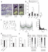
Figure 8. Increased mRNA levels of the K-ATP channel subunit SUR1 and high burst firing in SN DA neurons from Parkinson’s disease patients.
(a) Neuromelanin-positive SN neurons isolated via laser microdissection from cresyl violet-stained cryosections of human post mortem midbrain tissue (left). Individual neurons in control (middle) and Parkinson’s disease brain tissue (PD, right) before and after UV-LMD. (b) Selective transcriptional dysregulation of the K-ATP channel subunit SUR1 in SN DA neurons from human Parkinson’s disease midbrains. Cell-specific average mRNA levels of SUR1, calbindin and NR1 were significantly increased in Parkinson’s disease compared with control. mRNA levels of Kir6.2 or SUR2 were not different. *P < 0.05, ***P < 0.001. Data are presented as mean ± s.e.m. (N = number of brains, see Supplementary Table 4a). (c) Firing activity of a putative SN DA neuron recorded in a Parkinson’s disease patient presented as raster plot, ACH and ISI histogram. (d) Mean frequency and coefficient of variation of putative DA neurons recorded from Parkinson’s disease patients are plotted in comparison to SN DA neurons (n = 32, Fig. 1), awake rats (dark gray, dashed line) and awake monkeys (gray, dashed line). Mean frequencies were similar, but there was higher irregularity in neurons from Parkinson’s disease patients. Bars represent mean ± s.e.m. (n, number of single units). (e) High burst firing in human SN DA neurons of Parkinson’s disease patients. %SFB was 2.4-fold higher in Parkinson’s disease patients compared to rodents. (f) Dominance of bursty-oscillatory firing pattern in human Parkinson’s disease SN DA neurons revealed by ACH- and GLO-based classification (see Supplementary Table 4b).

Comment in

  • Bursting for exploration.
    da Silva JA, Costa RM. da Silva JA, et al. Nat Neurosci. 2012 Sep;15(9):1178-9. doi: 10.1038/nn.3198. Nat Neurosci. 2012. PMID: 22929910 No abstract available.

Similar articles

Cited by

References

    1. Bromberg-Martin ES, Matsumoto M, Hikosaka O. Dopamine in motivational control: rewarding, aversive and alerting. Neuron. 2010;68:815–834. - PMC - PubMed
    1. Schultz W. Multiple dopamine functions at different time courses. Annu. Rev. Neurosci. 2007;30:259–288. - PubMed
    1. Lammel S, et al. Unique properties of mesoprefrontal neurons within a dual mesocorticolimbic dopamine system. Neuron. 2008;57:760–773. - PubMed
    1. Lammel S, Ion DI, Roeper J, Malenka RC. Projection-specific modulation of dopamine neuron synapses by aversive and rewarding stimuli. Neuron. 2011;70:855–862. - PMC - PubMed
    1. Krebs RM, Heipertz D, Schuetze H, Duzel E. Novelty increases the mesolimbic functional connectivity of the substantia nigra/ventral tegmental area (SN/VTA) during reward anticipation: Evidence from high-resolution fMRI. Neuroimage. 2011;58:647–655. - PubMed

Publication types

MeSH terms

LinkOut - more resources