. 2012 Aug 14;22(2):250-62.
doi: 10.1016/j.ccr.2012.06.029.

Targeted disruption of heparan sulfate interaction with hepatocyte and vascular endothelial growth factors blocks normal and oncogenic signaling

Affiliations

Targeted disruption of heparan sulfate interaction with hepatocyte and vascular endothelial growth factors blocks normal and oncogenic signaling

Fabiola Cecchi et al. Cancer Cell. .

Abstract

Hepatocyte growth factor (HGF) and vascular endothelial cell growth factor (VEGF) regulate normal development and homeostasis and drive disease progression in many forms of cancer. Both proteins signal by binding to receptor tyrosine kinases and heparan sulfate (HS) proteoglycans on target cell surfaces. Basic residues comprising the primary HS binding sites on HGF and VEGF provide similar surface charge distributions without underlying structural similarity. Combining three acidic amino acid substitutions in these sites in the HGF isoform NK1 or the VEGF isoform VEGF165 transformed each into potent, selective competitive antagonists of their respective normal and oncogenic signaling pathways. Our findings illustrate the importance of HS in growth factor driven cancer progression and reveal an efficient strategy for therapeutic antagonist development.


Figures

Figure 1
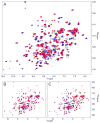
Figure 1. NMR analysis of NK1 proteins
1H-15N correlation spectra for substituted NK1 proteins (red) superimposed on NK1 WT spectra (blue): (A) NK1 3S (B) NK1 2S (C) NK1 1S. Spectra that are shifted in the substituted proteins are labeled in all panels. See also Figure S1 and Table S1.
Figure 2
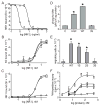
Figure 2. NK1 3S has reduced HS binding, normal Met binding, and fails to activate Met signaling
(A) Competitive displacement of Ru tagged NK1 WT from immobilized heparin by untagged NK1 WT (triangles) or 3S (circles). Saturation binding of Ru tagged NK1 3S (B) or NK1 WT (C) to Met ectodomain-Ig in the absence (triangles) or presence of HS oligomer (circles) or HS tetramer (squares). For A, B and C values are mean signal intensity (SI) +/− SD (n = 3). (D) Mean Met activation (phospho-Met [pM] per total protein [ug], n = 3) in 184B5 cells left untreated (“C”) or treated with HGF (1 nM), NK1 WT (5 nM), or NK1 3S (5 nM) for 20 min. Met content was equivalent in all samples. Asterisks indicate significant differences from control (p < 0.01). (E) Mean number of 184B5 cells (+/− SD; n = 3) migrating in response to treatment with NK1 WT or substituted NK1 proteins (7 nM each) in 24 h. Asterisks indicate significant differences from control (p < 0.01). (F) Mean DNA synthesis values (3H-thymidine incorporation; n = 3) in 184B5 cells after 16 h treatment with NK1 WT or substituted proteins at the indicated concentrations: WT (circles); 1S (triangles); 2S (squares); and 3S (inverted triangles). See also Figure S2 and Table S2.
Figure 3
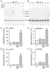
Figure 3. Long HS polymers promote NK1 clustering, Met activation and signaling
(A) Purified NK1 WT, or (B) NK1 3S proteins were incubated alone or with BS3 crosslinker in the absence or presence of heparin (Hep), HS oligomers (HSoligo), or HS tetramers (HStetra) before analysis by SDS-PAGE and immunoblotting. Arrows between the panels indicate the masses of NK1 monomer (24 kDa) and NK1 multimers. (C – F) The activation states of Met (mean phospho-Met/Met/total protein, n = 3; C), Akt (D), GSK3β (E) or p70S6K (F) in serum-deprived CHO 745 cells that were untreated (unfilled bars, “C”) or treated with NK1 WT (200 pM; gray bars) in the absence (−) or presence of HS tetramer (HStetra) or heparin (Hep). Values for panels D – F are mean signal intensity units +/− SD; n = 3. Asterisks indicate significant differences from control (p < 0.01).
Figure 4
Figure 4. NK1 3S is a potent antagonist of NK1 and HGF signaling
(A) Mean Met activation level (% maximum +/− SD, n = 3) in 184B5 cells treated with HGF (1 nM; circles) or NK1 WT (5 nM; squares) and with increasing concentrations of NK1 3S. HGF-stimulated cells were treated with PHA665752 (triangles) over the same dose range in parallel. (B) Mean DNA synthesis level (% maximum 3H-thymidine incorporation +/− SD; n = 3) in 184B5 cells treated with NK1 WT and NK1 3S (circles), or in cells treated with NK1 3S alone (squares) at the indicated doses. (C) Met-CD44 association in HT29 cells incubated with NK1 3S (5 nM), NK1 WT (5 nM) or HGF (1 nM) as indicated, in the presence of DTSSP prior to immunoprecipitation with anti-CD44, SDS-PAGE and immunoblotting with anti-Met (upper panel) or anti-CD44 (lower panel). (D) Met-CD44 association in PC3M cells treated with HGF (1 nM) and DTSSP in the absence or presence of NK1 3S or PHA665752 (PHA) at the indicated concentrations (nM) prior to immunoprecipitation with anti-CD44, SDS-PAGE and immunoblotting with anti-Met (upper panel) or anti-CD44 (lower panel). (E) Proliferation of U87 MG cells (mean cell number +/− SD, n = 3) expressing NK1 WT (squares), NK1 3S (triangles) or empty vector (circles). (F) NK1 3S antagonism of HGF-stimulated MDCK cell scatter. Left three panels: unstimulated control cells, or cells treated with HGF or NK1 3S at the indicated concentrations. Right three panels: HGF-stimulated cells treated with NK1 3S at the indicated concentrations. (G) Soft agar colony formation by U87 MG cells transfected with vector (control) or NK1 3S cDNA (3S plasmid), or control cells treated with NK1 3S protein or PHA665752 (PHA) at the indicated concentrations. See also Figure S3.
Figure 5
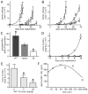
Figure 5. NK1 3S inhibits HGF-driven tumor growth, metastasis and Met kinase activation in vivo
(A) Mean tumor volume (+/− SD) in mice (n = 6/group) implanted with U87 MG transfectants expressing NK1 WT (squares), NK1 3S (triangles), or empty vector (circles) at the indicated days post-implantation. (B) Mean tumor volume (+/− SD) in mice (n = 6/group) implanted with clonal SK-LMS-1-derived cell lines expressing NK1 WT (squares) or NK1 3S (triangles), or empty vector (circles), at the indicated days post implantation. (C) Mean Met activation level (phospho-Met/Met/total protein +/− SD; n = 3) in SK-LMS-1 tumors derived from SK-LMS-1 cells expressing NK1 WT (black), empty vector (gray) and NK1 3S (unfilled). Asterisks indicate significant differences from vector control (p < 0.01). (D) Mean metastatic burden (total photon flux +/− SD) over time in mice (n = 10/group) injected via tail vein with B16-luc cells transfected with empty vector (circles) or NK1 3S (squares), or injected with empty vector cells and then treated on day 2 and thereafter with NK1 3S (50 mg/kg) by daily IP injection (triangles). (E) Mean metastatic burden (total photon flux +/− SD on day 27 post-implantation) in mice (n = 10/group) implanted subcutaneously with PC3M-luc cells and treated on day 5 and daily thereafter with NK1 3S protein by IP injection at 5 or 25 mg/kg, or treated with vehicle alone. (F) Mean plasma NK1 3S protein concentration (ng/ml, n = 2) in mice (n = 6) measured at the indicated times following a single IP injection of NK1 3S at 50 mg/kg. See also Figure S4.
Figure 6
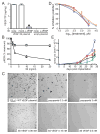
Figure 6. VEGF165 3S dimer binds KDR normally but does not signal
(A) Mean VEGF165 content (ng/mg total cell protein +/− SD; n = 3) in 24 h conditioned media prepared from 293/KDR cells transfected with empty vector (empty), VEGF165 WT (WT), or VEGF165 3S (3S). Asterisks indicate significant differences from vector control (p < 0.01). (B) VEGF165 3S (VEGF 3S; left) and VEGF165 WT (VEGF WT; middle) proteins in 24h conditioned media prepared from 293/KDR transfectants, and purified recombinant VEGF165 protein (VEGF 4 ng; right), after SDS-PAGE under non-reducing (NR) and reducing (R) conditions and immunoblotting with anti-VEGF. Migration of molecular mass standards (kDa) is indicated by arrows. (C) Saturation binding of KDR ectodomain-IgG fusion protein to VEGF165 WT (squares) or VEGF165 3S proteins (circles) in vitro. Values are mean KDR bound (ng/ml) +/− SD (n = 3). (D) Mean phospho-KDR level (signal intensity +/− SD; n = 3) in 293/KDR cells transfected with empty vector (empty; unfilled bar), VEGF165 WT (WT; light gray bar), VEGF165 3S (3S; dark gray bar), or empty vector cells treated with purified VEGF165 WT protein (2.5 nM) for 20 min (+VEGF; black bar). Asterisks indicate significant differences from empty vector control (p < 0.01). (E) Growth rate (mean cell number +/− SD, n = 3) of cultured 293/KDR cells transfected with empty vector (circles), VEGF165 WT (squares), or VEGF165 3S (triangles). (F) Soft agar colony formation by 239/KDR cells transfected with empty vector, (left), VEGF 3S (middle), or VEGF WT (right).
Figure 7
Figure 7. VEGF165 3S antagonizes VEGFR activation, colony formation and tumorigenesis
(A) VEGF165 content in media conditioned by 293/KDR cells transfected with VEGF165 3S (left) or empty plasmid (right), before (“none”) or after immunodepletion by anti-VEGF-A (“α-VEGF”) or an unrelated control antibody (“mock”), expressed as mean ng/mg total protein +/− SD (n = 3). Asterisk indicates significant difference from media prior to immunodepletion (p < 0.01). (B) Phospho-KDR levels (% maximum, +/− SD, n = 3) in serum-deprived 293/KDR cells treated with VEGF165 WT (10 ng/ml) in the presence of concentrated conditioned media from 293/KDR VEGF 3S transfectants that had been immunodepleted using a non-specific control antibody (triangles) or anti-VEGF (circles). The x-axis indicates VEGF165 3S concentration (ng/ml) in conditioned media before immunodepletion. (C) Soft agar colony formation by 239/KDR cells expressing VEGF165 WT (upper left panel), cells treated with the indicated concentrations of pazopanib (upper middle and right panels), or cells treated with media containing the indicated concentrations of VEGF165 3S (lower panels). (D) Dose-dependent inhibition of VEGF- or PlGF-induced Akt activation (phospho-Akt/total Akt) in EA.hy 926 cells. VEGF 3S blocked Akt activation by VEGF-A (dark blue circles) or PlGF (red squares) with potency similar to pazopanib (light blue triangles and yellow inverted triangles, respectively). (E) Mean tumor volume (mm3 +/− SD) in mice (n = 5/group) implanted with 293/KDR cells (3 × 10⁶ cells per animal) expressing VEGF165 3S (gray inverted triangles), VEGF165 WT (blue squares), or empty vector (black circles), at the indicated times post-implantation. Other groups were implanted with a mixture of empty vector cells and VEGF165 WT transfectants at 1.5 × 10⁶ cells each (red triangles), or a mixture of VEGF165 WT and VEGF165 3S transfectants at 1.5 × 10⁶ cells each (green diamonds). See also Figure S5.
Figure 8
Figure 8. VEGF165 3S antagonizes VEGF-driven tumor angiogenesis
Murine CD34 immunohistochemical analysis (low magnification above, higher magnification below) of tumors from animal groups as described in Figure 7. (A) 293/KDR/VEGF WT cell tumors; (B) 293/KDR/VEGF WT + control 293/KDR cell (1:1) tumors; (C) control 293/KDR cell tumors; (D) 293/KDR/VEGF WT + 293/KDR/VEGF 3S cell (1:1) tumors.

Similar articles

Cited by

References

    1. Appleton BA, Wu P, Maloney J, Yin J, Liang WC, Stawicki S, Mortara K, Bowman KK, Elliott JM, Desmarais W, Bazan JF, Bagri A, Tessier-Lavigne M, Koch AW, Wu Y, Watts RJ, Wiesmann C. Structural studies of neuropilin/antibody complexes provide insights into semaphorin and VEGF binding. EMBO Journal. 2007;26:4902–4912. - PMC - PubMed
    1. Backer MV, Gaynutdinov TI, Patel V, Bandyopadhyaya AK, Thirumamagal BT, Tjarks W, Barth RF, Claffey K, Backer JM. Vascular endothelial growth factor selectively targets boronated dendrimers to tumor vasculature. Molecular Cancer Therapeutics. 2005;4:1423–1429. - PubMed
    1. Boccaccio C, Comoglio PM. Invasive growth: a MET-driven genetic programme for cancer and stem cells. Nature Reviews Cancer. 2006;6:637–645. - PubMed
    1. Carmeliet P, Ng YS, Nuyens D, Theilmeier G, Brusselmans K, Cornelissen I, Ehler E, Kakkar VV, Stalmans I, Mattot V, et al. Impaired myocardial angiogenesis and ischemic cardiomyopathy in mice lacking the vascular endothelial growth factor isoforms VEGF164 and VEGF188. Nature Medicine. 1999;5:495–502. - PubMed
    1. Castagnino P, Lorenzi MV, Yeh J, Breckenridge D, Sakata H, Munz B, Werner S, Bottaro DP. Neu differentiation factor/heregulin induction by hepatocyte and keratinocyte growth factors. Oncogene. 2000;19:640–648. - PubMed

Publication types

MeSH terms

LinkOut - more resources