. 2012 Nov;86(21):11608-15.
doi: 10.1128/JVI.01260-12. Epub 2012 Aug 15.

Identification of the HIV-1 NC binding interface in Alix Bro1 reveals a role for RNA

Affiliations

Identification of the HIV-1 NC binding interface in Alix Bro1 reveals a role for RNA

Paola Sette et al. J Virol. 2012 Nov.

Abstract

HIV-1 recruits members of ESCRT, the cell membrane fission machinery that promotes virus exit. HIV-1 Gag protein gains access to ESCRT directly by binding Alix, an ESCRT-associated protein that promotes budding. The Alix Bro1 and V domains bind Gag NC and p6 regions, respectively. Whereas V-p6 binding and function are well characterized, residues in Bro1 that interact with NC and their functional contribution to Alix-mediated HIV-1 budding are unknown. We mapped Bro1 residues that constitute the NC binding interface and found that they are critical for function. Intriguingly, residues involved in interactions on both sides of the Bro1-NC interface are positively charged, suggesting the involvement of a negatively charged cellular factor serving as a bridge. Nuclease treatment eliminated Bro1-NC interactions, revealing the involvement of RNA. These findings establish a direct role for NC in mediating interactions with ESCRT necessary for virus release and report the first evidence of RNA involvement in such recruitments.


Figures

Fig 1
Fig 1
Mapping of Alix Bro1 residues involved in the interaction with HIV-1 and EIAV NC domains. (A and B) GST, GST-NCp6, and GST-NCp9 fusion proteins were expressed in E. coli, captured on glutathione-conjugated beads, and subsequently incubated with lysates from 293T cells expressing WT HA-tagged Bro1 domain or the indicated Bro1 mutants (Q8K, K2R, and Q8/K2R). Captured proteins and cell lysates were analyzed by SDS-PAGE and Western blotting. GST fusion proteins were visualized by Coomassie blue staining. (C) Mutations that compromise the NC binding interface in Bro1 do not affect Alix V domain interaction with HIV-1 p6. Pulldown assays were performed as described above, with the only difference being that full-length HA-tagged Alix WT and the indicated mutants were used instead of the isolated Bro1 domain.
Fig 2
Fig 2
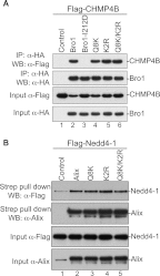
Alix Bro1 mutants retain binding to their natural cellular partners. (A) Alix Bro1 mutants bind CHMP4B. 293T cells were cotransfected with Flag-tagged CHMP4B alone (lane 1), in combination with HA-tagged WT Bro1 (lane 2), or with the indicated mutant (lanes 3 to 6). Cells were lysed in RIPA buffer, and cleared lysates were incubated with anti-HA antibody-conjugated beads. Both input and immunoprecipitated (IP) complexes were analyzed by SDS-PAGE and Western blotting (WB) using the indicated antibody. (B) Alix Bro1 mutants bind Nedd4-1. 293T cells were cotransfected with Flag-tagged Nedd4-1 alone (lane 1), in combination with Strep-tagged WT Alix (lane 2), or with the indicated mutant (lanes 3 to 5). Cells were lysed in RIPA buffer, and cleared lysates were incubated with Strep-Tactin Sepharose. The input and the purified complexes were probed with the anti-Flag and anti-Alix antibodies.
Fig 3
Fig 3
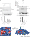
Alix Bro1 interface involved in NC binding plays a critical role in virus release. (A) The Alix Bro1 mutants with a compromised NC binding interface show a decreased ability to stimulate the release of the HIV-1 PTAP mutant. 293T cells were transfected with HIV-1 PTAP proviral DNA alone (lane 1), with WT HA-tagged Alix (lane 2), or with the indicated Alix mutants (lanes 3 to 8). Cells and viruses were collected 24 h posttransfection, and their protein content was analyzed by SDS-PAGE and Western blotting using the indicated antibodies. Release of infectious viral particles was quantified using HeLa TZM-bl assays from five independent experiments and expressed relative to WT Alix (lane 2). Error bars represent standard deviations (SD). (B) An intact NC binding interface is required for Alix function in EIAV release. 293T cells were transfected twice with Alix RNAi oligonucleotides at 36-h intervals. At the second transfection, cells were cotransfected with EIAV provirus alone (lanes 1 and 2), with an RNAi-resistant (RR) version of Alix WT (lane 3), or with the indicated mutants (lanes 4 to 6). Cells and viruses were collected 24 h posttransfection, and their protein content was analyzed by SDS-PAGE and Western blotting using the indicated antibodies. Release ratios were calculated as described in Materials and Methods from 3 independent experiments and expressed relative to WT Alix (lane 3), ±SD. (C) Alix Bro1 residues engaged in NC interaction define a new functional interface. Residues Q8, K11, K48, R51, R56, and K60 placed in the Alix Bro1 domain crystal structure (2OEW) cluster within a positively charged exposed surface (shown in red) on one side of the Bro1 domain. For reference, the Phe105 residue (required for Alix function in HIV-1 release [36]) and the CHMP4B binding interface (27) are shown in green and yellow, respectively.
Fig 4
Fig 4
Basic residues in NC are required for the interaction with the Alix Bro1 domain. (A) Schematic representation of the HIV-1 NC domain (amino acids 1 to 55). Lysine (K) and arginine (R) residues replaced with alanine in the RKI and RKII mutants are circled and underlined, respectively. (B) Mutation of basic residues at the N or C terminus of NC prevents NC-Bro1 interaction. GST, GST-NCp6, GST-NCp6RKI, or GST-NCp6RKII fusion protein was expressed in E. coli, captured on glutathione-conjugated beads, and subsequently incubated with lysates from 293T cells expressing the WT HA-Bro1 domain. Captured proteins and cell lysates were analyzed by SDS-PAGE and Western blotting using an anti-HA antibody. GST fusion proteins were visualized by Coomassie blue staining. (C) Alix function in HIV-1 release requires an intact NC domain. 293T cells were transfected with HIV-1 PTAP proviral DNA harboring a WT NC (lanes 1 and 2) or the indicated NC mutant (lanes 3 to 6) either alone (lanes 1, 3, and 5) or with WT HA-tagged Alix (lanes 2, 4, and 6). Cells and viruses were collected 24 h posttransfection, and their protein content was analyzed by SDS-PAGE and Western blotting using the indicated antibodies. Release of viral particles was quantified from two independent experiments.
Fig 5
Fig 5
RNA is involved in the Alix Bro1-NC interaction. (A) GST, GST-NCp6 (left), and GST-NCp9 (center) fusion proteins expressed in E. coli and captured on glutathione beads were incubated with lysates from 293T cells expressing HA-tagged WT Bro1 domain or APOBEC3G (right), followed by incubation with or without benzonase/nuclease. (B) GST and GST-NCp6 fusion proteins purified by glutathione beads were incubated with lysates from 293T cells expressing HA-tagged WT Bro1, followed by treatment with or without benzonase (lanes 3 and 4) or RNAse A (lanes 5 and 6). In both experiments, captured proteins and cell lysates were analyzed by SDS-PAGE and Western blotting. GST fusion proteins were visualized by Coomassie blue staining.
Fig 6
Fig 6
Schematic representation of a two-step model for Alix function in virus assembly and budding. In the first step, Alix is recruited by Gag to the plasma membrane during assembly through interaction between its V domain and the (L)YPXnL motif in p6 and Alix is thus anchored at the membrane (left). Whether NC binds Bro1 during this step or not is unclear, since it was insufficient to locate Alix to the membrane when the LYPXnL motif was disrupted (18). In the second step, we propose that the NC-Bro1 interaction occurs in the budding neck and is followed (or accompanied) by the recruitment of CHMP4B (right). Basic residues found in both NC and Bro1 proteins mediate binding, and RNA (in red) plays the role of the negatively charged factor that bridges this interaction.

Similar articles

Cited by

References

    1. Adachi A, et al. 1986. Production of acquired immunodeficiency syndrome-associated retrovirus in human and nonhuman cells transfected with an infectious molecular clone. J. Virol. 59:284–291 - PMC - PubMed
    1. Babst M, Katzmann DJ, Estepa-Sabal EJ, Meerloo T, Emr SD. 2002. Escrt-III: an endosome-associated heterooligomeric protein complex required for mvb sorting. Dev. Cell 3:271–282 - PubMed
    1. Babst M, Odorizzi G, Estepa EJ, Emr SD. 2000. Mammalian tumor susceptibility gene 101 (TSG101) and the yeast homologue, Vps23p, both function in late endosomal trafficking. Traffic 1:248–258 - PubMed
    1. Bello NF, et al. 2012. Budding of retroviruses utilizing divergent L domains requires nucleocapsid. J. Virol. 86:4182–4193 - PMC - PubMed
    1. Bieniasz PD. 2009. The cell biology of HIV-1 virion genesis. Cell Host Microbe 5:550–558 - PMC - PubMed

Publication types

MeSH terms

Substances