. 2012 Oct 5;287(41):33996-4008.
doi: 10.1074/jbc.M112.369603. Epub 2012 Aug 13.

The small GTPase RhoA regulates the contraction of smooth muscle tissues by catalyzing the assembly of cytoskeletal signaling complexes at membrane adhesion sites

Affiliations

The small GTPase RhoA regulates the contraction of smooth muscle tissues by catalyzing the assembly of cytoskeletal signaling complexes at membrane adhesion sites

Wenwu Zhang et al. J Biol Chem. .

Abstract

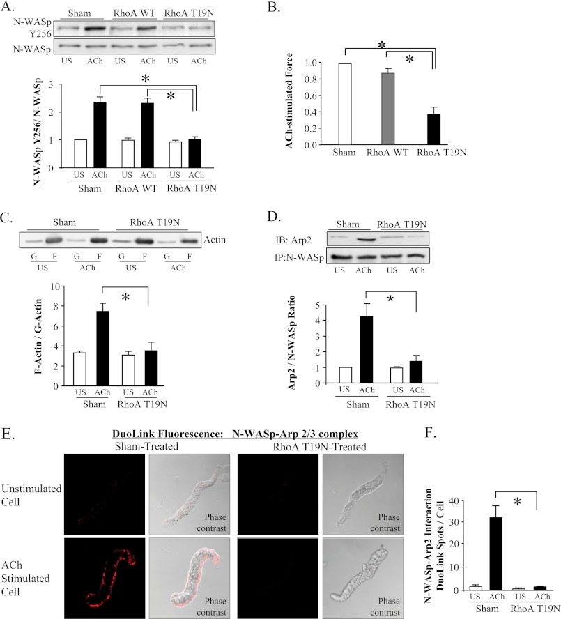
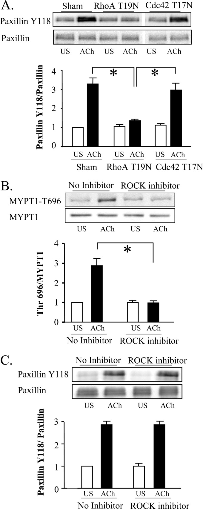
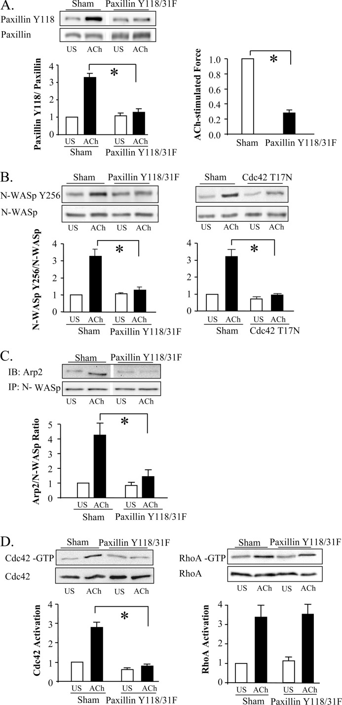
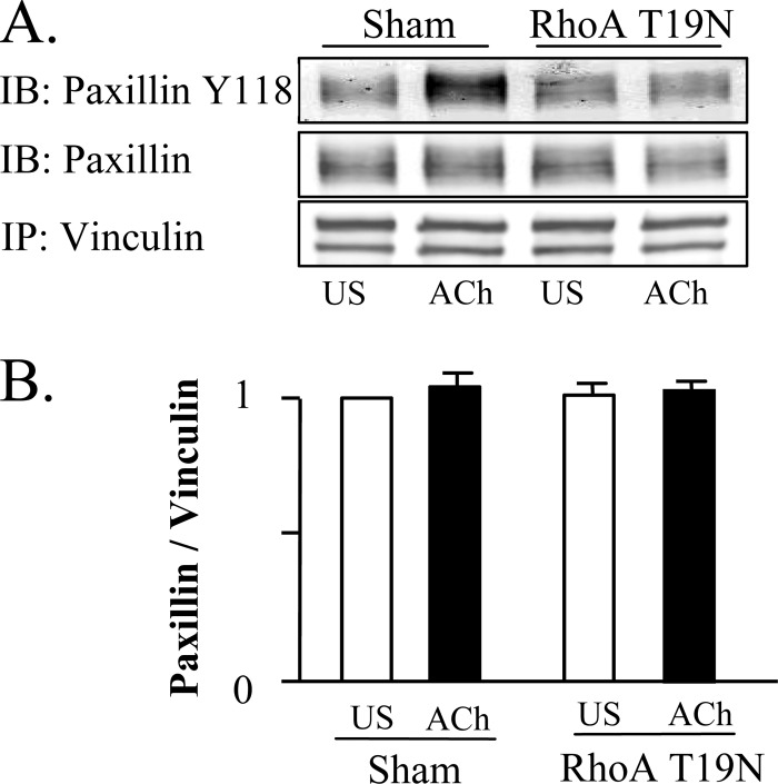
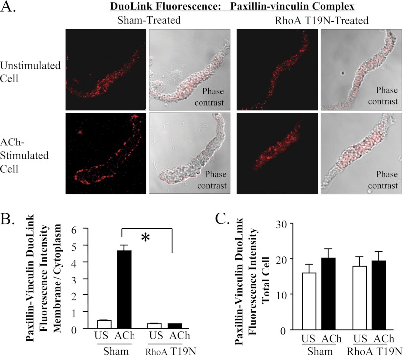
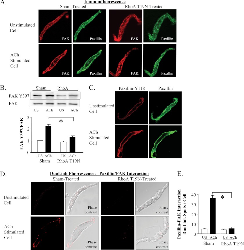
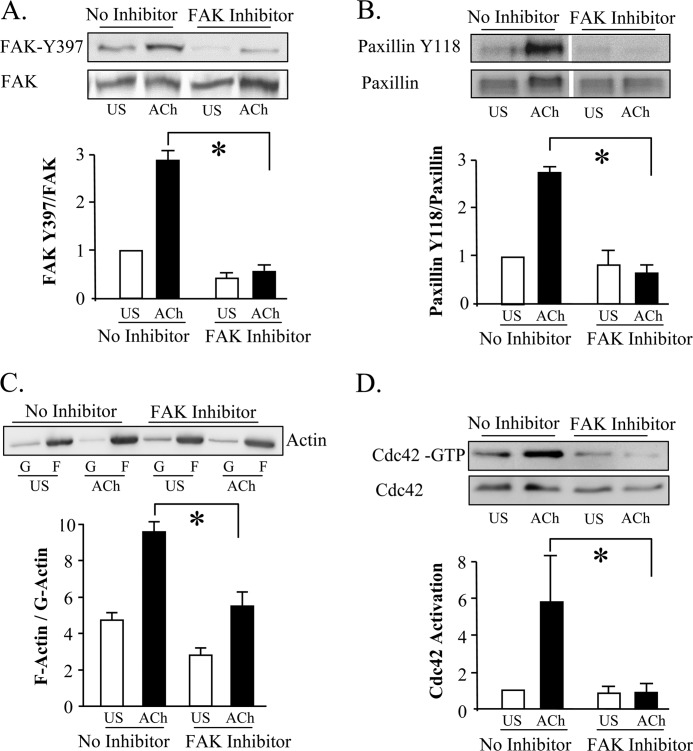
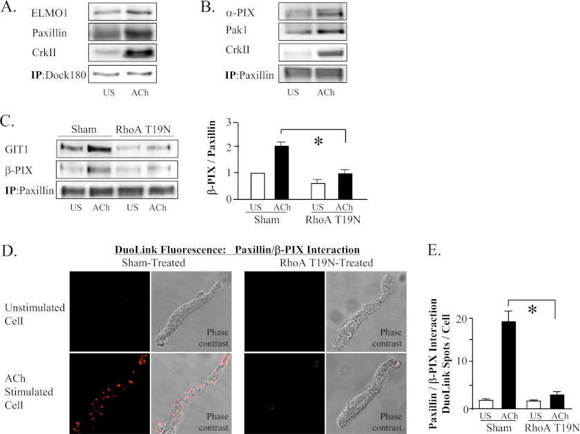
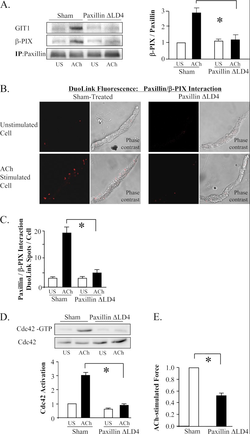
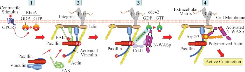
The activation of the small GTPase RhoA is necessary for ACh-induced actin polymerization and airway smooth muscle (ASM) contraction, but the mechanism by which it regulates these events is unknown. Actin polymerization in ASM is catalyzed by the actin filament nucleation activator, N-WASp and the polymerization catalyst, Arp2/3 complex. Activation of the small GTPase cdc42, a specific N-WASp activator, is also required for actin polymerization and tension generation. We assessed the mechanism by which RhoA regulates actin dynamics and smooth muscle contraction by expressing the dominant negative mutants RhoA T19N and cdc42 T17N, and non-phosphorylatable paxillin Y118/31F and paxillin ΔLD4 deletion mutants in SM tissues. Their effects were evaluated in muscle tissue extracts and freshly dissociated SM cells. Protein interactions and cellular localization were analyzed using proximity ligation assays (PLA), immunofluorescence, and GTPase and kinase assays. RhoA inhibition prevented ACh-induced cdc42 activation, N-WASp activation and the interaction of N-WASp with the Arp2/3 complex at the cell membrane. ACh induced paxillin phosphorylation and its association with the cdc42 GEFS, DOCK180 and α/βPIX. Paxillin tyrosine phosphorylation and its association with βPIX were RhoA-dependent, and were required for cdc42 activation. The ACh-induced recruitment of paxillin and FAK to the cell membrane was dependent on RhoA. We conclude that RhoA regulates the contraction of ASM by catalyzing the assembly and activation of cytoskeletal signaling modules at membrane adhesomes that initiate signaling cascades that regulate actin polymerization and tension development in response to contractile agonist stimulation. Our results suggest that the RhoA-mediated assembly of adhesome complexes is a fundamental step in the signal transduction process in response to agonist -induced smooth muscle contraction.


Figures

FIGURE 1.
FIGURE 1.
RhoA inactivation inhibits ACh induced N-WASp activation in smooth muscle tissues. A, N-WASp tyrosine 256 phosphorylation measured by immunoblot in extracts of 6 muscle tissues transfected with wild type RhoA (WT), RhoA T19N, or sham-treated. RhoA T19N significantly inhibited ACh-induced N-WASp phosphorylation relative to WT RhoA or sham-treated tissues (n = 5). B, RhoA T19N significantly inhibited ACh-induced tension development relative to WT RhoA or sham-treated tissues (n = 10). (All force measurements normalized to sham response.) C, immunoblot of soluble (G, globular) and insoluble (F, filamentous) actin in fractions from extracts of unstimulated or ACh-stimulated muscle tissues treated with RhoA T19N or with no treatment (sham). RhoA T19N significantly inhibited the increase in the F actin to G-actin ratio in response to 10−5 m ACh (n = 4). D, ACh induced a significant increase in Arp2 coimmunoprecipitation with N-WASp in sham-treated tissues but not in RhoA T19N-treated tissues (n = 4). IP: immunoprecipitate; IB, immunoblot. E, duolink in situ PLA shows the interaction of N-WASp and Arp2 in freshly dissociated differentiated canine tracheal smooth muscle cells stimulated with ACh, but not in unstimulated cells. Fluorescence and phase contrast images are shown for each cell. F, in cells from sham-treated tissues, mean DuoLink PLA spots were significantly higher in ACh-stimulated cells than in unstimulated cells (n = 27, 25). In cells from RhoA T19N-treated tissues, the mean number of DuoLink PLA spots was very small and did not change significantly with ACh-stimulation (n = 24, 32 respectively). (Cells dissociated from tissues obtained from three separate experiments.) All tissues and cells were incubated with ACh for 5 min. All values are means ± S.E. *, significantly different (p < 0.05).
FIGURE 2.
FIGURE 2.
RhoA inactivation inhibits the ACh-induced activation of cdc42 in canine tracheal smooth muscle tissues. A, activated cdc42 (cdc42-GTP) was affinity-precipitated from extracts of 3 unstimulated and 3 ACh-stimulated muscle strips, and the amount of activated cdc42 precipitated from each extract was quantified by immunoblot. Activated cdc42 was significantly higher in extracts from 10−5 m ACh stimulated sham-treated tissues than from ACh stimulated tissues expressing RhoA T19N or cdc42 T17N (n = 8). Blot shows non-contiguous lanes from a single gel. B, RhoA T19N and cdc42 T17N significantly inhibited tension development in response to ACh (n = 16). C, activated GTP-bound RhoA (RhoA-GTP) was affinity-precipitated from extracts of 6 unstimulated or ACh stimulated muscle strips and quantifitated by immunoblot. RhoA activation was significantly inhibited in tissues expressing RhoA T19N but was unaffected by the expression of cdc42 T17N (n = 6). Blot shows non-contiguous lanes from a single gel. All values are means ± S.E. *, significantly different, p < 0.05.
FIGURE 3.
FIGURE 3.
Rho regulates cdc42 and N-WASp activation by regulating paxillin phosphorylation. A, paxillin Tyr-118 phosphorylation in response to 10−5 m ACh stimulation was significantly inhibited in tissues expressing RhoA T19N, but not in tissues expressing cdc42 T17N (n = 10). Blot shows non-contiguous lanes from a single gel. B, Thr-696 phosphorylation of MYPT1, a substrate of activated ROCK, was measured by immunobot in tissue extracts from ACh stimulated tissues. MYPT1 phosphorylation was significantly lower in tissues treated with the ROCK inhibitor, H-1152P (1 μm) (n = 4). C, there are no significant effects of the ROCK inhibitor on paxillin phosphorylation in ACh-stimulated or unstimulated tissues (n = 4). All values are means ± S.E. *, significantly different, p < 0.05.
FIGURE 4.
FIGURE 4.
Paxillin tyrosine phosphorylation is required for cdc42 and N-WASP activation, but not for RhoA activation. A, increase of paxillin Tyr-118 phosphorylation in response to 10−5 m ACh was significantly lower in tissues expressing paxillin Y118/Y31F (n = 5). Blot shows non-contiguous lanes from a single gel. Paxillin Y118/31F significantly inhibited tension development relative to sham-treated tissues (n = 8). B, expression of paxillin Y118/31F (n = 4) or cdc42 T17N (n = 5) significantly inhibited N-WASp tyrosine 256 phosphorylation in response to ACh stimulation. C, increase in co-immunoprecipitation of Arp2 with N-WASp in response to ACh stimulation was significantly inhibited in tissues expressing paxillin Y118/31F (n = 3). Blot show non-contiguous lanes from a single gel. D, immunoblots against activated GTP-bound RhoA or GTP-bound cdc42 affinity-precipitated from extracts of 4 sham or 4 paxillin Y118/31F-treated muscle strips. Expression of paxillin Y118/31F did not significantly affect RhoA activation in response to 10−5 m ACh (n = 6), but it significantly inhibited cdc42 activation (n = 4). All values are means ± S.E. *, significantly different (p < 0.05).
FIGURE 5.
FIGURE 5.
The association between paxillin and vinculin is unaffected by ACh stimulation or RhoA inhibition. A and B, vinculin was immunoprecipitated from extracts from sham-treated and RhoA-treated muscles and immunoprecipitates were blotted for vinculin, paxillin, and paxillin Tyr-118. There were no significant differences between unstimulated and ACh-stimulated tissues, or between sham and RhoA T19N-treated tissues in the amount of paxillin that coprecipitated with vinculin (n = 4). Phospho-paxillin at Tyr-118 increased significantly in vinculin immunoprecipitates from 10−5 m ACh stimulated in sham-treated tissues, but not in RhoA T19N-treated tissues. All values are means ± S.E. *, significantly different (p < 0.05).
FIGURE 6.
FIGURE 6.
Rho regulates the paxillin-vinculin complex recruitment to the cell membrane in response to contractile stimulation. A, Duolink PLA was used to evaluate the interaction of paxillin and vinculin in freshly dissociated cells stimulated with 10−4 m ACh for 5 min or left unstimulated. DuoLink spots were observed throughout the cytoplasm of unstimulated cells but primarily at the cell periphery of ACh stimulated cells from sham treated tissues. In cells from RhoA T19N-treated tissues, DuoLink spots were distributed throughout the cytoplasm of both unstimulated and ACh-stimulated cells. B, localization of paxillin-vinculin complex was quantified by calculating ratio of fluorescence intensity between the cell periphery and the cell interior. (See supplemental Fig. S1 for details.) In cells from sham-treated tissues, ACh stimulation significantly increased the membrane to cytoplasm ratio. A significant increase in the membrane to cytoplasm ratio in response to ACh stimulation was not observed in cells treated with RhoA T19N (n = 14–23). C, there were no significant differences in the total cellular fluorescence intensity of paxillin-vinculin complexes in cells from sham-treated tissues and RhoA T19N-treated tissues with or without ACh (n = 14–23). All values are means ± S.E. *, significantly different, p < 0.05.
FIGURE 7.
FIGURE 7.
The phosphorylation of paxillin occurs at the cell membrane and requires the RhoA-dependent recruitment of FAK. A, cells freshly dissociated from sham-treated or RhoA T19N treated muscle tissues were stimulated with 10−4 m ACh or left unstimulated, fixed, and double-stained for FAK and paxillin. Both proteins were distributed throughout the cytoplasm of unstimulated cells and localized to the cell membrane of ACh-stimulated cells. Neither FAK nor paxillin significantly increased at the cell membrane in response to ACh stimulation in cells dissociated from RhoA T19N-treated tissues. B, FAK phosphorylation at tyrosine 397 was significantly increased by ACh in sham-treated tissues but not RhoA T19N-inhibited tissues (n = 11). C, Tyr-118 paxillin was observed at the cell membrane of both unstimulated and ACh-stimulated cells, but the intensity of paxillin Tyr-118 phosphorylation was higher in ACh-stimulated cells. D and E, interaction of paxillin and FAK in freshly dissociated tracheal smooth muscle cells was detected using the Duolink PLA. In cells from sham-treated tissues, few or no spots are detected in unstimulated cells; whereas many spots are seen at the membrane of the ACh-stimulated cell. The total number of DuoLink spots was significantly higher in ACh-stimulated smooth muscle cells than in unstimulated cells (cells from 5 separate experiments, n = 34, 30). In cells from RhoA T19N-treated smooth muscle tissues, few or no spots were detected in unstimulated cells or ACh-stimulated cells. Cells from three separate experiments, (n = 27, 15). All values are means ± S.E. *, significantly different (p < 0.05).
FIGURE 8.
FIGURE 8.
The inhibition of FAK prevents ACh-induced cdc42 activation and actin polymerization. A and B, treatment of tracheal smooth muscle tissues with 30 μm FAK inhibitor, FP 573228, inhibits phosphorylation of the FAK activation site Tyr-397 and paxillin phosphorylation at Tyr-118 in response to 10−5 m ACh (n = 4). Blot of paxillin phosphorylation shows non-contiguous lanes from a single gel C, FAK inhibitor, FP 573228, significantly inhibited the increase in the F actin to G-actin ratio in response to 10−5 m ACh (n = 4). Immunoblot of soluble (G, globular) and insoluble (F, filamentous) actin in fractions from extracts of unstimulated or ACh-stimulated muscle tissues treated with FAK inhibitor or with no inhibitor. D, treatment with FAK inhibitor, FP 573228 inhibits ACh- induced cdc42 activation (n = 3). For each assay, activated cdc42 (cdc42-GTP) was affinity-precipitated from extracts of 2 unstimulated and 2 ACh-stimulated muscle strips, and the amount of activated cdc42 precipitated from each extract was quantified by immunoblot.
FIGURE 9.
FIGURE 9.
RhoA activation regulates the association of paxillin with cdc42 GEF. A, co-immunoprecipitation of ELMO1, paxillin, and CrkII with DOCK180 increased in ACh-stimulated tissues. Result typical of six independent experiments. B, co-immunoprecipitation of α-PIX, PAK1 and CrkII with paxillin increased after contractile stimulation with ACh. Result typical of four independent experiments. C, co-immunoprecipitation of GIT1 and β-PIX with paxillin increased in ACh-stimulated tissues and was significantly inhibited in tissues expressing RhoA T19N (n = 4). D, Duolink PLA reveals interaction between β-PIX and paxillin at the membrane of muscle cells after contractile stimulation, whereas they are not observed in unstimulated muscle cells. RhoA T19N inhibited the formation of paxillin/β-PIX complexes. E, in sham-treated tissues, the mean number of DuoLink PLA spots was significantly higher in ACh-stimulated cells than in unstimulated cells. (Cells dissociated from tissues from 3 separate experiments (n = 19, 18 cells.) In cells from RhoA T19N-treated tissues, the mean number of DuoLink PLA spots was small in both unstimulated and ACh-stimulated cells and was not significantly different (n = 18). All values are means ± S.E. *, significantly different (p < 0.05).
FIGURE 10.
FIGURE 10.
The association of paxillin with β-PIX regulates cdc42 activation. A, co-immunoprecipitation of GIT1 and β-PIX with paxillin was significantly inhibited in tissues expressing paxillin ΔLD4 (n = 4). B, Duolink PLA reveals interaction between β-PIX and paxillin. Paxillin ΔLD4 inhibited the formation of paxillin/β-PIX complexes. C, in sham-treated tissues, the mean number of DuoLink PLA spots was significantly higher in ACh-stimulated cells than in unstimulated cells. (Cells dissociated from tissues from 3 separate experiments (n = 19, 18 cells.)) In cells from paxillin ΔLD4-treated tissues, the mean number of DuoLink PLA spots was small in both unstimulated and ACh-stimulated cells and was not significantly different (n = 18). All values are means ± S.E. *, significantly different (p < 0.05). D, expression of paxillin ΔLD4 inhibits ACh-induced cdc42 activation. (n = 4). For each assay, activated cdc42 (cdc42-GTP) was affinity-precipitated from extracts of 2 unstimulated and 2 ACh-stimulated muscle strips. E, paxillin ΔLD4 significantly inhibited tension development relative to sham-treated tissues (n = 8). All values are means ± S.E. *, significantly different (p < 0.05).
FIGURE 11.
FIGURE 11.
Model for proposed mechanism for the regulation of ACh induced RhoA activation on the assembly of an adhesome signaling complex in ASM. 1, ACh stimulation activates RhoA, which induces the independent recruitment of paxillin-vinculin complexes and FAK to cell adhesomes; 2, FAK and paxillin interact at the adhesome and activated FAK induces the phosphorylation of paxillin, which remains bound to activated vinculin; 3, phosphorylation of paxillin facilitates the formation of a complex containing paxillin and Crk II with DOCK180 and PIX GEFs. This complex induces the activation of cdc42. 4, cdc42 activation catalyzes the activation of N-WASp, which interacts with the Arp2/3 complex to induce actin polymerization in the cortical region of the smooth muscle cell. This enables tension generation by the smooth muscle contractile apparatus.

Similar articles

Cited by

References

    1. Allen W. E., Jones G. E., Pollard J. W., Ridley A. J. (1997) Rho, Rac, and Cdc42 regulate actin organization and cell adhesion in macrophages. J. Cell Sci. 110, 707–720 - PubMed
    1. Burridge K., Wennerberg K. (2004) Rho and Rac take center stage. Cell 116, 167–179 - PubMed
    1. DeMali K. A., Wennerberg K., Burridge K. (2003) Integrin signaling to the actin cytoskeleton. Curr. Opin. Cell Biol. 15, 572–582 - PubMed
    1. Ridley A. J. (1999) Rho family proteins and regulation of the actin cytoskeleton. Prog. Mol. Subcellul. Biol. 22, 1–22 - PubMed
    1. Ridley A. J. (2006) Rho GTPases and actin dynamics in membrane protrusions and vesicle trafficking. Trends Cell Biol. 16, 522–529 - PubMed

Publication types

MeSH terms

LinkOut - more resources