

Validation of Polo-like kinase 1 as a therapeutic target in pancreatic cancer cells
- PMID: 22892842
- PMCID: PMC3469479
- DOI: 10.4161/cbt.21412
Validation of Polo-like kinase 1 as a therapeutic target in pancreatic cancer cells
Abstract
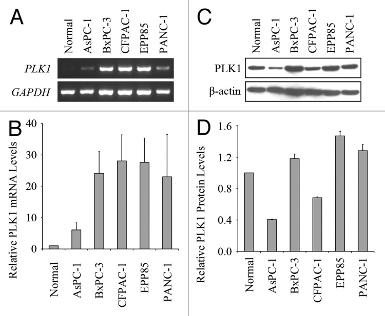
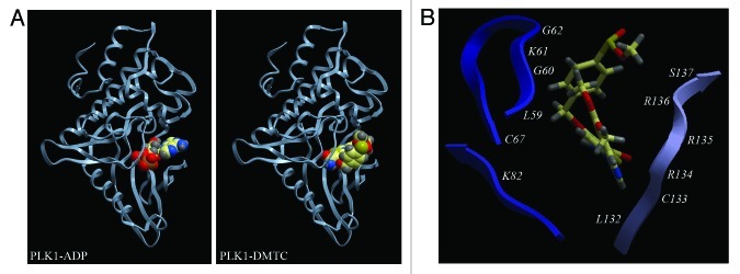
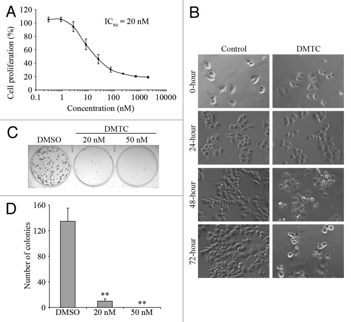
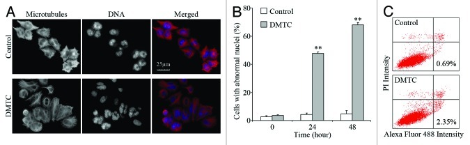
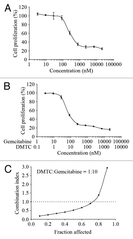
Polo-like kinase 1 (PLK1) is a serine/threonine protein kinase and plays a critical role in mitosis. PLK1 has also been regarded as a valuable target for cancer treatment, and several PLK1 inhibitors are currently undergoing clinical investigations. In this study, our data show that the expression level of PLK1 is upregulated in human pancreatic cancer cells. Molecular modeling studies indicate that DMTC inhibits PLK1 activity through competitive displacement of ATP from its binding pocket. Our data further show that DMTC suppresses the proliferation of pancreatic cancer cells and induces the formation of multinucleated cells, ultimately resulting in apoptosis. In addition, combination index analysis demonstrates that DMTC acts synergistically with the chemotherapeutic drug gemcitabine in inhibiting the proliferation of pancreatic cancer cells. These results thus suggest a potential of using PLK1 inhibitors for the treatment of pancreatic cancer.
Figures
Similar articles
-
RNA interference-mediated silencing of the polo-like kinase 1 gene enhances chemosensitivity to gemcitabine in pancreatic adenocarcinoma cells.J Cell Mol Med. 2008 Dec;12(6A):2334-49. doi: 10.1111/j.1582-4934.2008.00257.x. Epub 2008 Feb 5. J Cell Mol Med. 2008. PMID: 18266952 Free PMC article.
-
Drug sensitivity and resistance testing identifies PLK1 inhibitors and gemcitabine as potent drugs for malignant peripheral nerve sheath tumors.Mol Oncol. 2017 Sep;11(9):1156-1171. doi: 10.1002/1878-0261.12086. Epub 2017 Jul 5. Mol Oncol. 2017. PMID: 28556483 Free PMC article.
-
Plk1 inhibition enhances the efficacy of gemcitabine in human pancreatic cancer.Cell Cycle. 2016;15(5):711-9. doi: 10.1080/15384101.2016.1148838. Cell Cycle. 2016. PMID: 26890815 Free PMC article.
-
Polo-like Kinase 1 as an emerging drug target: structure, function and therapeutic implications.J Drug Target. 2021 Feb;29(2):168-184. doi: 10.1080/1061186X.2020.1818760. Epub 2020 Sep 14. J Drug Target. 2021. PMID: 32886539 Review.
-
Non-mitotic functions of polo-like kinases in cancer cells.Biochim Biophys Acta Rev Cancer. 2021 Jan;1875(1):188467. doi: 10.1016/j.bbcan.2020.188467. Epub 2020 Nov 7. Biochim Biophys Acta Rev Cancer. 2021. PMID: 33171265 Review.
Cited by
-
Hyaluronate-coated perfluoroalkyl polyamine prodrugs as bioactive siRNA delivery systems for the treatment of peritoneal cancers.Biomater Adv. 2022 May;136:212755. doi: 10.1016/j.bioadv.2022.212755. Epub 2022 Mar 17. Biomater Adv. 2022. PMID: 35813988 Free PMC article.
-
Liver kinase B1 regulates the centrosome via PLK1.Cell Death Dis. 2014 Apr 10;5(4):e1157. doi: 10.1038/cddis.2014.135. Cell Death Dis. 2014. PMID: 24722282 Free PMC article.
-
Disulfiram, a drug widely used to control alcoholism, suppresses the self-renewal of glioblastoma and over-rides resistance to temozolomide.Oncotarget. 2012 Oct;3(10):1112-23. doi: 10.18632/oncotarget.604. Oncotarget. 2012. PMID: 23047041 Free PMC article.
-
PLK1, A Potential Target for Cancer Therapy.Transl Oncol. 2017 Feb;10(1):22-32. doi: 10.1016/j.tranon.2016.10.003. Epub 2016 Nov 24. Transl Oncol. 2017. PMID: 27888710 Free PMC article. Review.
-
Impact of RUNX2 on drug-resistant human pancreatic cancer cells with p53 mutations.BMC Cancer. 2018 Mar 20;18(1):309. doi: 10.1186/s12885-018-4217-9. BMC Cancer. 2018. PMID: 29558908 Free PMC article. Review.
References
-
- Sunkel CE, Glover DM. polo, a mitotic mutant of Drosophila displaying abnormal spindle poles. J Cell Sci. 1988;89:25–38. - PubMed
-
- Burkard ME, Randall CL, Larochelle S, Zhang C, Shokat KM, Fisher RP, et al. Chemical genetics reveals the requirement for Polo-like kinase 1 activity in positioning RhoA and triggering cytokinesis in human cells. Proc Natl Acad Sci USA. 2007;104:4383–8. doi: 10.1073/pnas.0701140104. - DOI - PMC - PubMed
Publication types
MeSH terms
Substances
LinkOut - more resources
Full Text Sources
Medical
Miscellaneous